Parasitology. Cestodes
 Call Kai
 Learn
Learn Practice Test
 Spaced Repetition
 Match
Match Flashcards
 Knowt Play
1/87

Earn XP
Description and Tags
Cestodes. Intestinal Tapeworms. Tissue Tapeworms
Name | Mastery | Learn | Test | Matching | Spaced | Call with Kai | Chat |
|---|
No analytics yet
Send a link to your students to track their progress
88 Terms
General Characteristics
flat and ribbon-like
hermaphroditic
no digestive track - absorbed nutrients from host food
produce little pathology
General Morphology
scolex
neck
proglottids
Scolex
orientation of worm
biological holdfast
not for support or feeding
hooks, rostellum, suckers
Neck
ares for growth of worm
area where proglottids bud off
undifferentiated stem cells
Proglottids
segments that are sexually immature, sexually mature, or gravid
gravid = are egg-filled
Strobilia
body of tapeworm minus scolex
chain of proglottids
How to tapeworms absorb nutrients?
across tegument
pinocytosis
diffusion/facilitated diffusion
active transport
Microtriches
microvilli that covers the tegument of cestodes and assist with nutrient absorption
Dwarf tapeworm
Hymenolepis nana
Hymenolepis nana Morphology
~1-3 inches
scolex - 4 suckers and hooks on rostellum
proglottids disintegrate in gut
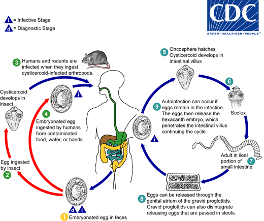
Hymenolepis nana Life Cycle (direct)
ingestion of mouse pills or embryonated eggs (contaminated food/water)
oncosphere hatches→ cysticercoid
adults develop in small intestine
gravid proglottis disintegrates → eggs in gut
eggs leave through feces
Hymenolepis nana Life Cycle (indirect)
egg is ingested by insect
cysticerrcoid in insect
human/rodent host consume insect
(normal cycle continuous in host)
Hymenolepis nana Life Cycle (auto infection)
occurs if eggs remain and hatch in intestine
Hymenolepis nana Epidemiology
person to person contact
common in children
mouse pills infect humans or beetles
tenebrious beetle can be intermediate
Does Hymenolepiis nana need an intermediate host?
No
Hymenolepis nana Pathology
asymptomatic or mild symptoms
irritation to gut wall
allergic reaction to worm metabolites (diarrhea, headache, nervousness, dizzy)
Hymenolepis nana Diagnosis
eggs in stool
Hymenolepis nana Prevention
personal hygiene
mass chemotherapy
What is the reservoir host for Hymenolepis nana?
mice
When is Hymenolepis nana infective?
egg stage
Hymenolepis nana (LC pic)
Taenia saginata has _____ pathology, while Taenia solium has _____ pathology
little, high
Taenia saginata has _____ and Taenia sodium has _____
no hooks, hooks
Beef tapeworm
Taenia saginata
Taenia saginata Morphology
5-10 meters (but can grow up to 25)
scolex - 4 suckers, no hooks
proglottids - mobile and whole in stool; more than 15 uterine branches; migrate out of anus
Taenia saginata Life Cycle
cattle infected by contaminated vegetation
eggs hatch into oncospheres
oncospheres migrate to muscle
oncosphere → cyctercerci in muscle
humans consume raw/undercooked meat
scolex attaches to intestine
adult worm in small intestine
eggs or proglottids in feces
Taenia saginata Epidemiology
in feed lots → infected by lot workers
in grazing areas → polluted by sewage
endemic in all beef eating areas
Taenia saginata Pathology
little pathology; no weight loss
potential intestinal blockage
reaction to worm metabolites → diarrhea
Taenia saginata Diagnosis
proglottids in stool
Taenia saginata Prevention
inspect beef
cook beef @ 56°C → denatures protein
no sewage in grazing areas
import canned, not fresh beef
When is Taenia saginata infective for humans?
cysticerci stage
When is Taenia saginata infective for cattle?
egg stage
What is the intermediate host for Taenia saginata?
cattle/cows
Cysticercus bovis
intermediate larval stage of Taenia saginata in cattle; not in humans

Taenia saginata (LC pic)
Pork Tapeworm
Taenia solium
Taenia solium Morphology
8-10 meters
scolex - 4 suckers; hooks
proglottids - less than 15 uterine branches
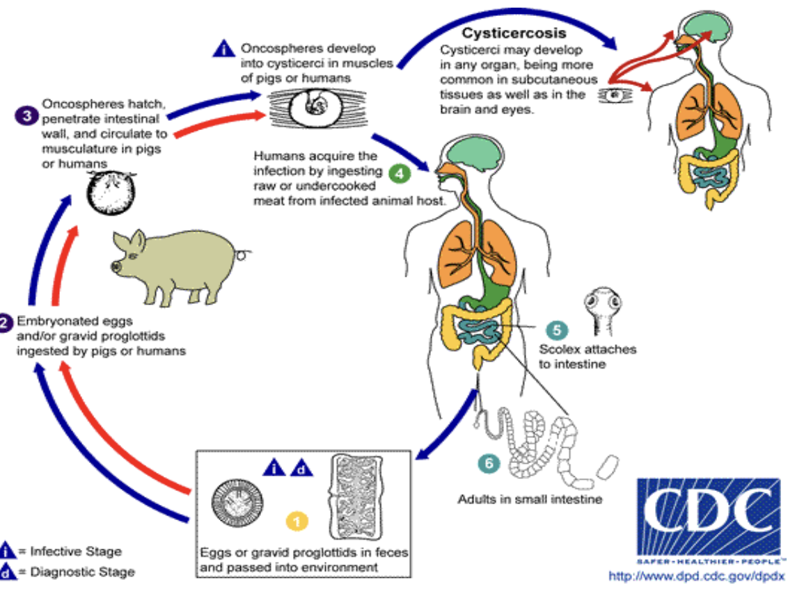
Taenia solium Life Cycle
humans ingest raw/undercooked meat
scolex attaches to intestine
adults in small intestine
eggs/proglottids in feces
pigs ingest contaminated food/water
oncospheres hatch and migrate to muscle
oncosphere → cysticerci in pig muscle
*can cause cysticercosis in humans if eggs ingested*
Taenia solium Epidemiology
areas with high pork consumption
areas with lax inspection
Taenia solium Pathology (intestinal)
asymptomatic - mild symptoms
allergic reaction to worm metabolites → diarrhea
Taenia solium Pathology (cysticercus cellulosae)
encystment → calcification → dead end (intramuscular)
no encystment → grapelike cluster → Jacksonian epilepsy (brain)
floater in vitreous humor, no encystment → enters sub retinal space (eye)
death of organism → inflammation → loss of eye (eye)
encystment (soft tissues)
Cysticercus cellulosae
ingestion of eggs → onchosphere hatches in gut → enters circulation → travel to any organ
Taenia solium Diagnosis
proglottids in feces for adult worms (intestinal)
X-ray, CT scans, MRI (cysticercus cellulosae)
Taenia solium Prevention
proper disposal of feces
cook/freeze pork
inspect pork
What type of host are humans for Taenia solium?
definitive - consumption of cysticerci in uncooked/raw pork
accidentally intermediate - autoinfection or consumption of eggs
What is the intermediate host for Taenia solium?
pigs
When is Taenia solium infective for pigs?
egg stage
When is Taenia solium infective for humans?
egg stage (cysticercus cellulosae)
cysticerci stage (taeniasis)
Taenia solium (LC pic)
Fish tapeworm
Diphyllobothrium latum
Diphyllobothrium latum Morphology
5-10 meters
long lived (25-30 years)
scolex - bothrium (leaf-like suckers)
Diphyllobothrium latum Life Cycle
unembryonated eggs in feces
embryonate in water
eggs hatch into coracidia
copepod ingests coracidium
coracidium → procercoid larvae in body cavity
infected copepod eaten by small fish
procercoid → plerocercoid larvae in small fish
small infected fish eaten by larger predator
humans consume undercooked/raw fish
adults in small intestine
proglottids release immature eggs
unembryonated eggs in feces
Diphyllobothrium latum Life Cycle Stages
eggs
coracidium
procercoid larvae
plerocercoid larvae
adult worm
Diphyllobothrium latum Epidemiology
temperate regions with fresh water fish consumption
Great Lakes & Pacific Northwest
Finland, Scandinavia, Baltic region
Diphyllobothrium latum Pathology
mild attachment damage
allergic reaction to metabolites
has affinity for vitamin B-12 → pernicious anemia
Diphyllobothrium latum Diagnosis
eggs in stool
Diphyllobothrium latum Prevention
cook fish
prevent sewage from contaminating water
When is Diphyllobothrium latum infective to humans?
plerocercoid stage
What are the intermediate hosts for Diphyllobothrium latum?
copepods → small fish → large predatory fish

Diphyllobothrium latum (LC pic)
Unilocular Hydatid Disease
Echinococcus granulosus
Echinococcus granulosus Morphology
very small - ¼ inch long
scolex - neck - 3 proglottids (immature, mature, gravid)
Enchinococcus granulosus Life Cycle
embryonated eggs in feces
sheep (or humans) ingest eggs
oncosphere hatches
penetrate intestinal wall and migrate
hydatid cyst in organs (heart, lung, etc)
dog ingests cysts from sheep meat/organs
cyst → protoscolex
scolex attaches to intestine
adults in small intestine
eggs in feces
Echinococcus granulosus Life Cycle Stages
egg
oncosphere
hydatid cyst
protoscolex
adult worm
Enchinococcus granulosus Epidemiology
any place with sheep
Echinococcus granulosus Pathology
cysts expand uniformly
mechanical damage & pressure atrophy (liver)
cysts in brain/heart valves/lungs
allergic reaction to hydatid fluid → anaphylaxis
Echinococcus granulosus Diagnosis
observation
X-ray, CT scan, MRI
Echinococcus granulosus Prevention
treat dogs
cook sheep organs before feeding to dogs
What is the definitive host of Echinococcus granulosus?
dogs
What is the intermediate host of Echinococcus granulosus?
sheep
What type of host are humans to Echinococcus granulosus?
accidental intermediate hosts; dead-end hosts
When is Echinococcus granulosus infective to definitive host?
cyst stage
When is Echinococcus granulosus infective to intermediate host?
egg stage

Echinococcus granulosus (LC pic)
Alveolar hydatid disease
Echinococcus multilocularis
Echinococcus multilocularis Morphology
same as E. granulosus
very small
scolex - neck - 3 proglottids (immature - mature - gravid)
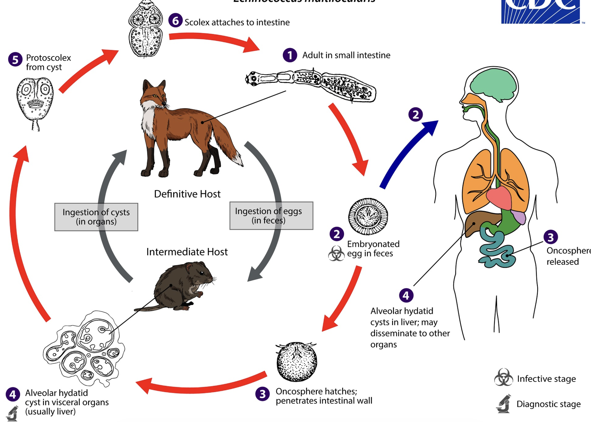
Echinococcus multilocularis Life Cycle
eggs in feces
rodents (or humans) ingest eggs
oncosphere hatches
penetrate intestinal wall and migrate
multi-chambered hydatid cyst in liver (sometimes other organs)
dog/fox ingests cysts from rodent meat/organs
cyst → protoscolex
scolex attaches to intestine
adults in small intestine
eggs in feces
Echinococcus multilocularis Epidemiology
northern hemisphere (Europe, China, Russia, Japan)
eating wild blackberries/blueberries/mushrooms contaminated with eggs
Northern U.S.
Echinococcus multilocularis Pathology
hydatid cyst that expands to create multiple chambers → multi-locular
slow - 5 to 15 years
mainly in liver, can affect other organs
cavitation → organ is replaced by cyst
Echinococcus multilocularis Diagnosis
indirect - X-ray, CT scan, MRI
liver function tests
observation
Echinococcus multilocularis Prevention
treat/control infected dogs
wash fruits
What are the intermediate hosts for Echinococcus multilocularis?
moles, voles, rodents
What are the definitive hosts for Echinococcus multilocularis?
dog, fox
What type of hosts are humans for Echinococcus multilocularis?
accidental intermediate hosts
Echinococcus multilocularis (LC pic)
Why do E. multilocularis and E. granulosus manifest differently?
multilocularis: weak/thin cyst wall → pressure makes it bulge out → multilocular
granulosus: strong/thick cyst wall → grows uniformly → unilocular
When is Echinococcus multilocularis infective to intermediate host?
egg stage
When is Echinococcus multilocularis infective to definitive host?
cyst stage