Prosthetic Heart Valves
1/45
There's no tags or description
Looks like no tags are added yet.
Name | Mastery | Learn | Test | Matching | Spaced | Call with Kai |
|---|
No analytics yet
Send a link to your students to track their progress
46 Terms
Indications for valve replacement:
valvular stenosis
valvular regurgitation
prolapse
perforated leaflet
aortic dissection w/ severe AI
2 types of prosthetic valves:
Bioprosthetic
Mechanical
Types of Bioprosthetic valves:
Auto-graft
Homograft / Allograft
Heterograft / Xenograft
Auto-graft
a bioprosthetic self-transplant valve
valve moved from one position to another within the same pt
most common is the “Ross Procedure”
Homograft / Allograft
bioprosthetic transfer from one human to another
valve is removed from another person (after death) and transplanted into the recipient
Heterograft / Xenograft
bioprosthetic transfer from animal to human
tissue from an animal of a different species
“hetero” = different
“xeno” = foreign
Types of Heterografts / Xenografts:
Porcine
Bovine
Porcine Xenograft
Bioprosthetic
a pig’s AoV is placed on stents and attached to a sewing ring
most commonly used for MV replacement
will have 3 leaflets
Manufacturers:
Hancock
Carpentier-Edwards
Intact
Bovine Hetrograft
bioprosthetic
a cow’s pericardium made into a tri-leaflet valve and mounted on stents and sewing ring
most commonly used for AoV replacement
Manufacturers:
Carpentier-Edwards
Mitroflow
Ionescu-Shiley
taken off market
Hetrograft AKA:
Xenograft
Ross Procedure
PV placed in AoV position of same pt
auto-graft used for high pressure side
homograft used for low pressure side
usually performed on children
must be <40 y/o
Advantage of Ross Procedure:
pt’s valve will grow w/ rest of body
Disadvantage of Ross Procedure:
if there’s a congenital AoV deformity
PV may be deformed too
Complications of Bioprosthetic Valves:
calcification / degeneration
30% of porcine replaced after 15 years
dehiscence
“bursting open” / falling apart
regurg
perivalvular leak / regurg
stenosis
infective endocarditis
thrombus
pseudoaneurysm
ring abscess
found at annulus
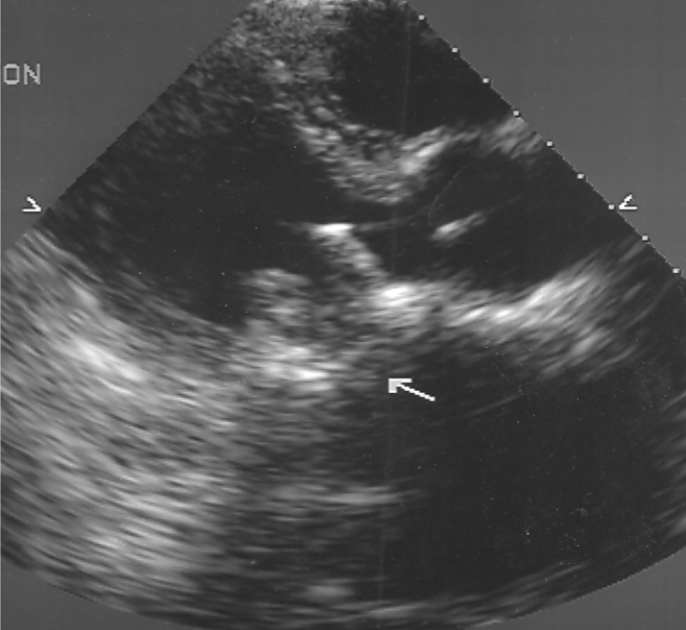
Bioprosthetic valves s/p cardiac surgery:
septal motion should be paradoxical
normal septal motion <6 months s/p cardiac surgery may indicate sig MR or AI
Bioprosthetic Valve M-Mode findings:
usually limited
used for timing
opening/closure of prosthesis
coarse flutter of leaflets
abnormal if seen
2D findings of Bioprosthetic Valves:
identify:
leaflet thickness
>3mm is abnormal
leaflet motion
thrombus
calcification
dehiscence
vegetation
evaluate LV function
Drawbacks of Mechanical Valves:
produce an audible click
increased risk of thrombosis + vegetation
using an un-natural and metal material
limited anticoagulant properties
pt required to take Coumadin for life
Benefits of Mechanical Valves:
Will last longer than the patient
Basic parts of Mechanical Valves:
ball
cage
houses / catches ball
disc
strut
arm that attaches disc to apparatus
sewing ring
sutures anchor the sewing ring of prosthesis to the native annulus
Types of Mechanical Valves:
Ball and Cage
Tilting Disc
Bileaflet Tilting Disc
Ball and Cage Valve
mechanical valve
first type implanted in humans
not used anymore
Starr-Edwards
Tilting Disc Valve
mechanical valve
single leaflet
Medtronic Hall
Omniscience
Bjork-Shiley
no longer sold in US d/t arm breakage
Bileaflet Tilting Disc Valve
90% of mechanical valves used today
St. Jude is leading manufacturer
Carbomedics
Mechanical Valve M-mode findings:
ring down artifact
Mechanical Valves 2D findings:
produce shadowing and reverberation
appearance will vary d/t:
angle of insertion by surgeon
type of valve used
TTE can be difficult
take extra time to evaluate and image
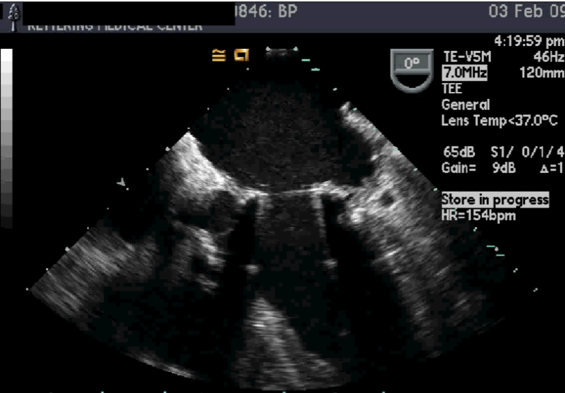
TEE Evaluation of Mechanical Valves:
method of choice
assess regurg
may be masked by prosthesis on TEE
assess anatomic detail:
LA appendange
thrombus / vege
ring abscess
pseudoaneurysm
intraoperative monitoring for:
perivalvular leak
regurg
dysfunction
Symptoms of Mechanical Valve Malfunction:
may be asymptomatic
CHF
fatigue
SOB d/t pulmonary emboli
Structural changes of Mechanical Valve Malfunction:
ball or disc may crack
“Variance”
occurs when the ball or disc loses shape and no longer fits within the struts or cage
strut’s arm has been noted to break off
Mechanical Valve Malfunction may cause:
thrombus formation
abscess formation
endocarditis
regurg
may require surgical correction
perivalvular leak
dehiscence
Thrombus formation w/ Mechanical Valve malfunction:
most common dysfunction of mechanic valves
usually located at sewing ring
can cause PE or stroke
may result in regurg or stenosis
reason single disc is no longer used
Abscess formation w/ Mechanical Valve malfunction:
may occur near the sewing ring
sonolucent center
Endocarditis w/ Mechanical Valve malfunction:
increased risk b/c valve is foreign
vegetation may form
may restrict valve closure
causes regurg
test blood once a month
“Dehiscence” w/ Mechanical Valve malfunction:
rupture of one or more of the sutures that anchor the sewing ring of prosthesis to annulus
“rocking of the valve” appearance
usually occurs soon after surgery
may be d/t infection eroding valve
may be associated w/ abscess formation
Doppler of Prosthetic Valves
prosthetic valves are mildly stenotic by nature
doppler values are different
degree of obstruction depends on:
type
size
location
baseline study w/ Dp values
important to obtain s/p surgery
usually within 30 days
make note of best trdx location for accurate d/u exams
At least 5 beats should be utilized/recorded to obtain hemodynamic Dp measurements
Prosthetic Mitral Valves
obtain optimal MV Dp signal from apex
valve clicks/slaps should be seen
determine peak velocity
>2.5 m/s = abnormal
determine peak and mean pressure gradients
mpg >10 mmHg = abnormal
determine MVA using P½T method
PHT >180 ms = abnormal
MVA <1.8 cm² = abnormal
determine presence + severity of MR
Prosthetic Aortic Valves
obtain optimal AoV Dp signal
pedoff probe
usually apical or RPS
determine peak and mean pressure gradient
ppg >45 mmHg = abnormal
mpg >15 mmHg = abnormal
determine AVA using Continuity Equation
if LVOT diameter is difficult to determine, use:
2.0 cm for M
1.8 cm for F
not accurate w/ co-existent AI
some labs may not use AVA
use only peak/ mean grad’s
Prosthetic Tricuspid Valves
tissue valves most com’ly used
reduces incidence of thrombosis + pulmonary emboli risk
Assess for complications
Prosthetic Tricuspid Valve Doppler Findings:
determine TV Area
perform as would MVA
find peak and mean pressure gradient
determine presence and severity of TR
Prosthetic Pulmonic Valves
rarely replaced
bioprosthetic used
assess for complications
Prosthetic Pulmonic Valve Doppler Findings:
determine peak velocity
>2.5 m/s = abnormal
determine presence + severity of PI
Cloth Covered Rings
Form of Ring Prosthesis
used for valve repair w/o replacement
usually b/c annulus has been stretched out
usually Teflon / Dacron material
always note repair / replacement on Hx
What position are Cloth Covered Rings most commonly used in?
MV / TV position
ring is sutured into position + will add support to annulus
Cloth Covered Rings Echo appearance:
may be similar to MAC
MAC rarely affects AMVL
TAVR / TAVI
transcatheter aortic valve replacement
transcatheter aortic valve implantation
newest procedure for valve replacement
Post TAVR Auscultation
“Honking” murmur → like a goose