Looks like no one added any tags here yet for you.
Internal surfaces are covered by what two types of membrane?
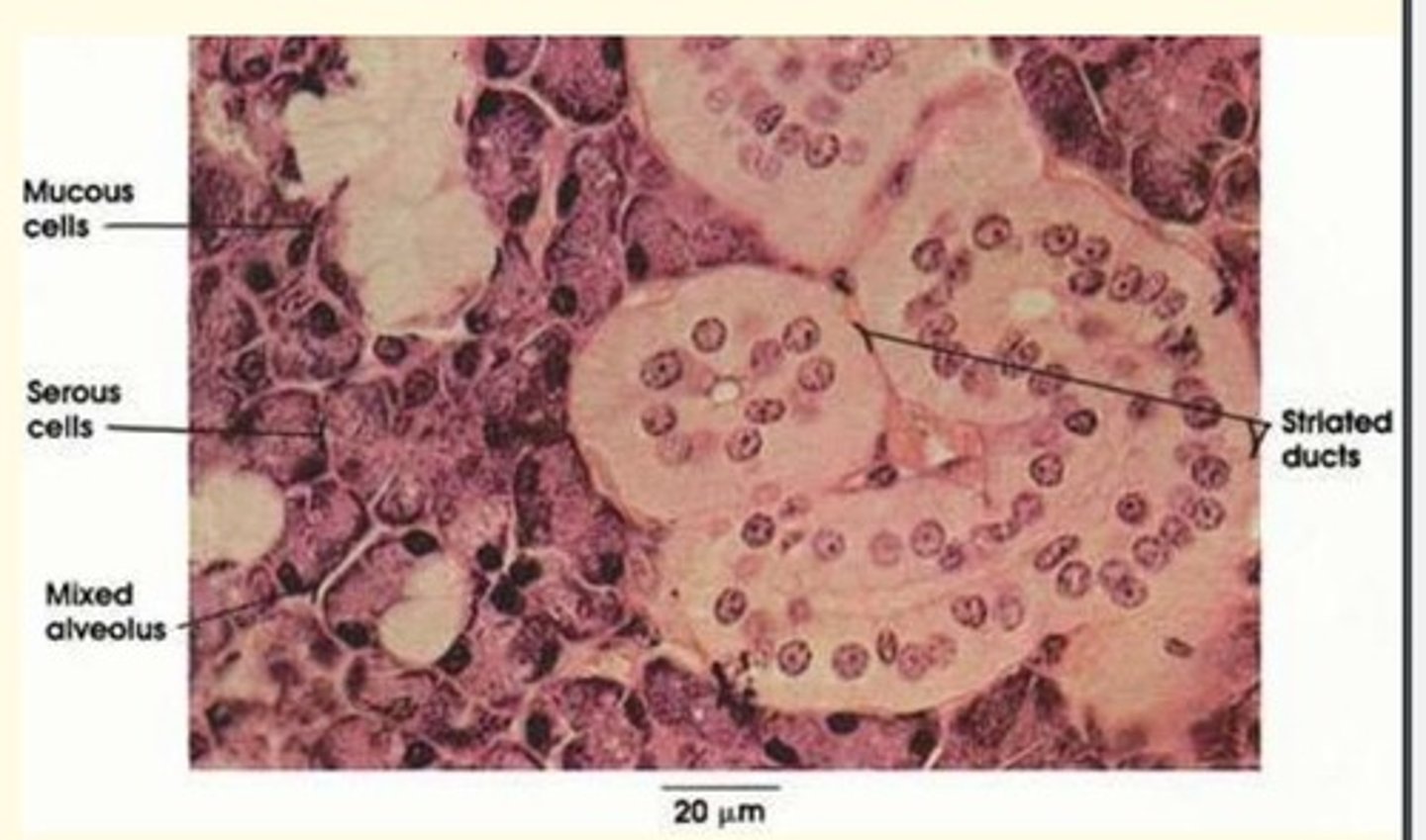
- Mucous
- Serous
Mucous membranes
Line internal tubes that open to the exterior
Examples of locations of mucous membrane
- Alimentary tract (GI tract)
- Respiratory tract
- Urinary tract
- Reproductive tract
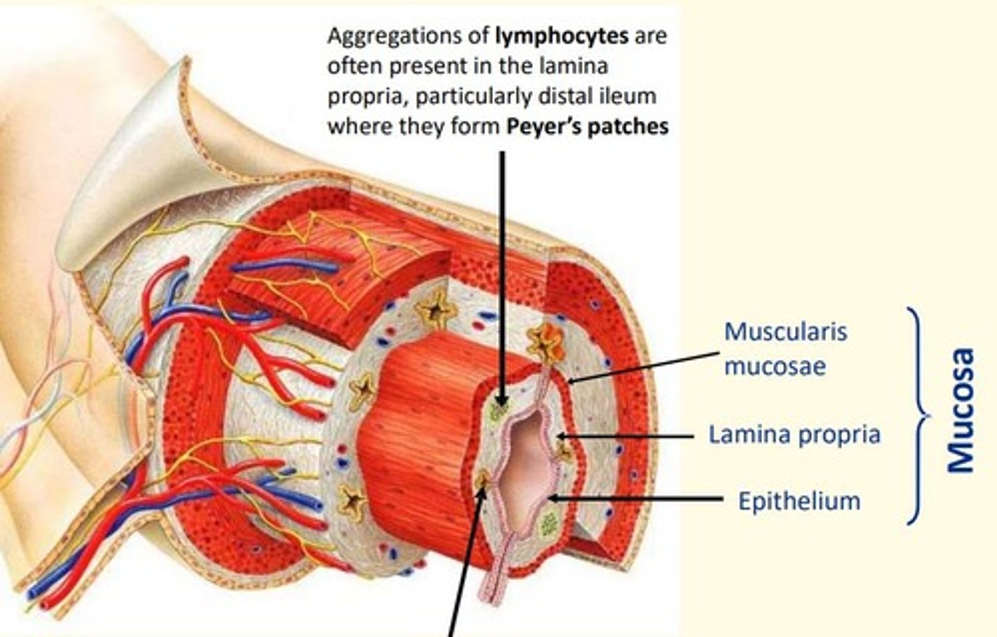
The layers of a mucous membrane
1. epithelial layer
2. connective tissue layer (lamina propria)
3. smooth muscle layer (muscularis mucosae)
Serous membranes
Thin, two-part membranes that line certain closed body cavities (spaces that do not open to the exterior)
Organs that do not move freely e.g., the oesophagus, are surrounded by a connective tissue layer (an ____) that adheres the organ to the surrounding structures
adventitia
During embryonic development, which three organs develop next to a bag-like cavity into which they invaginate?
- Heart
- Lungs
- Digestive tract
Which three organs are enveloped by serous membranes?
- Heart = pericardial sac
- Lungs = pleural sacs
- Digestive tract = peritoneum
Peritoneum
membrane that lines the abdominal cavity
pleural sacs
surround the lungs
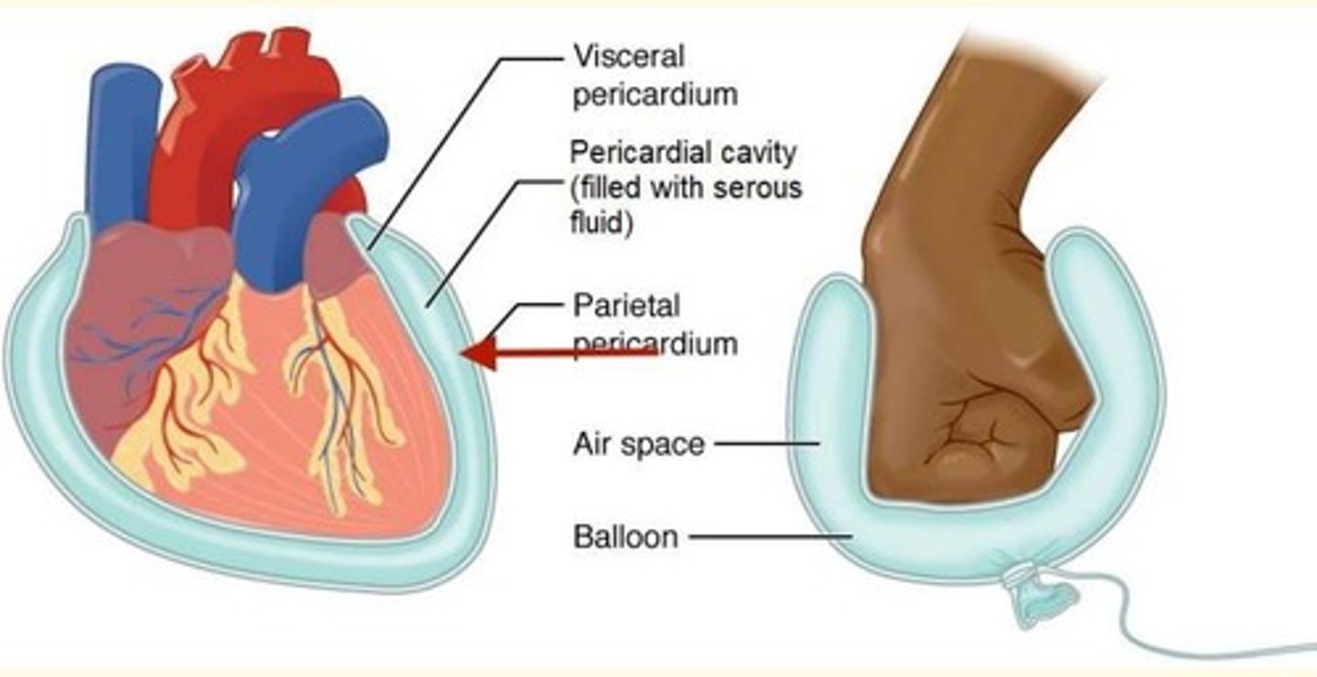
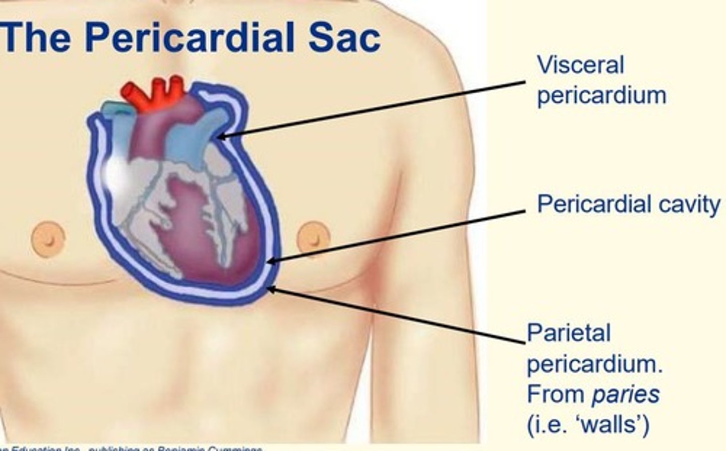
pericardial sac
surrounds the heart and helps prevent overfilling.
Serous membranes surrounding the peritoneum, pleural sacs and pericardial sacs secrete a ____ fluid that promotes relatively friction-free movement of the structures that they surround
lubricating
The layers within the pericardial sac
1. Visceral pericardium = inner
2. Pericardial cavity = middle
3. Parietal pericardium = outer
Most digestive organs lie within the ____ cavity
abdominopelvic
Mesentery
Double layer of peritoneum extending from the posterior body wall, supplying blood vessels, lymphatics and nerves to the intestines. It is also a site of fat storage
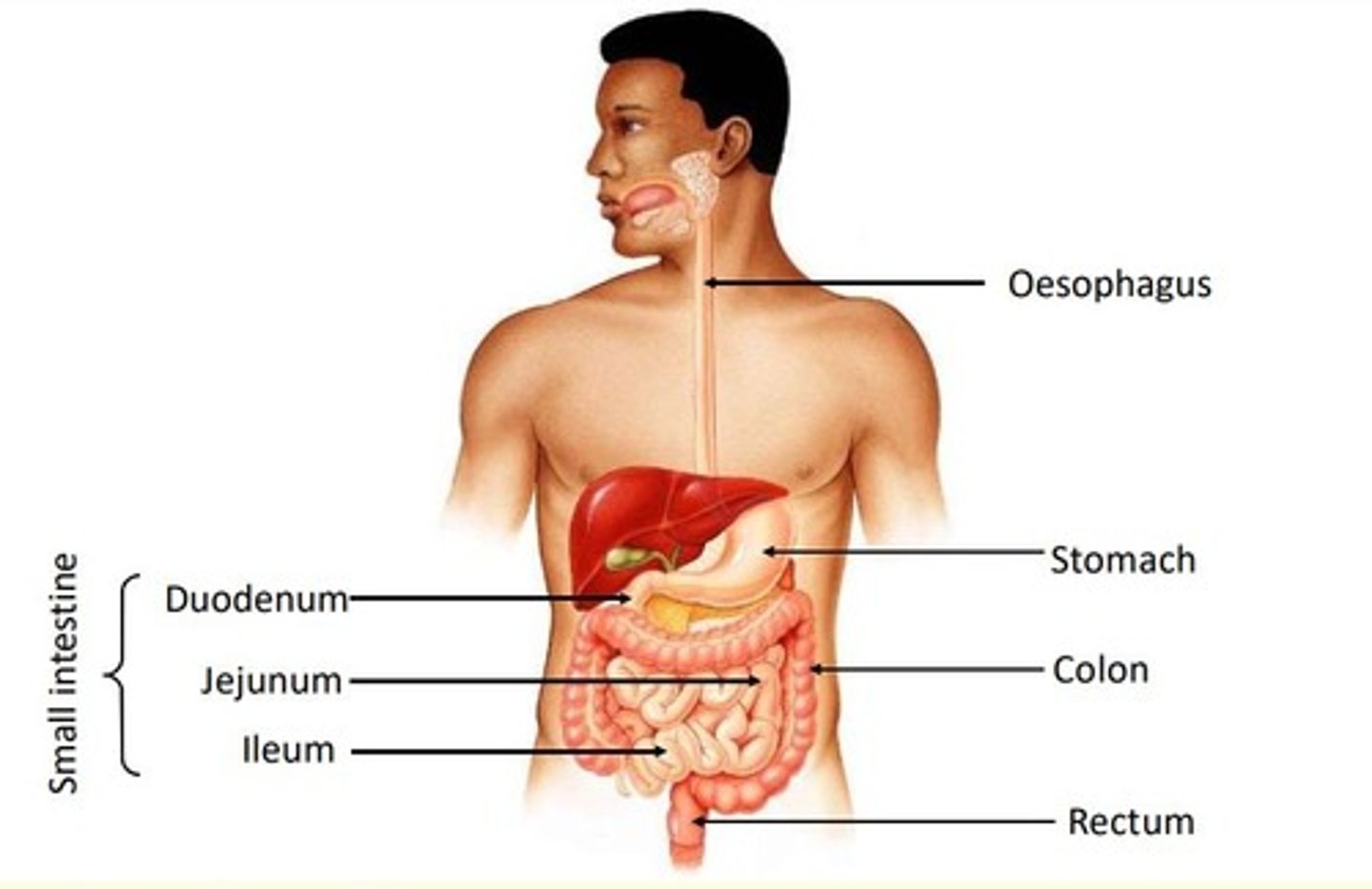
The gastrointestinal tract sections
1. Oesophagus
2. Stomach
3. Colon
4. Rectum
5. Small intestine = duodenum, jejunum, ileum
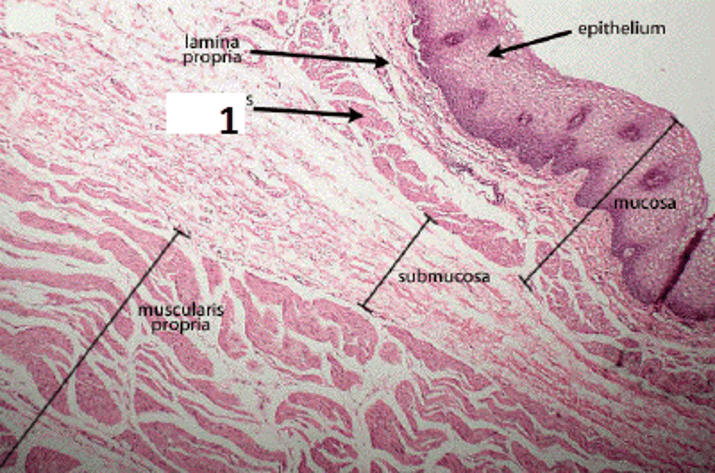
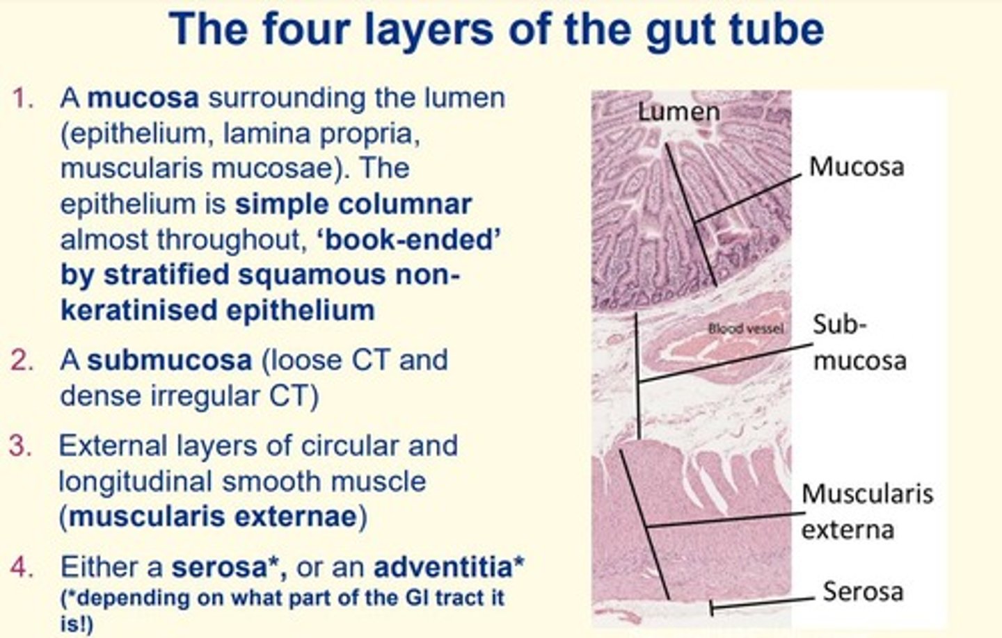
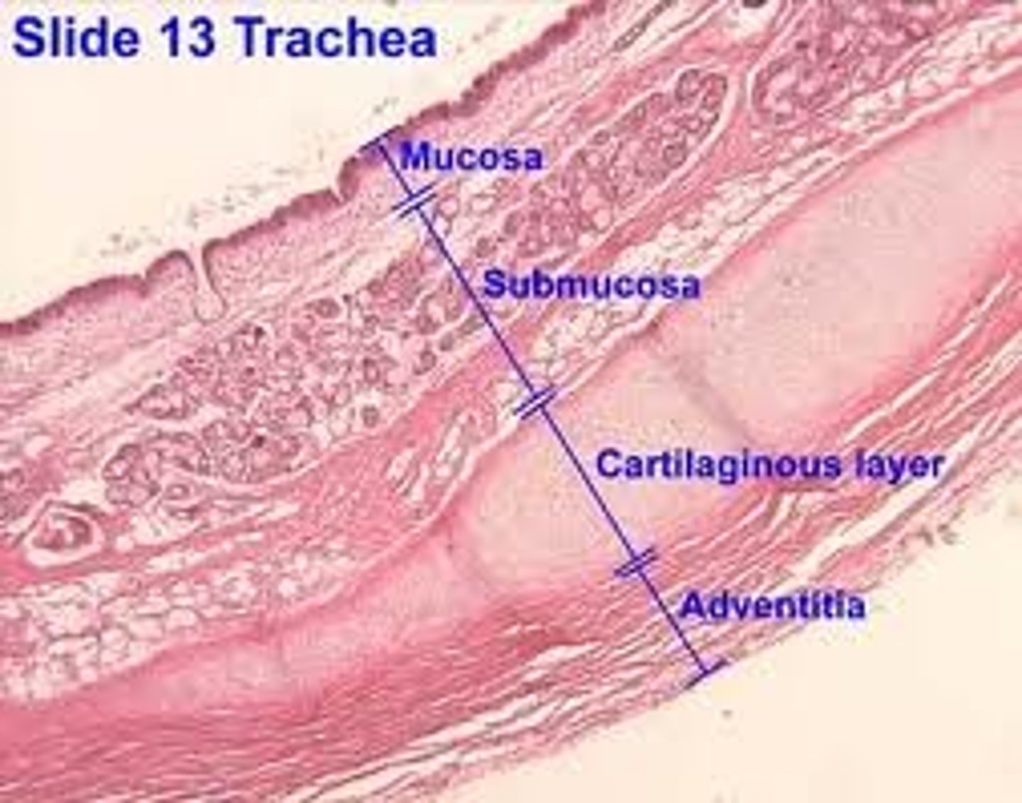
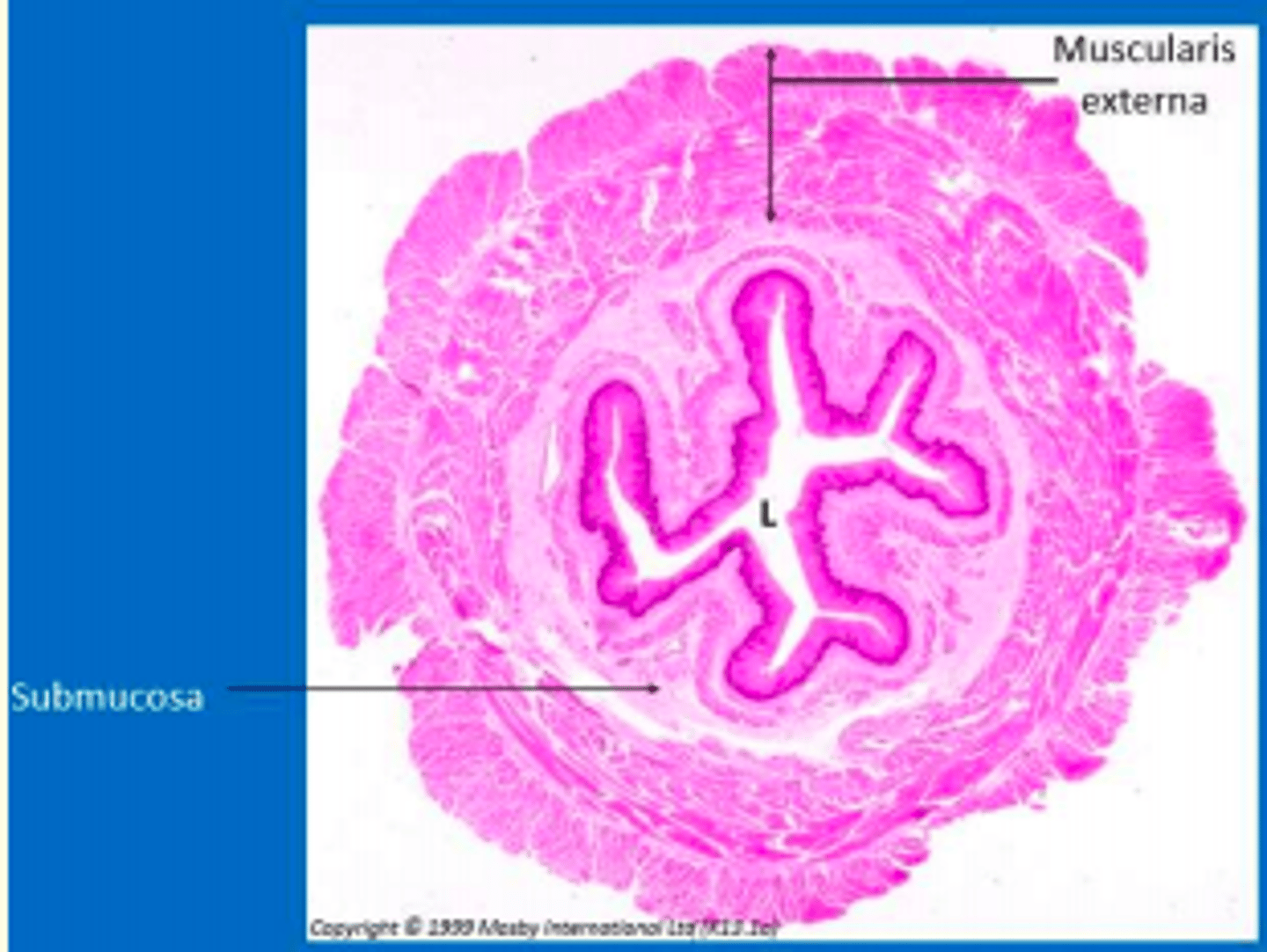
The four layers of the gut tube
1) Mucosa
2) Submucosa
3) Muscularis externae
4) Serosa or adventitia
Mucosa
The innermost layer of the human digestive tract; in some parts of the digestive system, it contains mucus-secreting cells and glands that secrete digestive enzymes
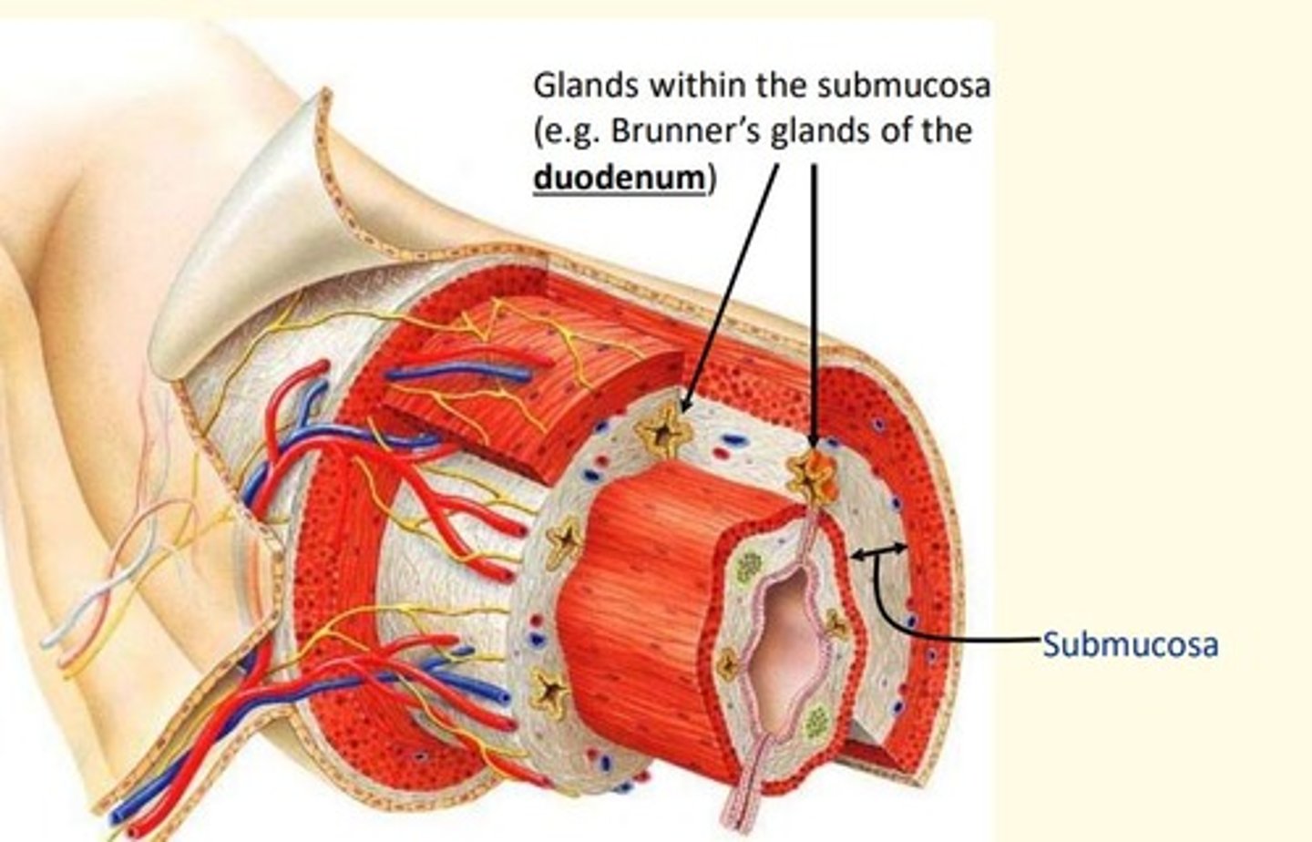
Submucosa
The layer of connective tissue directly under the mucosa of an open body cavity.
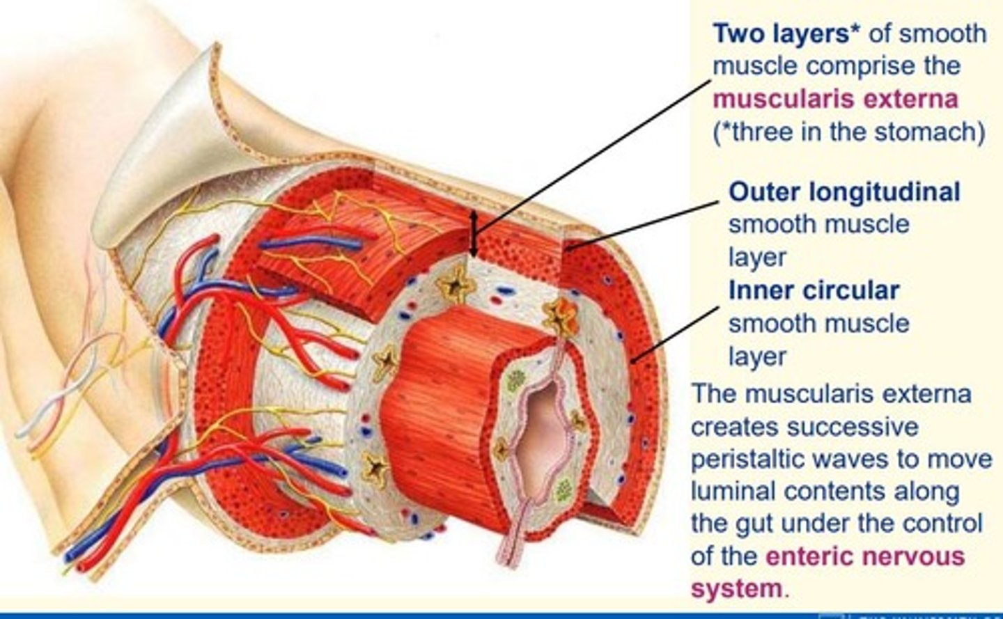
Muscularis externae
Usually smooth muscle layers (Skeletal in part of the esophagus), commonly arranged in an inner circular and an outer longitudinal layer (GI Tract)
Serosa
outermost layer; serous fluid eliminates friction
Adventitia
A thin layer of loose connective tissue that binds an organ to surrounding tissues or organs
Aggregations of lymphocytes present in the lamina propria, particularly distal ileum where they form...
Peyer's patches
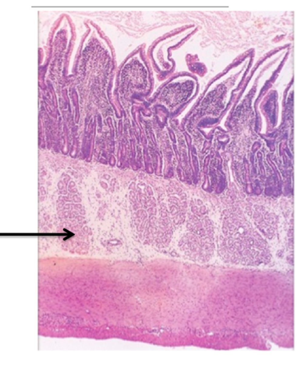
What glands can you find in the submucosa of the gut wall (duodenum)?
Brunner's glands
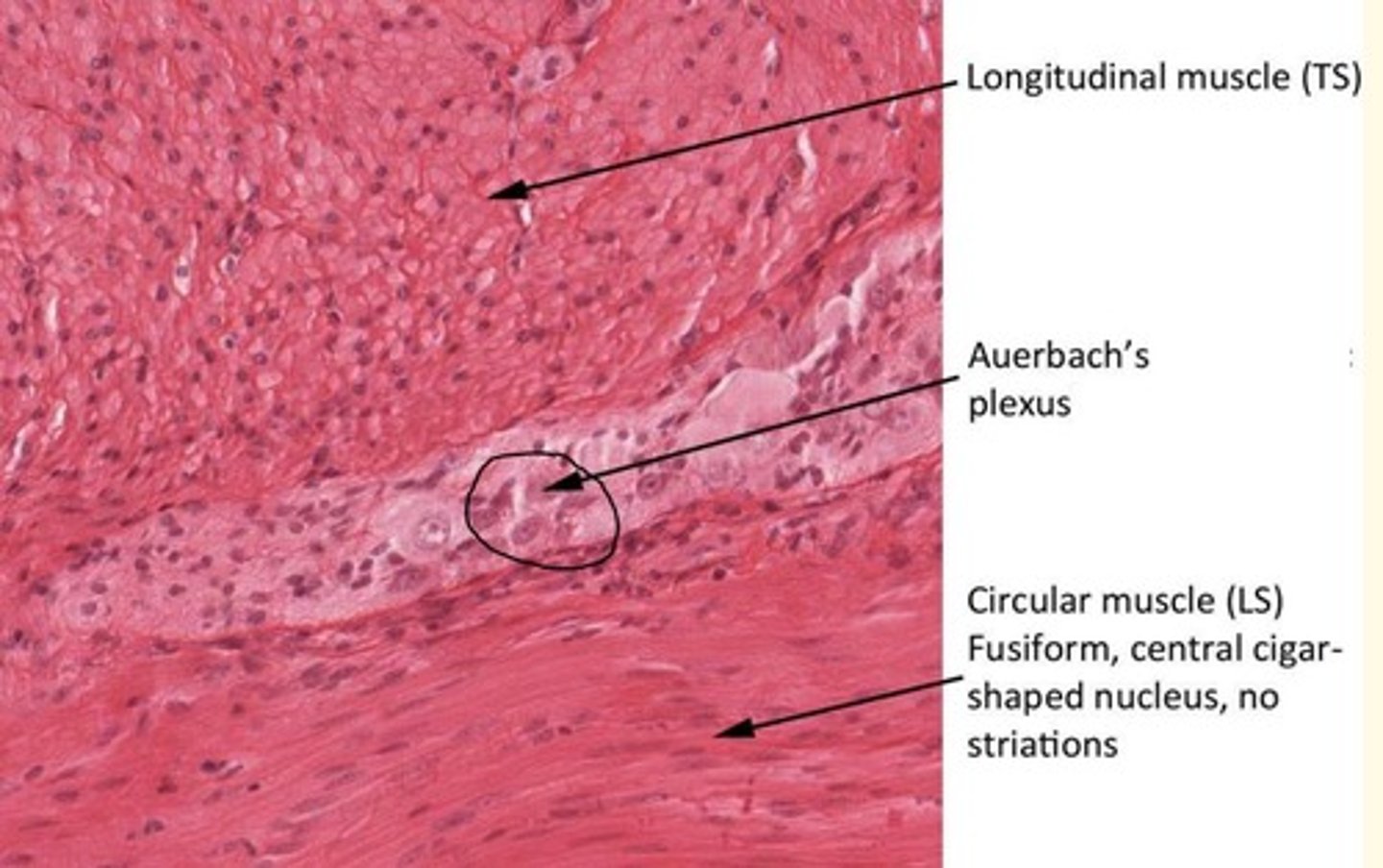
External muscle of gut wall (muscularis externa)
Two layers of smooth muscle
1) Outer longitudinal
2) Inner circular
What is the purpose of the muscularis externa lining the gut wall?
create successive peristaltic waves to move luminal contents of the gut along the gut via control by enteric nervous system
Muscularis externa of gut wall nerves
- Auerbach's (or myenteric) plexus of nerves
The mesentery is a double layer of ___
Peritoneum
containing arteries, veins and nerves
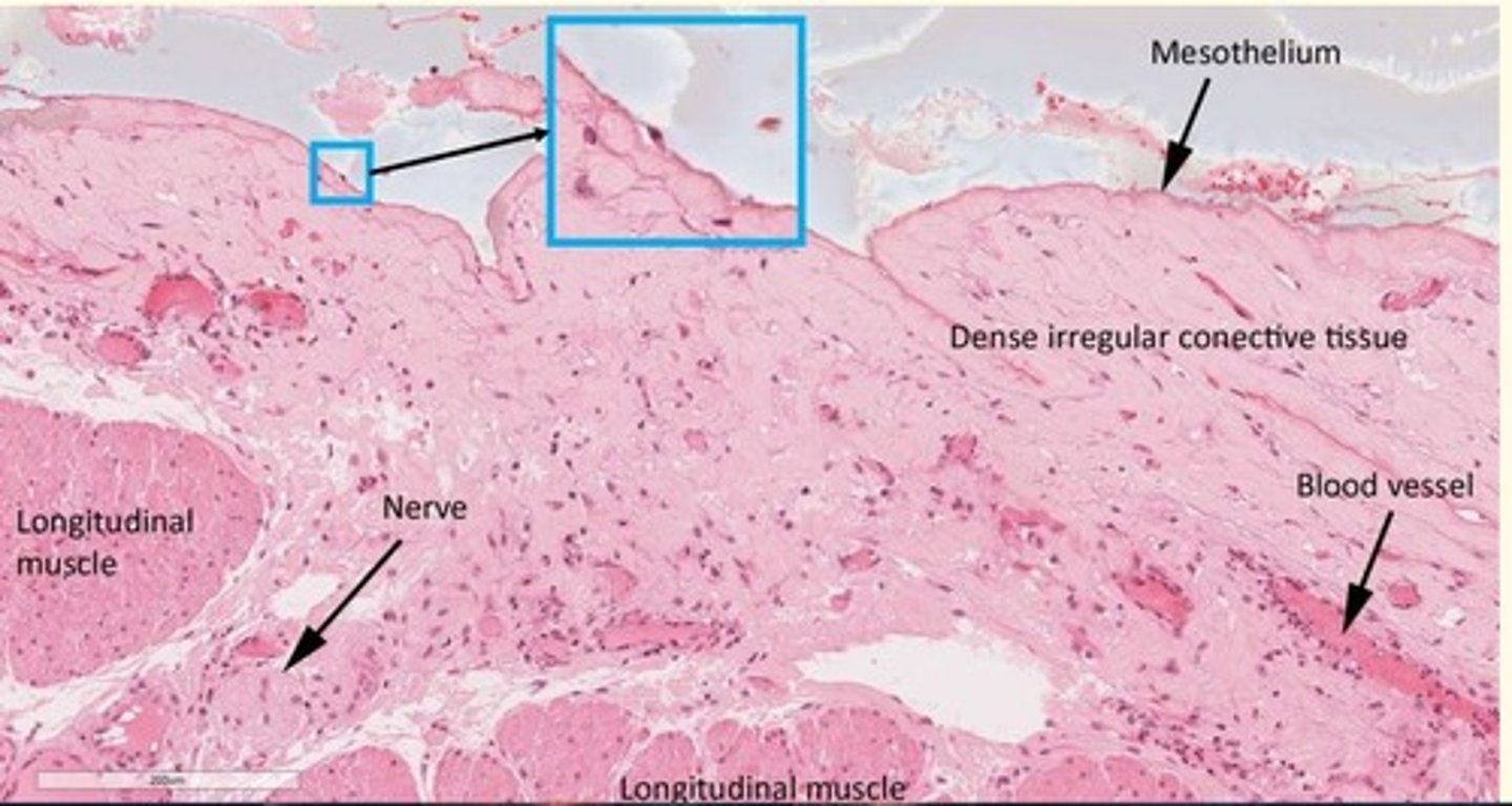
Histology of the serosa of the ileum
Serosa
- External simple squamous epithelium (mesothelium)
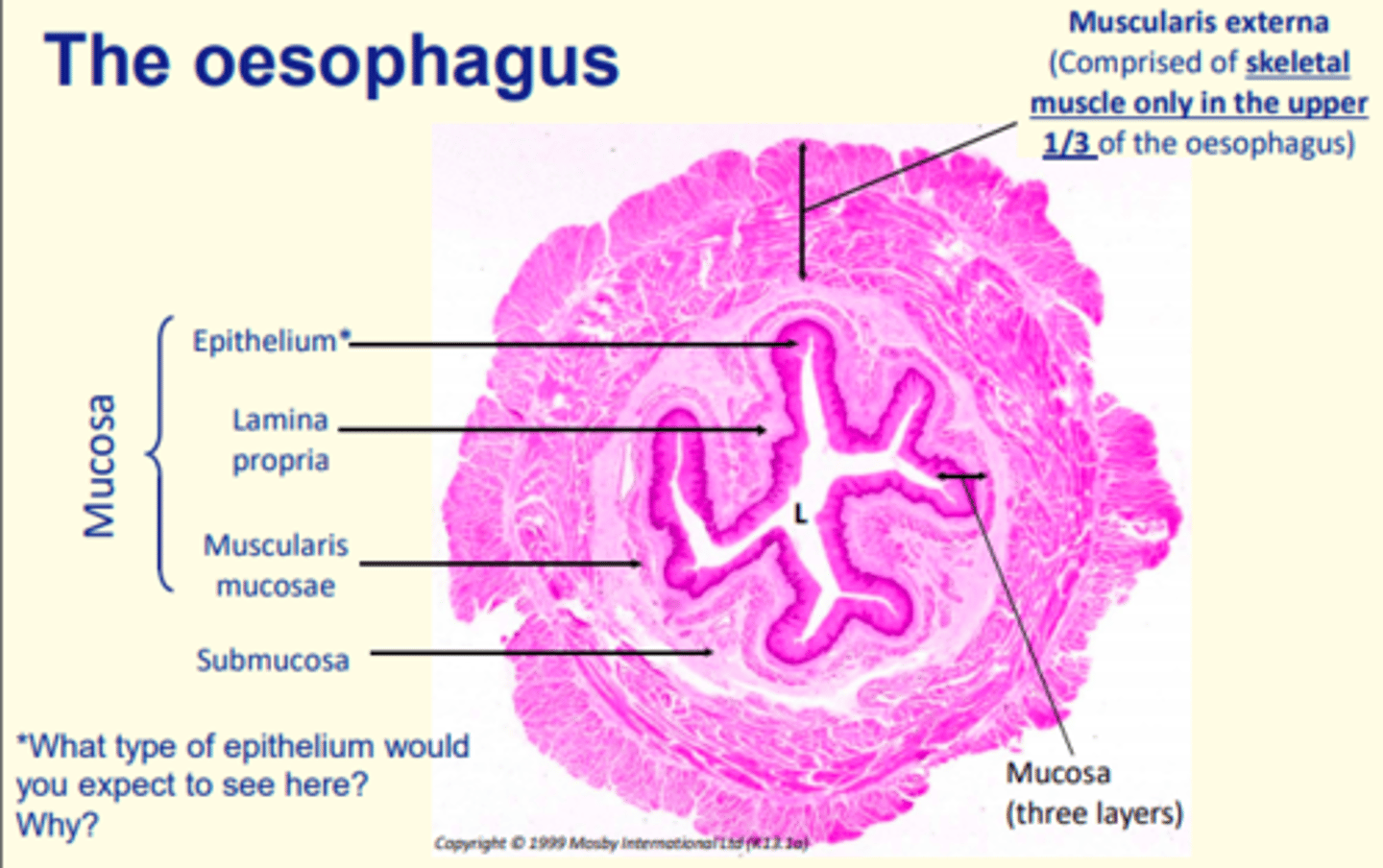
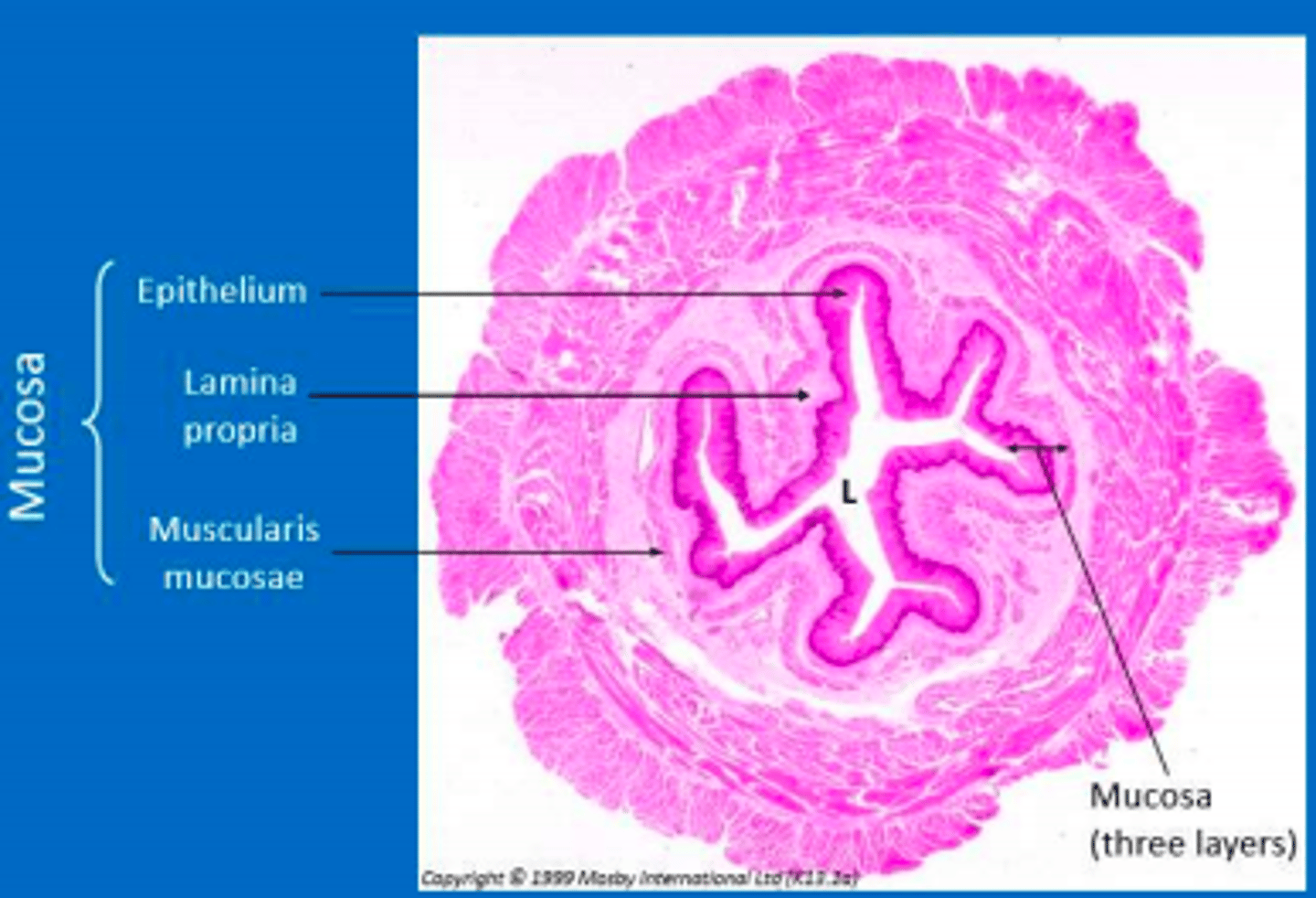
Oesophagus histology
Oesophageal mucosa
Epithelium = stratified squamous non-keratinized (withstands abrasion)
Lamina propria = loose connective bearing blood and lymph vessels, some smooth muscle cells and many cells of immune system
Muscularis mucosae = thin layer of smooth muscle
Outer layers of the oesophagus
Submucosa = subtending layer of connective tissue containing mucus-secreting glands
Muscularis externa = mix of smooth and skeletal muscle layers (inner - circular; outer - longitudinal) that move food by peristalsis
Adventitia = thin outermost of connective tissue to anchor organ
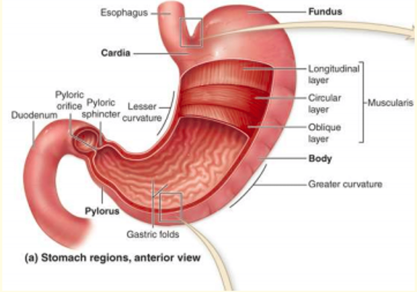
Four regions of the stomach
cardia, fundus, body and pylorus
The mucosa and submucosa are folded into ___ when empty
rugae
The contractions of the three layers of muscle in the stomach are useful for what function?
Mixing of ingested food with enzymes and mucus to form chyme
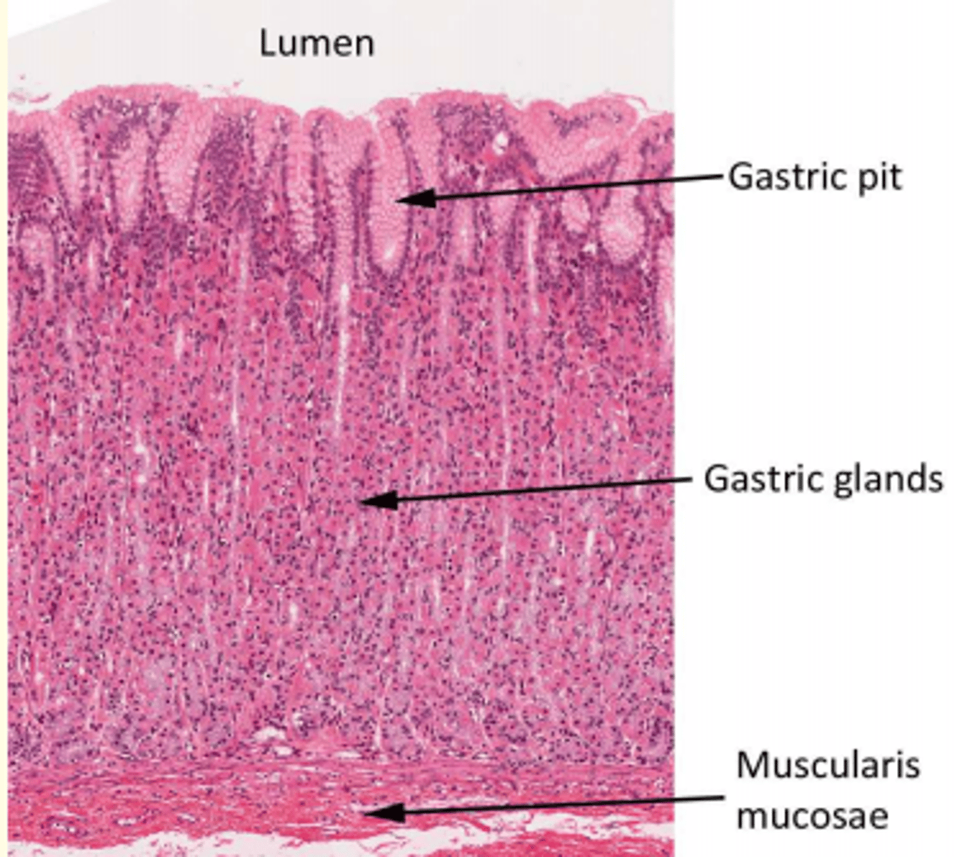
Gastric glands in the mucosa of the stomach and what they secrete
Simple branched tubular glands
Secrete mucus, HCl, digestive enzymes and digestive hormones
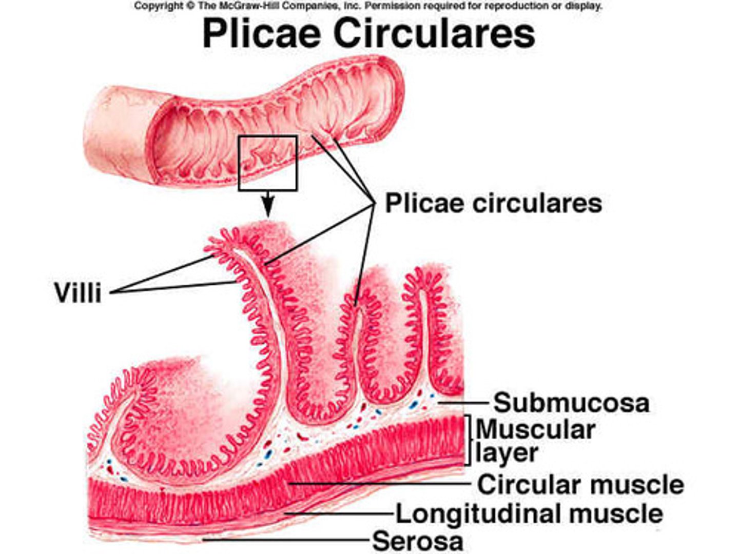
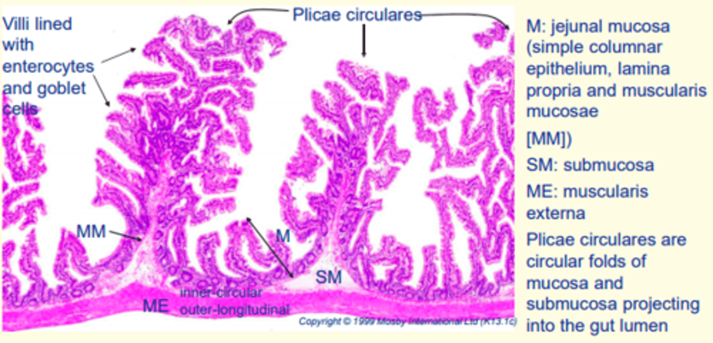
Adaptation of the small intestine (jejunum) which amplifies surface area of the organ - promoting efficient nutrient absorption
- Plicae circulares
- Villi
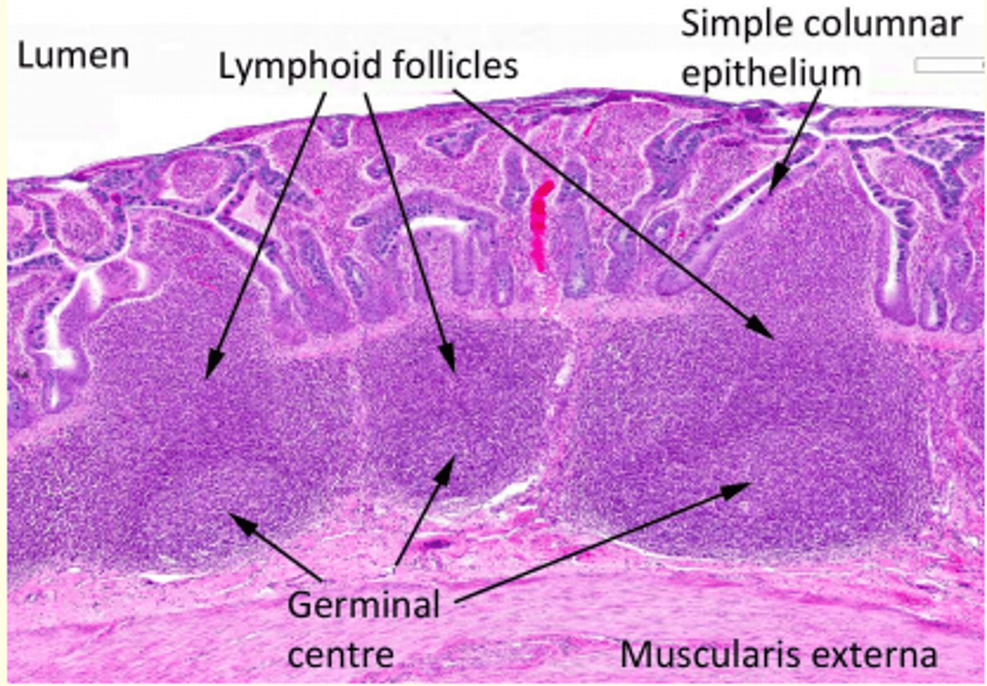
Small intestine (jejunum) histology
Large aggregations of lymphoid follicles found in the ileum
Peyer's patches
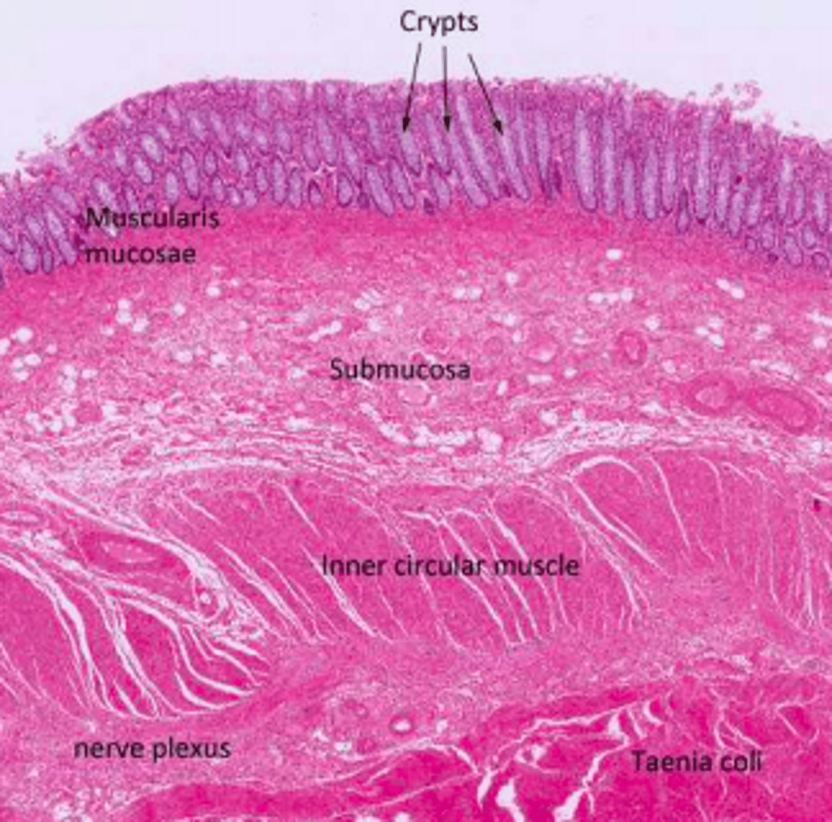
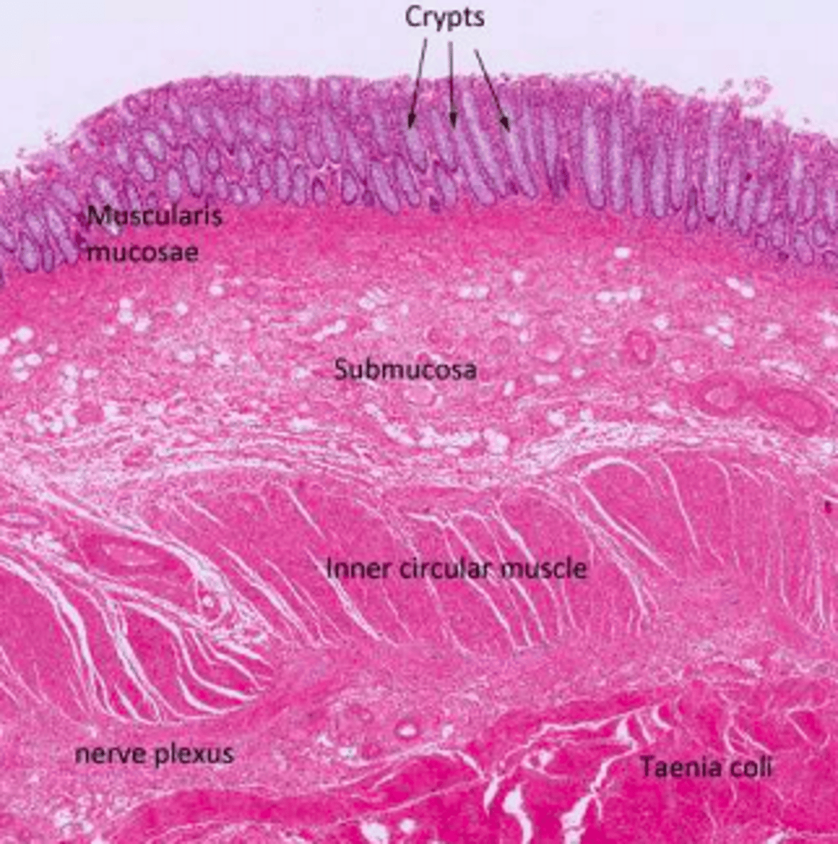
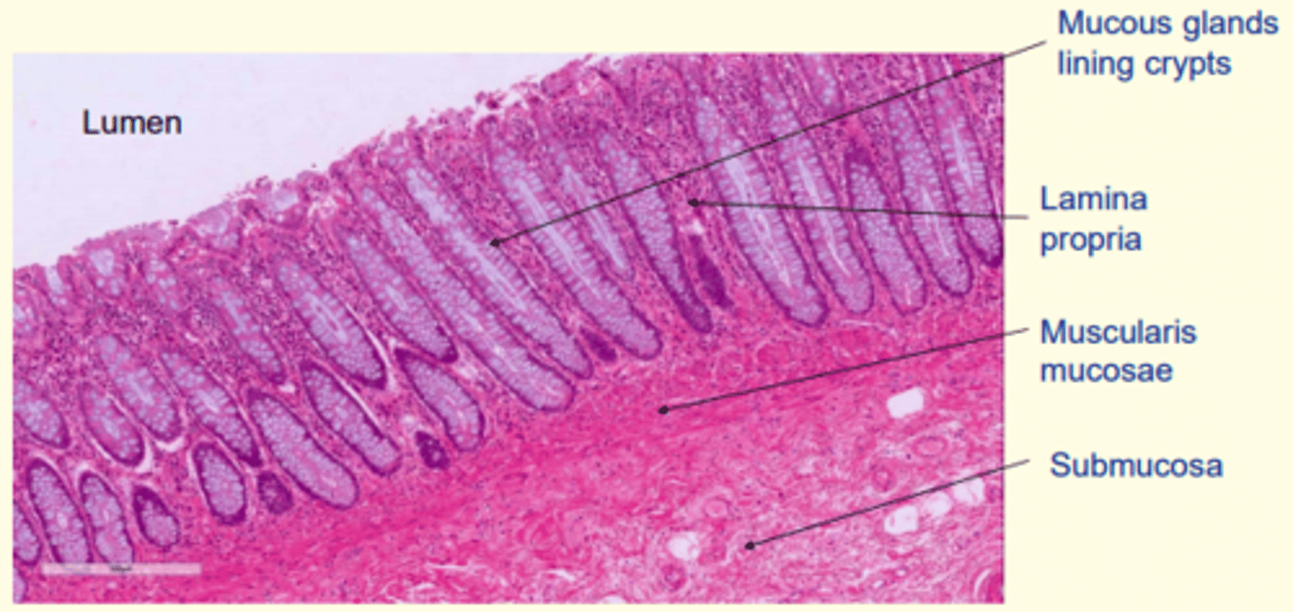
What types of cells are found in the crypts of Lieberkühn of the colon - that secrete mucus?
Goblet cells
The organisation of the muscularis externa of the colon changes. The longitudinal muscle reorganises into three bands known as ___ ___
Taeniae coli
The mucosa of the colon contains ___ ___ epithelium
simple columnar epithelium
Crypts of Lieberkuhn
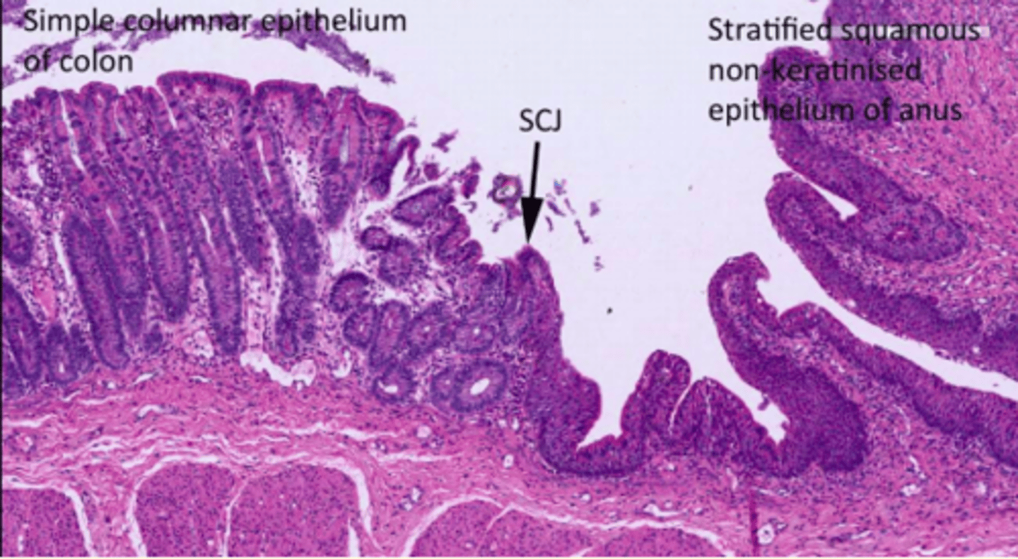
Colon epithelium
Simple columnar epithelium
Anus epithelium
Stratified squamous non-keratinised epithelium
A hotspot for malignant transformation in the colon where frequent metaplasia occurs (change of one differentiated cell type to another)
Recto-anal junction or the 'squamocolumnar junction' (SCJ)
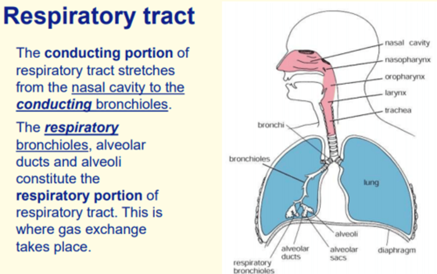
Respiratory tract - conducting portion
Nasal cavity to terminal bronchioles
Respiratory tract - respiratory portion
Respiratory bronchioles, alveolar ducts, alveoli = site of gaseous exchange
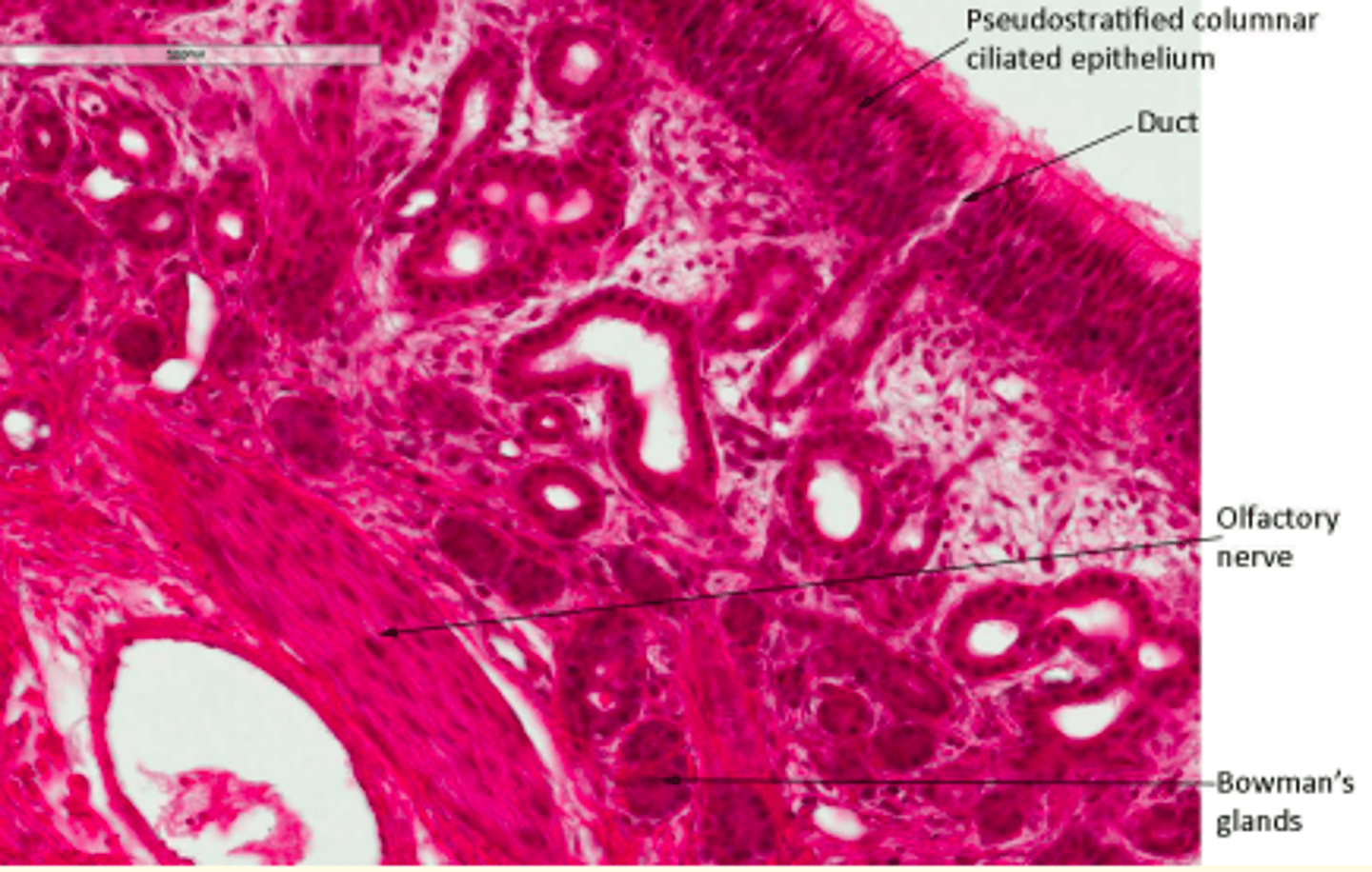
What type of epithelium is found in the roof of the nasal cavity?
Ciliated olfactory receptor cells (olfactory epithelium)
What types of glands can be found in the olfactory epithelium in the roof of the nasal cavity?
Bowman's glands = produce fluid to dissolve odiferous substances
Structure of Bowman's glands in the nasal cavity?
Branched tubuloalveolar
The lamina propria in the nasal cavity is richly vascularised with ___ ___
Venous plexuses
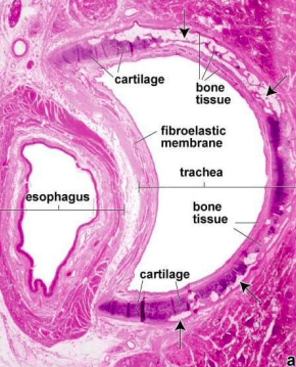
What type of cartilage can be seen in the trachea?
C-shaped hyaline cartilage
The fibroelastic membrane in the trachea contains the ___ muscle which relaxes when you swallow and contracts when you cough
trachealis muscle
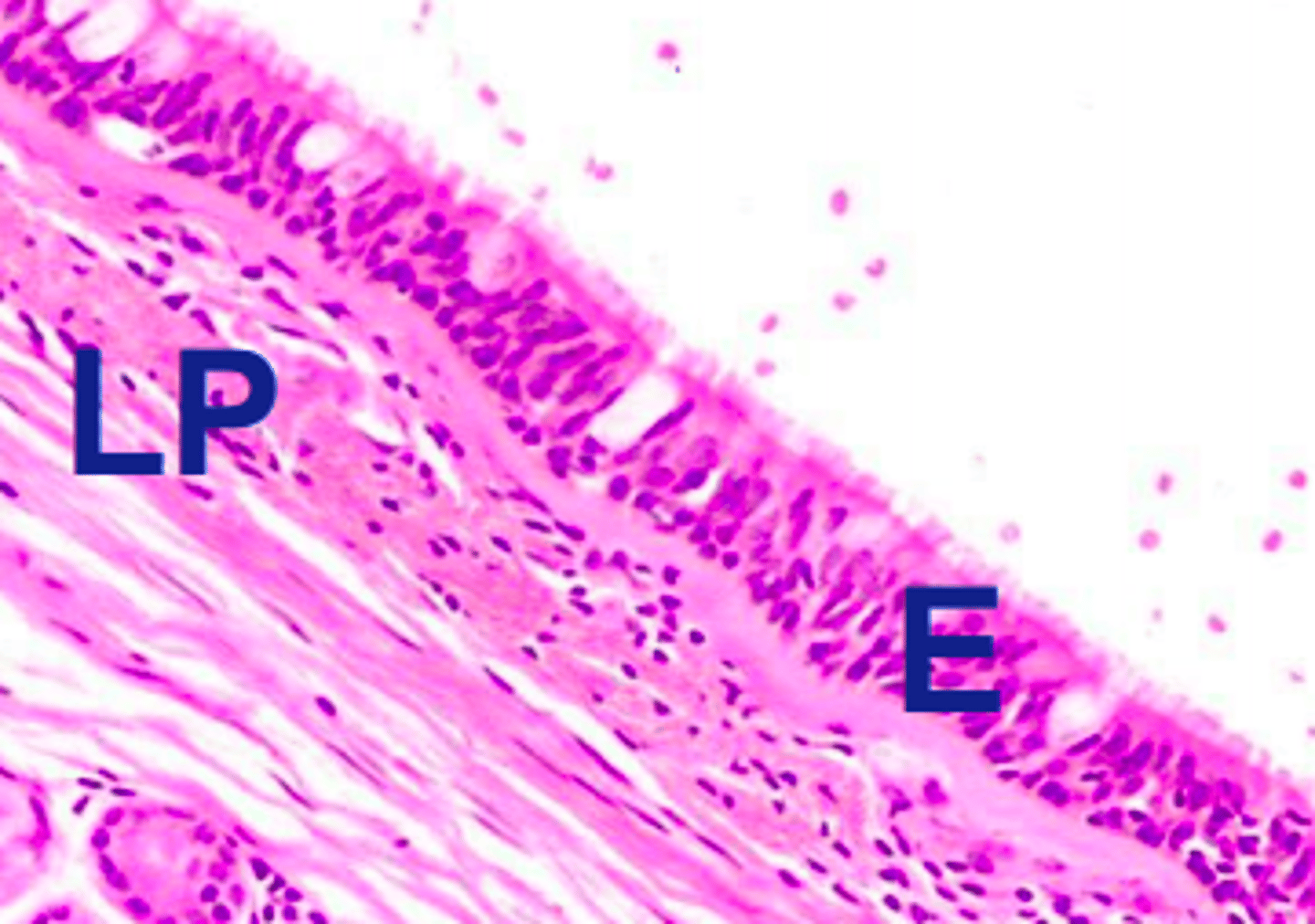
What type of epithelia can be found in the wall of the trachea?
Pseudostratified ciliated columnar epithelium
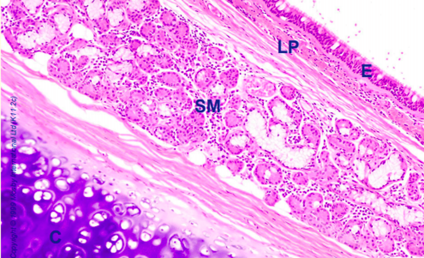
What types of glands can be found in submucosa of the trachea?
Seromucous (tubuloacinar) glands
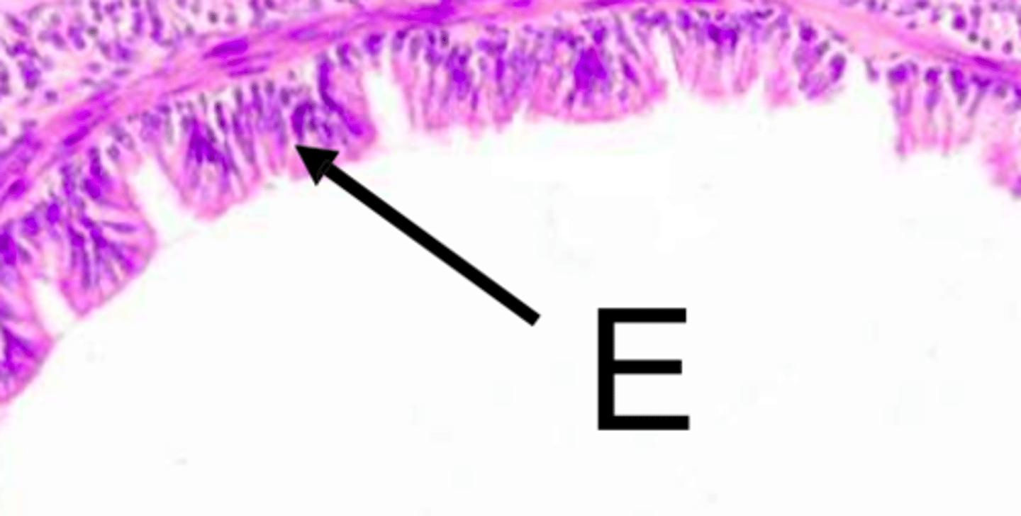
Wall of the trachea
E = Epithelium (pseudostratified ciliated columnar)
LP = Lamina propria
C = C-shaped hyaline cartilage
SM = Submucosa with seromucous (tubuloacinar) glands
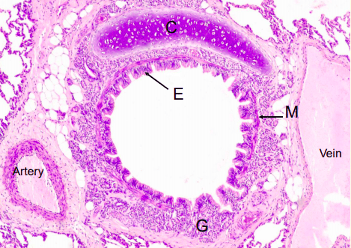
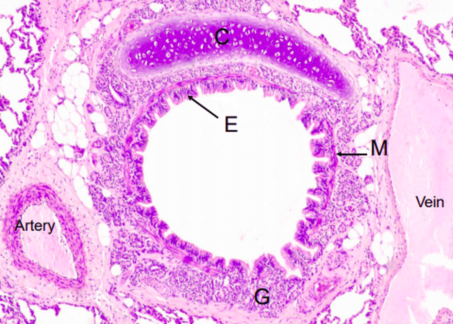
What is this image showing?
A bronchus
What type of epithelium can be found in the bronchus?
Pseudostratified ciliated columnar epithelium
Bronchus
E = Epithelium (pseudostratified ciliated columnar)
M = Smooth muscle
G = Glands in submucosa
C = Crescent-shaped hyaline cartilage
What types of epithelium lines smaller respiratory bronchioles?
Cuboidal epithelium
Difference between bronchus and bronchiole structure
Bronchiole = no cartilage to keep the lumen open
Bronchus = crescent-shaped hyaline cartilage to keep lumen open
Smaller, respiratory bronchioles have no smooth muscle and are lined with ___ epithelium
cuboidal
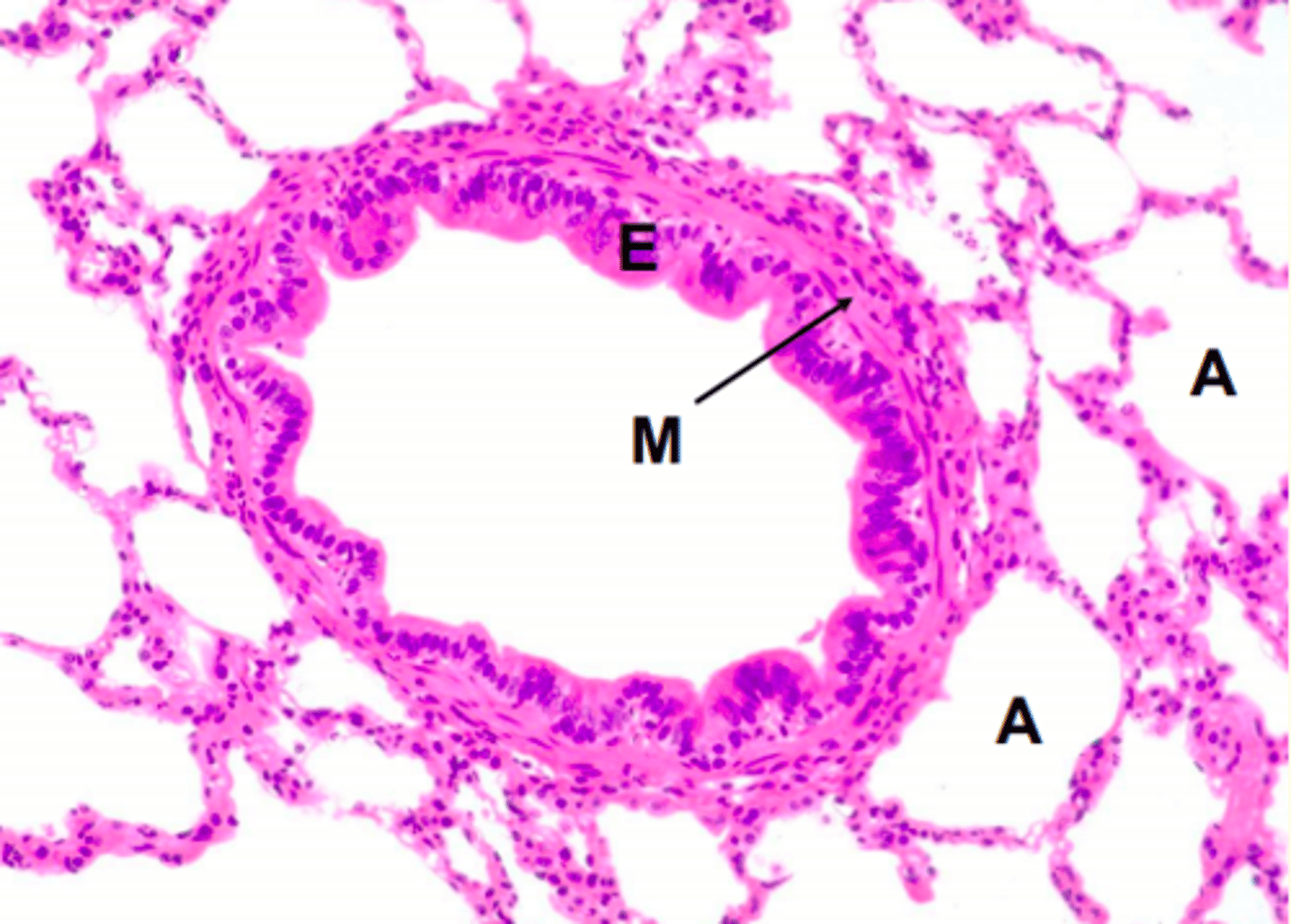
A respiratory bronchiole
E = Epithelium (simple columnar: varies from ciliated to cuboidal)
M = Smooth muscle
A = Alveoli
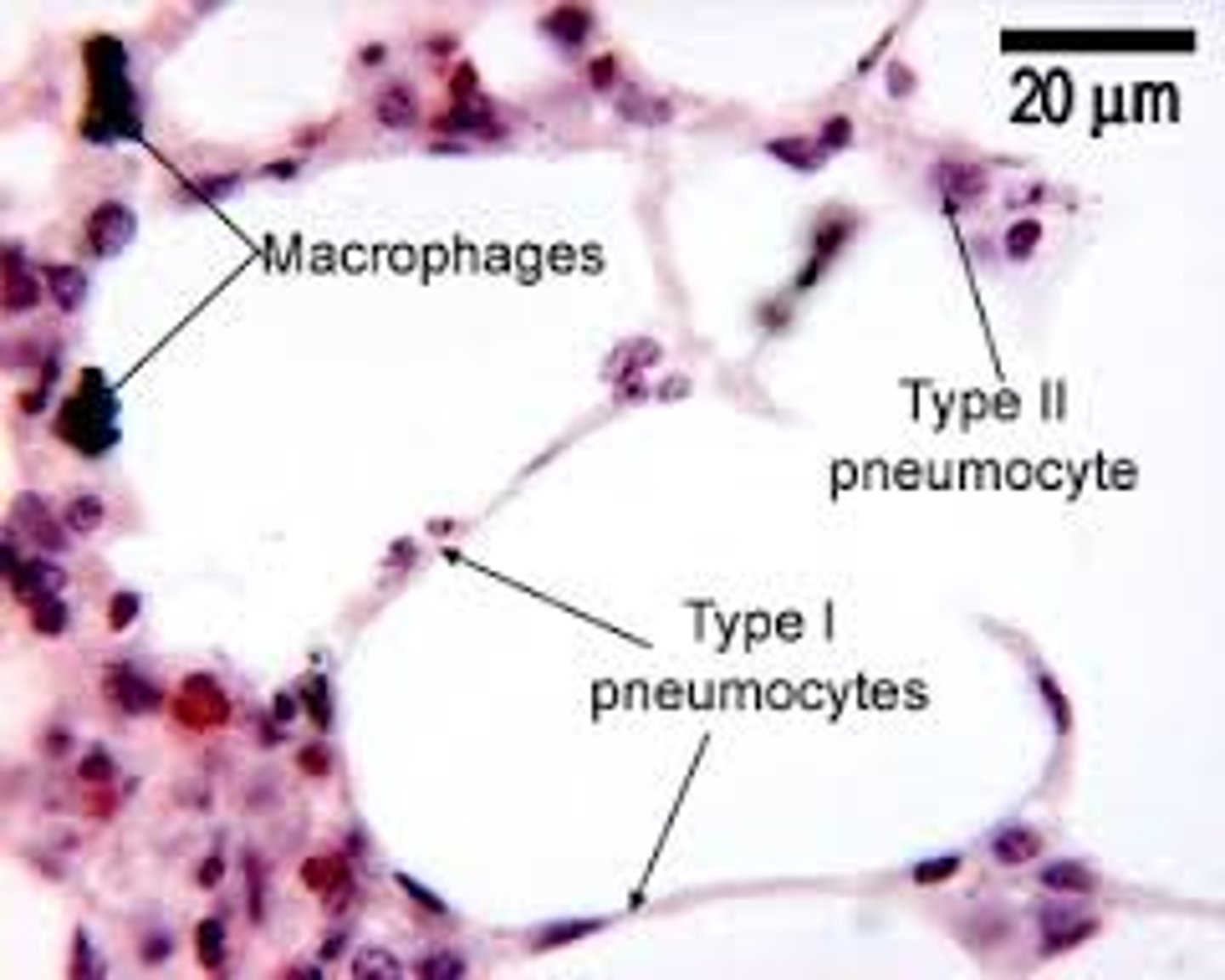
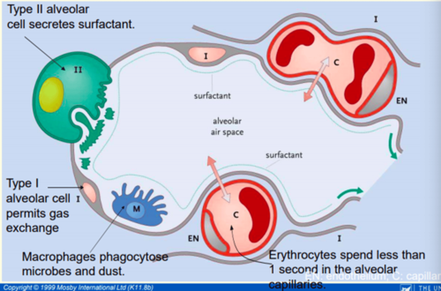
a) Type I pneumocytes function
b) Type of epithelium found in type I pneumocytes
a) Extremely thin alveolar cells that are adapted to carry out gas exchange
b) Simple squamous epithelial cells
a) Type II pneumocytes function
b) Type of epithelium found in type II pneumocytes
a) Production of alveolar surfactant
b) Cuboidal epithelial cells
Macrophages found lining the alveolar surface which phagocytose foreign particles
Dust cells
Type I pneumocytes account for ___% of alveolar cell population but cover ___% of surface area of the alveoli
Type I pneumocytes account for 40% of alveolar cell population but cover 95% of surface area of the alveoli
Type II pneumocytes account for ___% of the alveolar cell population, but cover only ___% of the alveolar surface
Type iI pneumocytes account for 60% of the alveolar cell population, but cover only 5% of the alveolar surface
Cells of the alveolus
- Simple squamous (type I) alveolar cells
- Cuboidal (type II) alveolar cells
- Alveolar macrophages (dust cells)
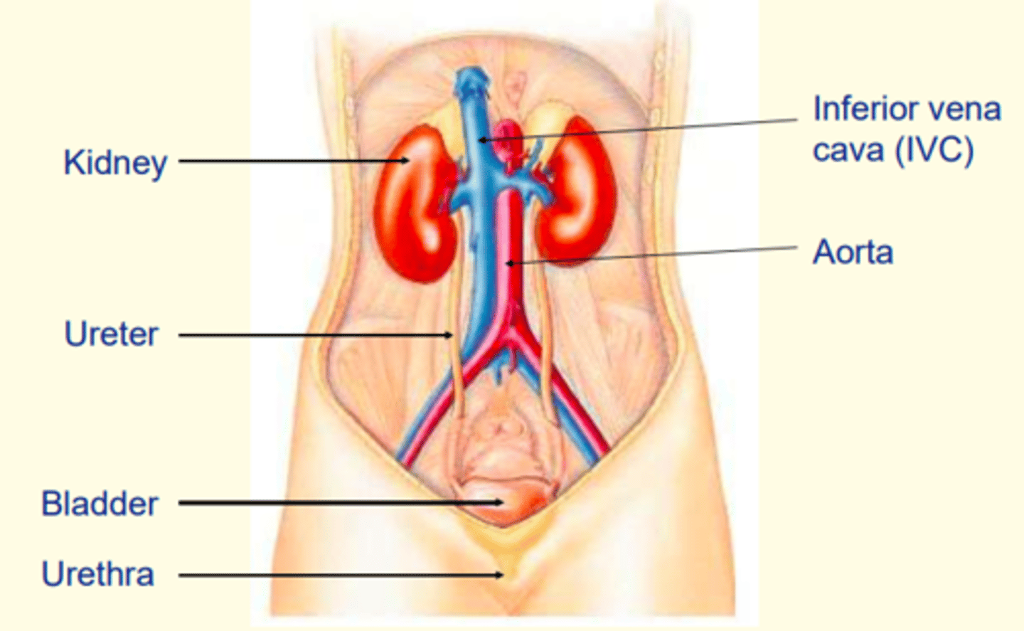
Urinary tract
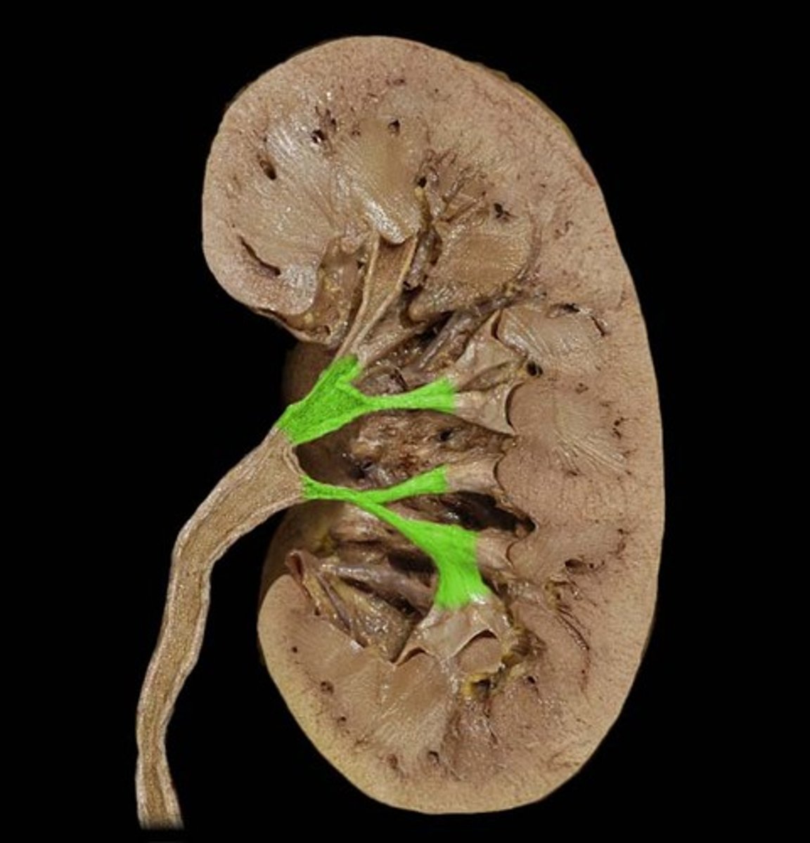
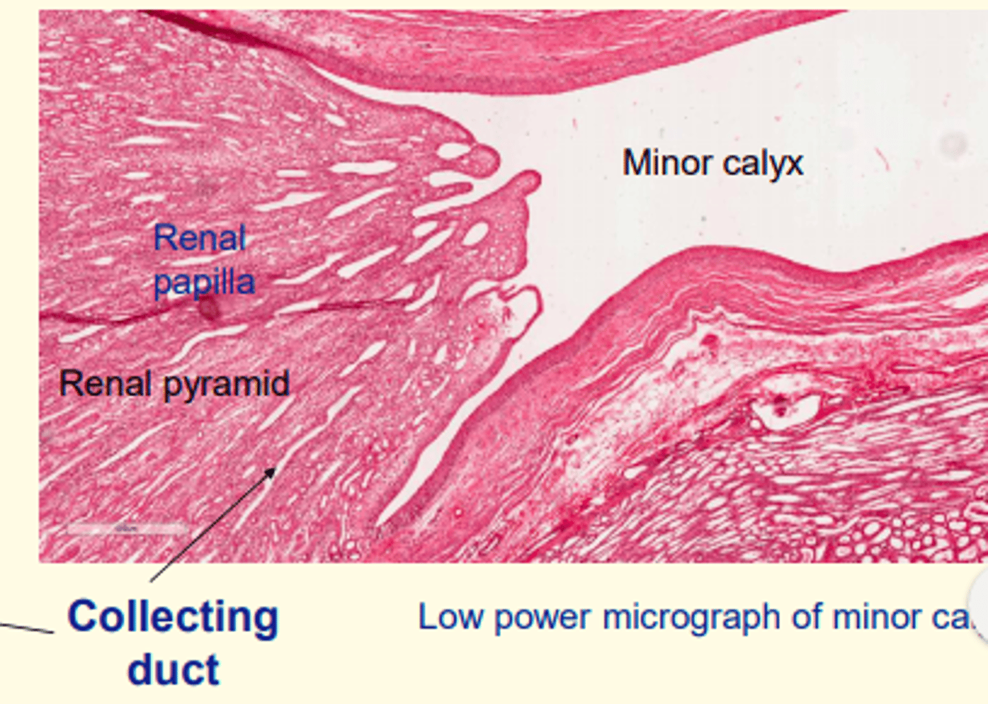
Renal calyces
Cup-like cavities that collect urine and empty into the renal pelvis
The renal calyces are lined with ___ epithelium
Transitional
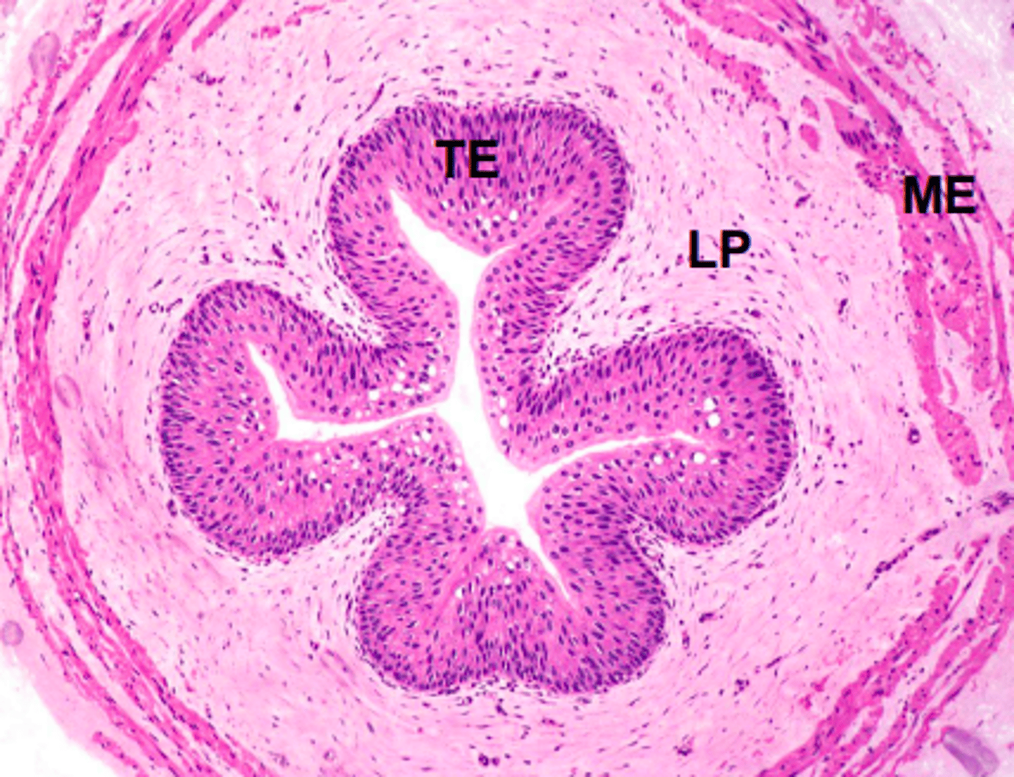
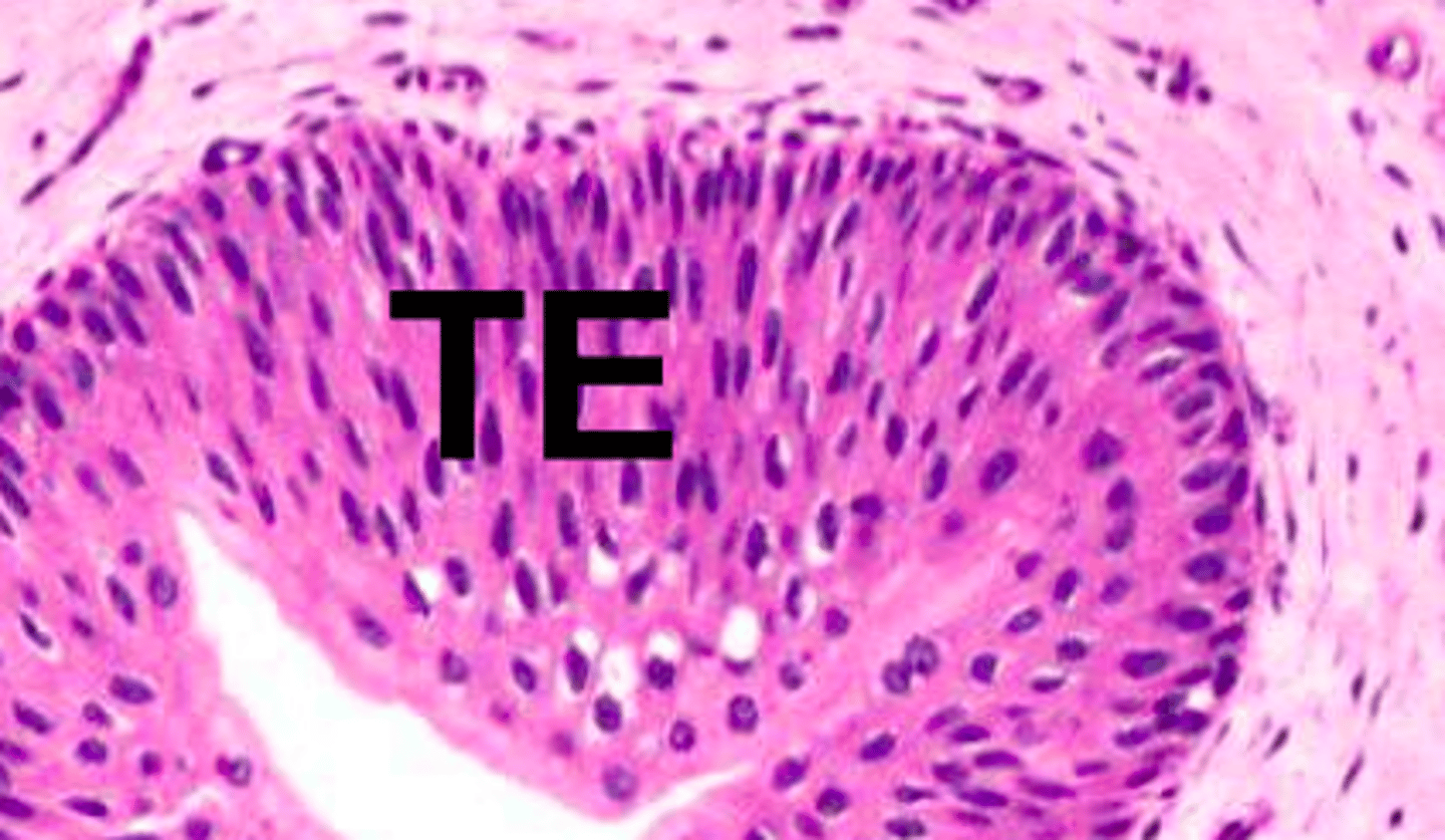
The ureter
TE = Transitional epithelium
LP = Fibroelastic lamina propria
ME = Muscularis externa
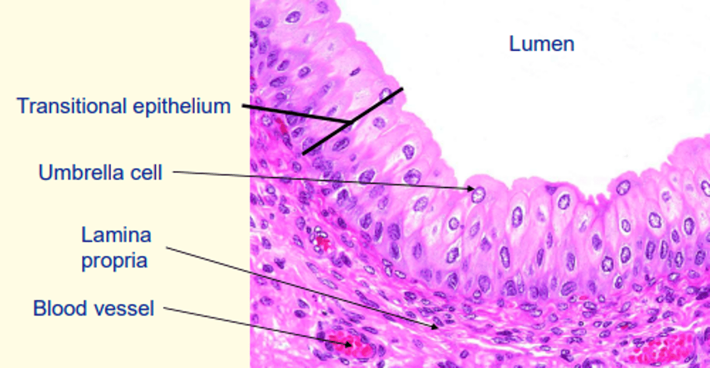
Importance of transitional epithelium in the urinary tract?
- Distensible = tissues can expand when bladder fills with urine
- Protection = (multiple layers thick) from cytotoxic effects of urine
The muscularis (smooth muscle) in the bladder wall is comprised of three poorly-delineated layers forming the ___ muscle
detrusor
Transitional epithelium (urothelium) of the bladder
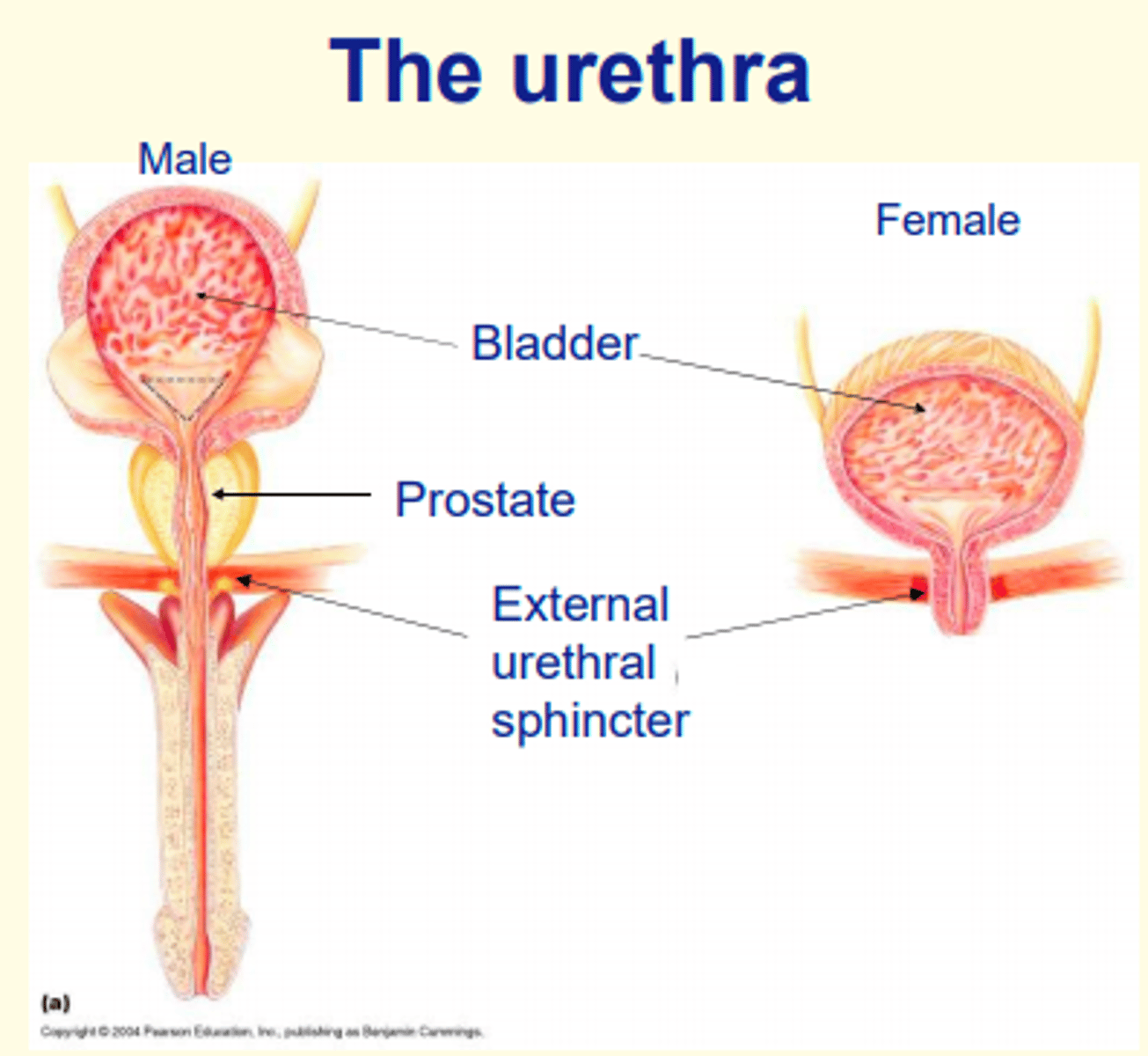
The proximal region of the urethra is lined by ___ epithelium
Transitional
The female urethra is entirely ___
The male urethra has both ___ and ___ roles
The female urethra is entirely urinary
The male urethra has both urinary and reproductive roles
Distal regions of the urethra (which open to the outside) are lined with what type of epithelium?
Stratified squamous, non-keratinised epithelium
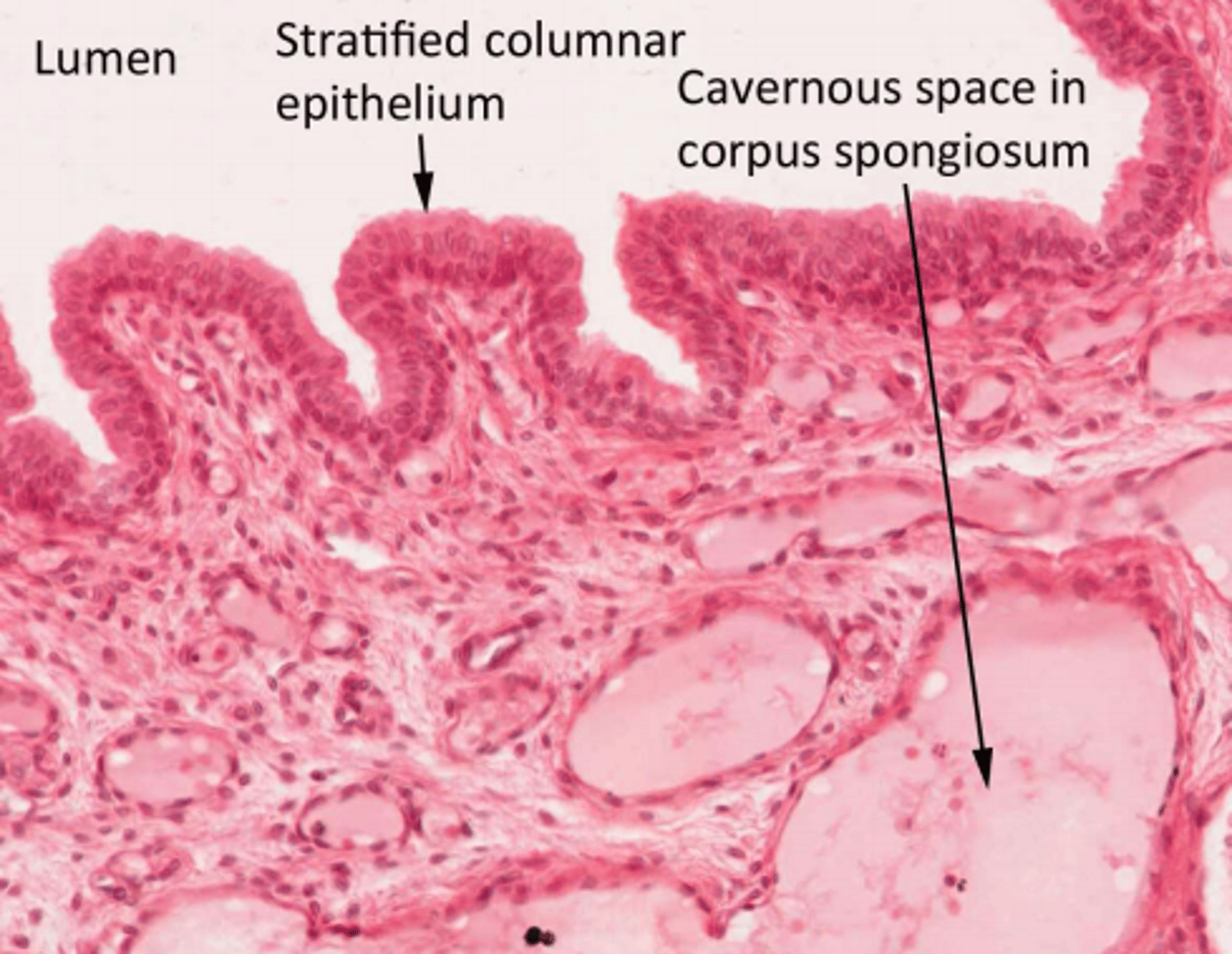
The penile urethra is enclosed by the __________.
Corpus spongiosum
What type of epithelium can be found in the penile urethra?
Stratified columnar epithelium
Does the peritoneal cavity connect to exterior?
Body cavities tubes and ducts that are enclosed and make no connection with the exterior of the body
Does the respiratory tract connect to exterior?
Body cavities tubes and ducts that ultimately connect with the exterior of the body
Does the pleural cavity connect to exterior?
Body cavities tubes and ducts that are enclosed and make no connection with the exterior
Does the pericardial cavity connect to exterior?
Body cavities tubes and ducts that are enclosed and make no connection with the exterior
Does the alimentary canal connect to exterior?
Body cavities tubes and ducts that ultimately connect with the exterior of the body
Does the cardiovascular system connect to exterior?
Body cavities tubes and ducts that are enclosed and make no connection with the exterior
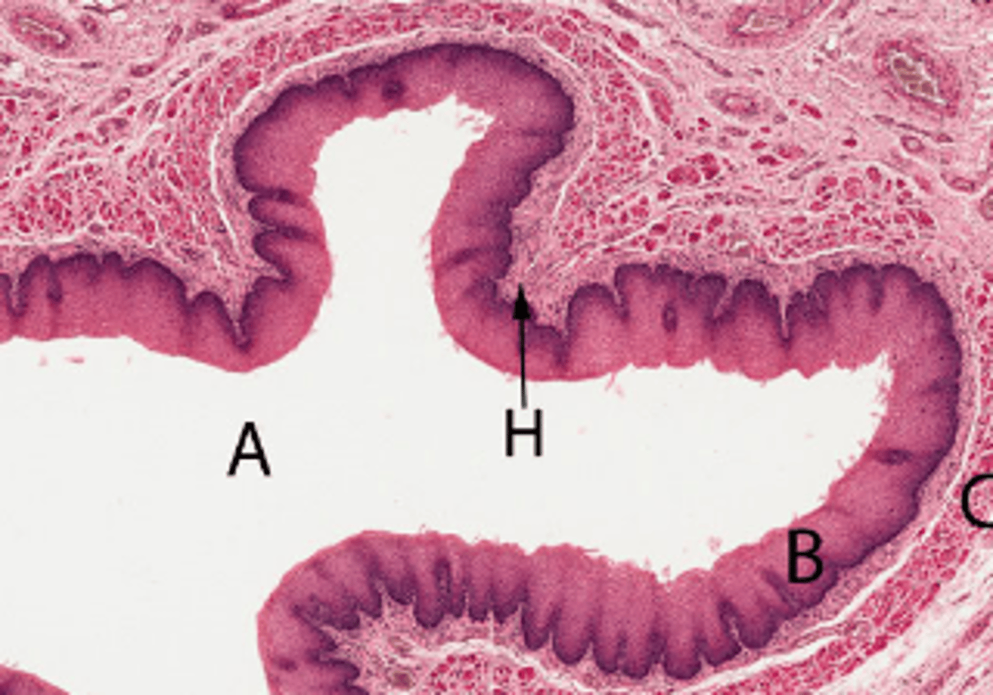
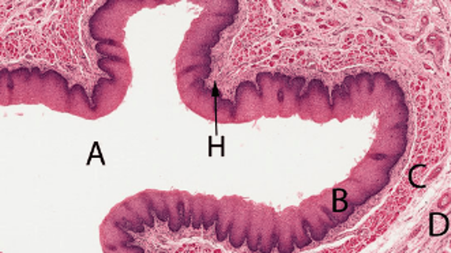
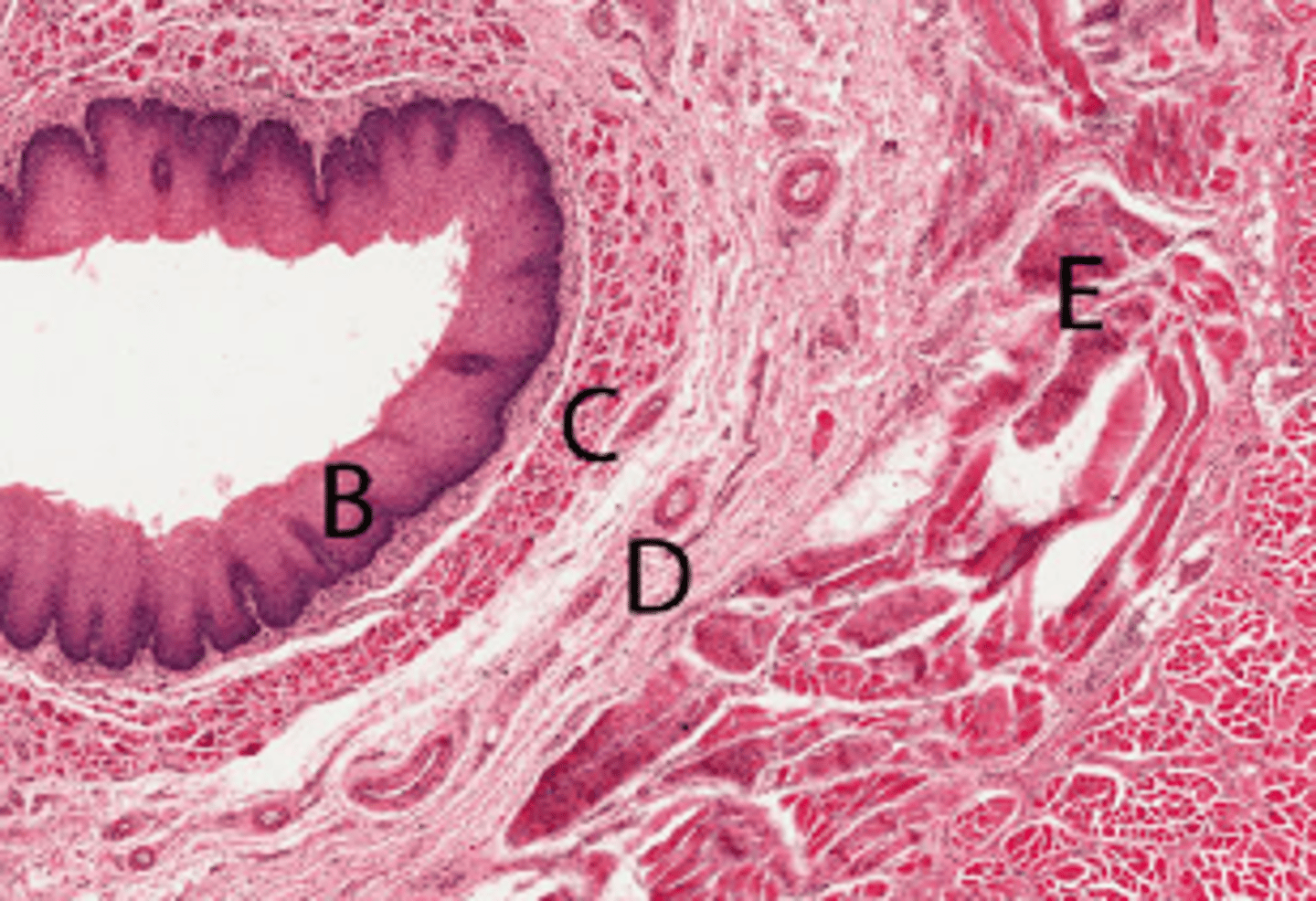
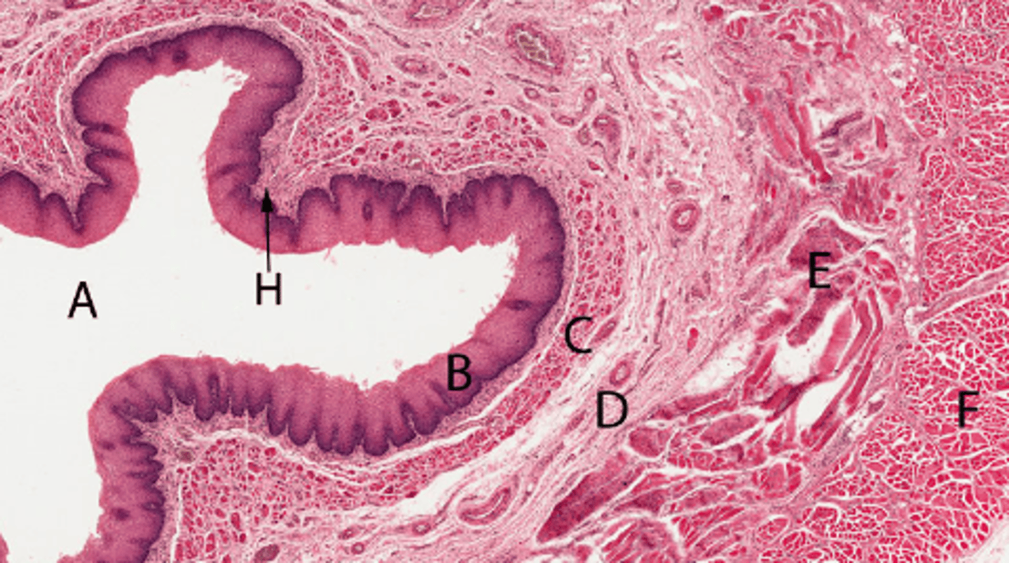
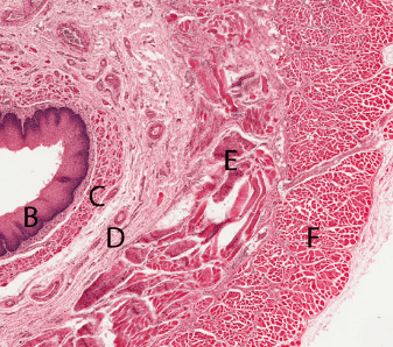
Micrograph of oesophagus.
Identify B
Stratified squamous non-keratinised epithelium
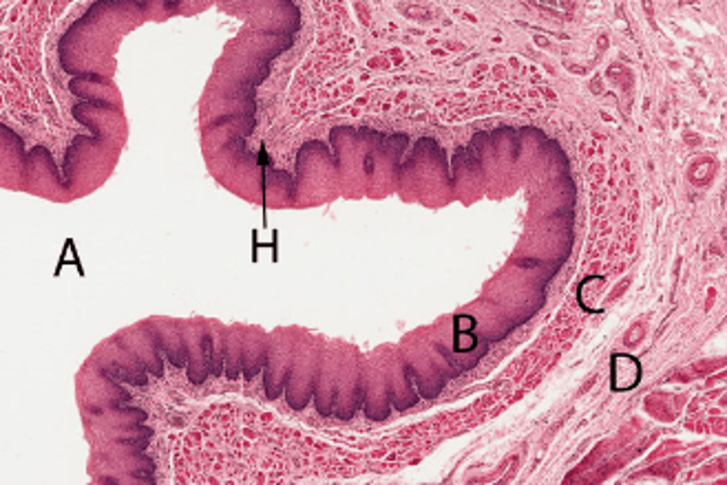
Micrograph of oesophagus.
Identify D
Submucosa
Micrograph of oesophagus.
Identify A
Oesophageal lumen
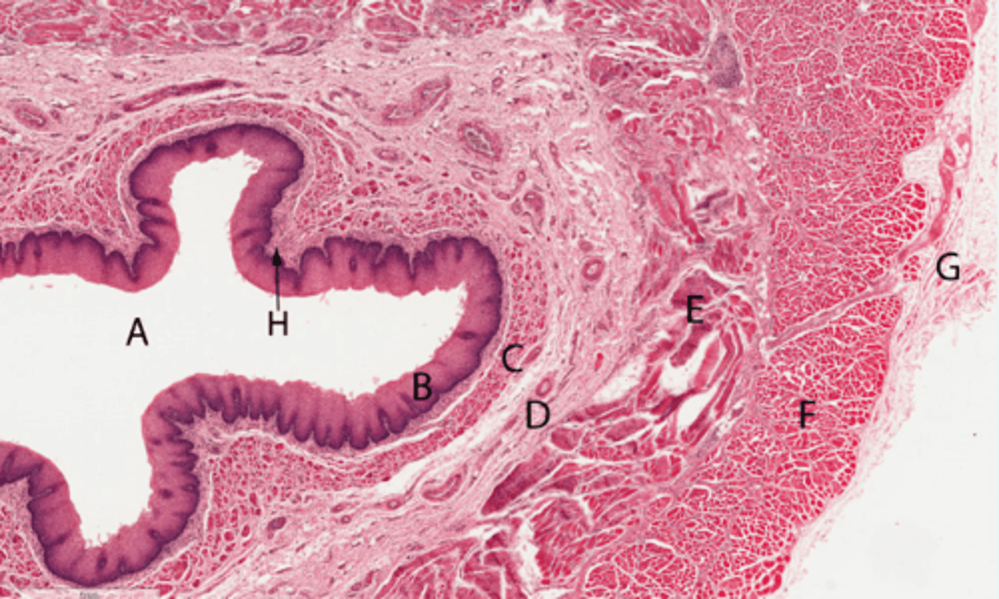
Micrograph of oesophagus.
Identify G
Adventitia
Micrograph of oesophagus.
Identify H
Lamina propria
Micrograph of oesophagus.
Identify C
Muscularis mucosae
Micrograph of oesophagus.
Identify E
Mix of skeletal and smooth muscle of circular layer
Micrograph of oesophagus.
Identify longitudinal muscle layer
F
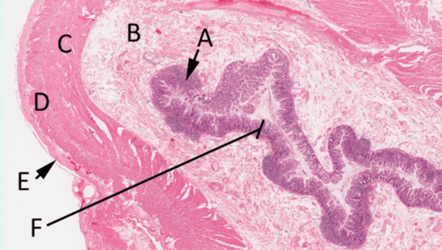
Histological section of small intestine
Identify structures
A =
B =
C =
D =
E =
F =
A = MALT
B = Submucosa
C = Circular muscle
D = Longitudinal muscle
E = Mesothelium (simple squamous epithelium of the serosa)
F = Mucosa
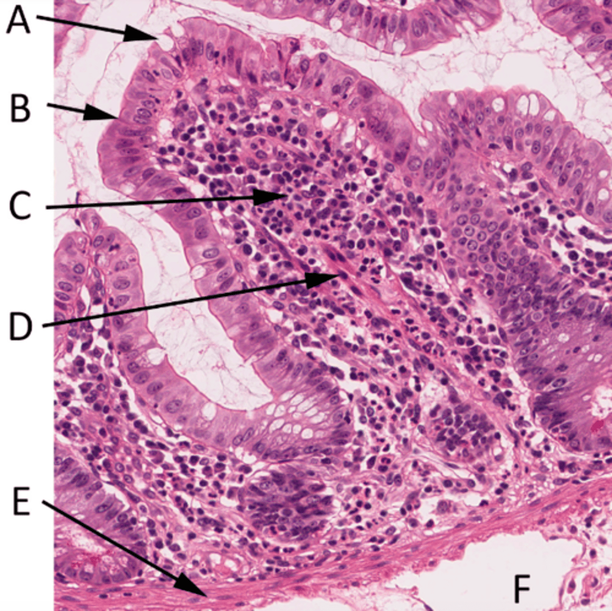
Histological section of villus
Identify structure
A =
B =
C =
D =
E =
F =
A = Goblet cell
B = Enterocytes (simple columnar epithelium)
C = Lamina propria
D = Isolated smooth muscle fibres
E = Muscularis mucosa
F = Blood vessels in submucosa
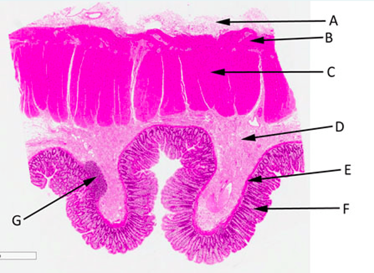
Histological section of region of colon
Identify structure
A =
B =
C =
D =
E =
F =
G =
A = Mesothelium comprised of connective tissue and simple squamous epithelium
B = Outer longitudinal smooth muscle
C = Inner circular smooth muscle
D = Region of loose or dense irregular connective tissue containing glands, adipose, nerve, immune cells and blood vessels
E = Structure that allows local movement of the mucosa
F = Mucous membrane comprised of simple columnar epithelium with supporting lamina propria and smooth muscle
G = Region of lymphoid tissue
Pathophysiology of Crohn's disease
Chronic inflammatory disorder affecting any region of the digestive tract, leading to fissuring (deep) ulceration of the gut wall