Physio
0.0(0)
Studied by 1 personCard Sorting
1/96
There's no tags or description
Looks like no tags are added yet.
Last updated 3:52 PM on 10/16/22
Name | Mastery | Learn | Test | Matching | Spaced | Call with Kai |
|---|
No analytics yet
Send a link to your students to track their progress
97 Terms
1
New cards
Non pacemaker cells
Stable resting potential; prolonged depolarization sustained by Ca++ influx
2
New cards
Cardiac output (CO)
- % distribution of blood?
- % distribution of blood?
CO LV = CO RV (in steady state)
- distributed among various organs via parallel arteries
25% --> GI
25% --> MSK
25% --> renal
15% --> cerebral
5% --> coronary
5% --> skin
- distributed among various organs via parallel arteries
25% --> GI
25% --> MSK
25% --> renal
15% --> cerebral
5% --> coronary
5% --> skin
3
New cards
Cardiac arrhythmias
- 3 causes
- 3 causes
caused by altered impulse formation, altered impulse conduction, or both altered impulse conduction /formation.
4
New cards
Heart failure
- caused by
- caused by
caused by defects in mechanical component (pump not functioning)
5
New cards
Ectopic foci
- results from
- results from
when other cells become pacemakers
- can be due to disease --> if they fire faster than the SA node ==> rapid abnormal heart rate
- can also be slow (ex. Purkinje fibers can fire APs but heart will beat much slower)
- can be due to disease --> if they fire faster than the SA node ==> rapid abnormal heart rate
- can also be slow (ex. Purkinje fibers can fire APs but heart will beat much slower)
6
New cards
Tachyarrhythmia
- results in:
- results in:
blood pressure can not be maintained → syncope, sudden death
7
New cards
Cardiomyocyte
- structure/features
- structure/features
striated, + by AP, troponin, involuntary, intercalated discs & gap junctions
8
New cards
Effect of hyperkalemia on heart rate
- Decrease HR
high extracellular K+ depolarizes the cell & decreases the full activation of funny channels
high extracellular K+ depolarizes the cell & decreases the full activation of funny channels
9
New cards
Electrical activity in cardiac cells
- What is it?
- How is it transmitted?
- What is it?
- How is it transmitted?
movement of ions & current flow
- transmitted to neighboring cells via intercalated disks
- transmitted to neighboring cells via intercalated disks
10
New cards
AV node conduction velocity
Slowest of the pacemakers which allows for ventricular filling
~ 0.5 m/s
~ 0.5 m/s
11
New cards
AV bundle
only tissue continuous between atria and ventricles (everywhere else surrounded by fibrous tissue)
12
New cards
Beta blockers affect on heart rate
Decrease heart rate
13
New cards
Non pacemaker cells
________: Stable resting potential; prolonged depolarization sustained by Ca++ influx.
14
New cards
Steady state
CO= VR
- Cardiac output of LV= cardiac output of RV
- Venous return equal in L and R heart
- Cardiac output of LV= cardiac output of RV
- Venous return equal in L and R heart
15
New cards
AV fibrous tissue function
barrier between atria & ventricles
- acts as insulator (prevents passage of impulse between them except through AV bundle)
- acts as insulator (prevents passage of impulse between them except through AV bundle)
16
New cards
Venous return (VR) in study state
VR of L heart = VR of R heart in steady state
17
New cards
Arterioles
come off arteries
- blood pressure regulation
- blood pressure regulation
18
New cards
Pacemaker cells
- Resting potential
- Comparison to cardiac muscle
- Resting potential
- Comparison to cardiac muscle
- Unstable resting potentials
- More negative than cardiac muscle
- Spontaneous depolarization/repolarization
- More negative than cardiac muscle
- Spontaneous depolarization/repolarization
19
New cards
Non-pacemaker cells
- Resting potential
- Resting potential
Stable resting potential
Prolonged depolarization sustained by Ca++ influx
Prolonged depolarization sustained by Ca++ influx
20
New cards
SANS effect on HR
NE → + B1 adrenergic R (Gs/GPCR)→ increase cAMP
- Increased HR, increased rate of conduction through AV node
- Increased HR, increased rate of conduction through AV node
21
New cards
PANS effect on HR
ACh → +M2 receptors (Gi/GPCR) → decrease cAMP 2 effects
1. decrease HCN → decrease Na+ influx & rate of phase 4 depolarization
2. decrease Ca+ channel phosphorylation → less Ca+ → AP threshold moves away from Vm
OVERALL: decreased HR, decreased rate of conduction through AV node
1. decrease HCN → decrease Na+ influx & rate of phase 4 depolarization
2. decrease Ca+ channel phosphorylation → less Ca+ → AP threshold moves away from Vm
OVERALL: decreased HR, decreased rate of conduction through AV node
22
New cards
Anti-arrhythmic drugs mechanism of action
can block Na+, K+, or Ca++ channels
23
New cards
Excitation-contraction coupling in cardiac muscle
Calcium mediated calcium release (Ca++ enters via L type VG Ca++ channels → triggers release of Ca++ from SR → muscle contraction)
24
New cards
Tachyarrhythmia
blood pressure can not be maintained → syncope, sudden death
25
New cards
Physiologic basis for conduction
local currents & gap junctions
26
New cards
Purkinje system conduction velocity
1.5 to 4 m/sec
Fastest!
- gap junctions
- syncytium (ventricular muscle mass contracts together)
Fastest!
- gap junctions
- syncytium (ventricular muscle mass contracts together)
27
New cards
Electrical component
regulates timing of mechanical component
28
New cards
AV nodal blocks
most clinically significant heart block (conduction block can also occur @ Bundle of His or at left or right bundle branches)
29
New cards
Pulmonary veins
- number
- pathway
- number
- pathway
4 (2 from each lung) --> bring oxygenated blood from the lungs to the LA
30
New cards
Pulmonary arteries
- number
- pathway
- number
- pathway
2; deoxygenated blood travels from right ventricle through the pulmonary semilunar valve into the lungs
31
New cards
Capillaries
- features
- features
where venules & arterioles meet
- single endothelial cell thick
- site of substance exchange
-very high surface area
- single endothelial cell thick
- site of substance exchange
-very high surface area
32
New cards
Passive mechanisms which maintain Em
1. Permeability of K+ >>> Na+ at rest (more K+ channels)
2. K+ concentration gradient
3. Non diffusible intracellular anions from negatively charged proteins
2. K+ concentration gradient
3. Non diffusible intracellular anions from negatively charged proteins
33
New cards
Active mechanisms which maintain Em
Na+-K+-ATPase
34
New cards
SA node
- Intrinsic rate
- Em
- Intrinsic rate
- Em
60-100 BPM
-55 to -60 mV
NATIVE PACEMAKER --> overdrive suppression
-55 to -60 mV
NATIVE PACEMAKER --> overdrive suppression
35
New cards
Intrinsic rate of:
AV node
AV node
40-60 BPM
36
New cards
Intrinsic rate of:
Bundle of His
Bundle of His
40 BPM
37
New cards
Intrinsic rate of:
Purkinje fibers
Purkinje fibers
15-20 BPM
38
New cards
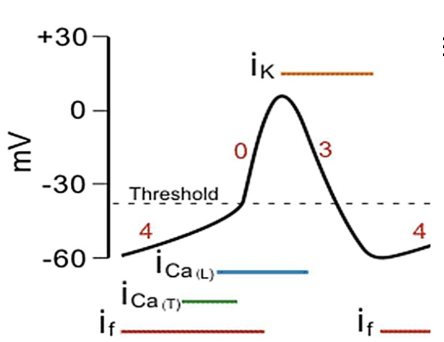
Non pacemaker cell action potential phases
0: Rapid depolarization (VG Na+ open & Na+ influx)
1: Initial repolarization (VG Na+ channels inactivate & K(to) open w/ K+ efflux)
2: Plateau (VG Ca++ open & Ca++ influx) --> ventricles contract
3: Repolarization (VG K+ open, K+ efflux)
4: Rest: outward K+ current
1: Initial repolarization (VG Na+ channels inactivate & K(to) open w/ K+ efflux)
2: Plateau (VG Ca++ open & Ca++ influx) --> ventricles contract
3: Repolarization (VG K+ open, K+ efflux)
4: Rest: outward K+ current

39
New cards
What is responsible for the plateau & why is it important?
- Phase 2: VG calcium channel opens and calcium enters the cell
- Ventricles contract
- Ventricles contract

40
New cards
Pacemaker cells action potential phases
4: Diastolic/spontaneous depolarization: Cation (typically Na+) influx via funny channels
0: Slow depolarization: VG Ca++ channels, Ca++ influx
3: Repolarization: K+ efflux ==> Maximum diastolic potential (-65 mV)
0: Slow depolarization: VG Ca++ channels, Ca++ influx
3: Repolarization: K+ efflux ==> Maximum diastolic potential (-65 mV)
41
New cards
Effect on HR:
Beta adrenergic agonists
Beta adrenergic agonists
Increase HR
42
New cards
Effect on HR:
Beta adrenergic antagonists
Beta adrenergic antagonists
Decrease HR
43
New cards
Effect on HR:
Hyperthyroidism
Hyperthyroidism
(Elevated T3, T4)
Increase HR
Increase HR
44
New cards
Effect on HR:
Hypothyroidism
Hypothyroidism
(decreased T3, T4)
Decrease HR
Decrease HR
45
New cards
Effect on HR:
Catecholamines
Catecholamines
Epinephrine primary one
- Increase HR
- Increase HR
46
New cards
Effect on HR:
Digoxin
Digoxin
Decreases HR, but makes it pump harder
47
New cards
Propagation & spread of heart cell depolarization
1. Ca++ triggers contraction: spreads through intercalated disks & connexons
2. Excitation-contraction coupling
--> calcium mediated Ca++ release ==> contracted sarcomere
2. Excitation-contraction coupling
--> calcium mediated Ca++ release ==> contracted sarcomere
48
New cards
Bradyarrhythmia
- Death of a pacemaker
Blood pressure cannot be maintained --> sudden death
Blood pressure cannot be maintained --> sudden death
49
New cards
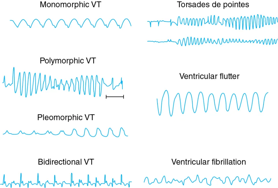
Types of tachyarrhythmia
- V tach
- V fib
- Torsades de pointes
- V fib
- Torsades de pointes
50
New cards
Intercalated disks contain:
- Gap junctions
- Connexins (channels formed by proteins in gap junctions)
- Desmosomes (firm mechanical attachments)
- Connexins (channels formed by proteins in gap junctions)
- Desmosomes (firm mechanical attachments)
51
New cards
Myocyte conduction pathway
SA node --> Atria --> AV node --> Bundle of His --> Purkinje fibers --> Ventricles
52
New cards
Conduction through the atria
- Ends of SA node fibers --> directly connect w/ surrounding atrial muscle fibers
- Velocity: 0.3 m/s
- Some fibers are faster & are located in the internodal pathways & interatrial band to the left atrium
- Velocity: 0.3 m/s
- Some fibers are faster & are located in the internodal pathways & interatrial band to the left atrium
53
New cards
Conduction through the AV node
AV DELAY = allow for ventricular filling & coronary circulation
- occurs due to decreasing number of gap junctions (increases resistance to flow of ions)
- approx. 0.16 seconds between origin in SA node before traveling to Bundle of His
- occurs due to decreasing number of gap junctions (increases resistance to flow of ions)
- approx. 0.16 seconds between origin in SA node before traveling to Bundle of His
54
New cards
Conduction through the Bundle of His
Delay of 0.4 seconds
- AV bundle: only tissue continuous between atria & ventricles
- AV bundle: only tissue continuous between atria & ventricles
55
New cards
Conduction through ventricles
Rapid conduction thanks to gap junctions & Purkinje system
Once impulse reaches the end of the Purkinje fibers --> must travel through the ventricular muscle mass & slows down to 0.3 - 0.5 m/s
Once impulse reaches the end of the Purkinje fibers --> must travel through the ventricular muscle mass & slows down to 0.3 - 0.5 m/s
56
New cards
Types of cardiac arrhythmias
- Re-entry/circus movements
- Conduction blocks
- Accessory pathways
- Conduction blocks
- Accessory pathways
57
New cards
Circus movements
Re-entry movements - type of arrhythmia
Normal tissue:
Purkinje twig splits into 2 --> AP traveling down each branch (can cancel each other out in the middle)
Diseased tissue: retrograde impulse can travel through diseased/weak tissue & re-excite the tissue
Normal tissue:
Purkinje twig splits into 2 --> AP traveling down each branch (can cancel each other out in the middle)
Diseased tissue: retrograde impulse can travel through diseased/weak tissue & re-excite the tissue
58
New cards
Causes of conduction block
- Ischemia
- Scarred tissue (disease)
- Refractory tissue (disease)
- Scarred tissue (disease)
- Refractory tissue (disease)
59
New cards
Drug treatment for conduction delay & mechanism:
symptomatic bradycardia
symptomatic bradycardia
Atropine can treat symptomatic bradycardia
- block ACh @ M2 receptors on nodal tissues
- block ACh @ M2 receptors on nodal tissues
60
New cards
ECG:
P wave
P wave
Atrial depolarization
61
New cards
ECG:
PR interval
PR interval
atria contract, AV node excitation
** AV delay
** AV delay
62
New cards
ECG:
QRS complex
QRS complex
Ventricular depolarization & atrial repolarization
- pushing blood out of the heart
- pushing blood out of the heart
63
New cards
ECG:
QT interval
QT interval
Covers the:
QRS complex - ventricular depolarization
ST segment - start of ventricular repolarization
QRS complex - ventricular depolarization
ST segment - start of ventricular repolarization
64
New cards
ECG:
ST segment
ST segment
- Correlates w/ phase 2
- Ventricles contracting & emptying (depolarized) = isoelectric
Start of ventricular repolarization
- Ventricles contracting & emptying (depolarized) = isoelectric
Start of ventricular repolarization
65
New cards
ECG:
T wave
T wave
Ventricular repolarization
66
New cards
ECG:
TP interval
TP interval
Ventricles relaxing & filling
67
New cards
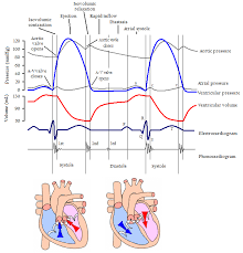
Cardiac cycle
see photo
68
New cards
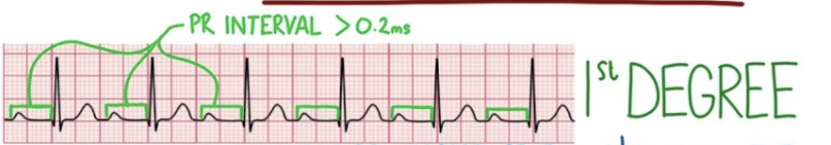
1st degree AV block
- Delayed conduction thru AV
- Still sinus rhythm
ECG: prolonged PR interval
- Still sinus rhythm
ECG: prolonged PR interval
69
New cards
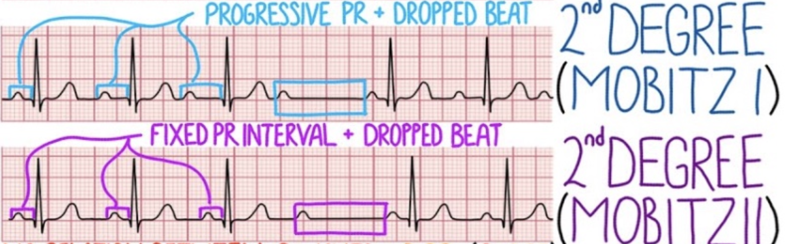
2nd degree AV block
Some AP do not proceed to ventricles --> ventricular bradycardia
Mobitz type 1: Progressive PR + dropped beats
Mobitz type 2: Fixed PR + dropped beats
* can be treated w/ antiarrhythmics
Mobitz type 1: Progressive PR + dropped beats
Mobitz type 2: Fixed PR + dropped beats
* can be treated w/ antiarrhythmics
70
New cards
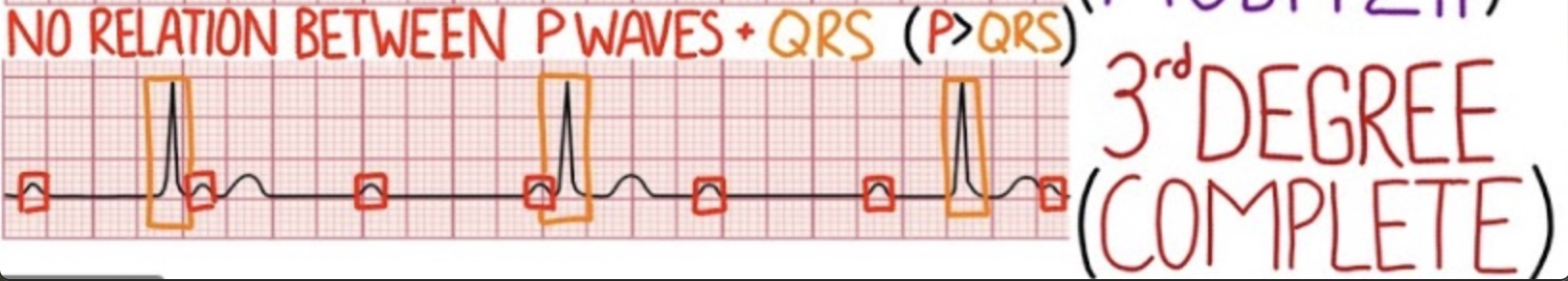
3rd degree AV block
- P > QRS
- complete dissociation between atria & ventricles
- latent pacemakers (Purkinje fibers) could take over --> ventricular bradycardia
- complete dissociation between atria & ventricles
- latent pacemakers (Purkinje fibers) could take over --> ventricular bradycardia
71
New cards
Ventricular depolarization sequence
1. depolarization of the heart from left to right in septum (small negative deflection ~ Q)
2. depolarization towards apex of the heart (~ R upstroke)
3. depolarization of the left ventricle (back to baseline, R downstroke )
2. depolarization towards apex of the heart (~ R upstroke)
3. depolarization of the left ventricle (back to baseline, R downstroke )
72
New cards
MEA
mean electrical axis; net vector of depolarization
73
New cards
effect of ventricular mass on deflection size
larger mass --> larger deflection
74
New cards
LVH on EKG
larger amplitudes
75
New cards
EKG
- U wave
- U wave
only seen in pathology
ex. could be seen in hypokalemia
ex. could be seen in hypokalemia
76
New cards
Normal intervals:
P wave
P wave
0.08 - 0.10 seconds
77
New cards
Normal intervals:
PR interval
PR interval
0.12 - 0.20 seconds
78
New cards
Normal intervals:
QRS
QRS
0.06 - 0.10 seconds
79
New cards
Normal intervals:
QTc
QTc
QT/square root of RR
less than or equal to 0.44 seconds
less than or equal to 0.44 seconds
80
New cards
Bipolar standard limb leads
Leads I, II, III
81
New cards
Unipolar augmented leads
aVR, aVL, aVF
82
New cards
unipolar chest leads
& placement
& placement
V1-V6
V1 R 4th IC space
V2 L 4th IC space
V4 mid clavicular line 5th IC
V6 mid axillary line 5th IC
V1 R 4th IC space
V2 L 4th IC space
V4 mid clavicular line 5th IC
V6 mid axillary line 5th IC
83
New cards
Lateral leads
& coronary circulation
& coronary circulation
I, aVL, V5, V6
LCx
LCx
84
New cards
Inferior leads
& coronary circulation
& coronary circulation
II, III, aVF
RCA and/or LCx
RCA and/or LCx
85
New cards
Anterior leads
& coronary circulation
& coronary circulation
V3, V4
LAD
LAD
86
New cards
Septal leads
& coronary circulation
& coronary circulation
V1, V2
LAD
LAD
87
New cards
Normal intervals
MEA of:
- aVR
MEA of:
- aVR
P = negative
QRS = negative, exaggerated R
T = inverted
QRS = negative, exaggerated R
T = inverted
88
New cards
Normal intervals
MEA of:
- aVL
MEA of:
- aVL
P = small
QRS = biphasic
T = normal
QRS = biphasic
T = normal
89
New cards
Normal intervals
MEA of:
- aVF
MEA of:
- aVF
P = normal
QRS = positive, exaggerated R, small S
T = normal
QRS = positive, exaggerated R, small S
T = normal
90
New cards
MEA moves __ hypertrophy
MEA moves towards hypertrophy
91
New cards
MEA moves __ heart attack
MEA moves away from heart attack
92
New cards
Rhythms which lack a P wave
Atrial fibrillation
Atrial flutter
Sinus arrest with escape rhythm
Atrial flutter
Sinus arrest with escape rhythm
93
New cards
EKG characteristics of:
Atrial fibrillation
Atrial fibrillation
Lacking P wave
Atrial fibrillatory waves
Decreased amplitude & increased frequency
Atrial fibrillatory waves
Decreased amplitude & increased frequency
94
New cards
EKG characteristics of:
Atrial flutter
Atrial flutter
Saw tooth pattern
Coarse fibrillatory waves
Irreg. irreg.
Coarse fibrillatory waves
Irreg. irreg.
95
New cards
EKG characteristics of:
Sinus arrest w/ escape rhythm
Sinus arrest w/ escape rhythm
retrograde atrial stimulation
P and QRS synchronized
- Small QRS & no P
Bradycardia
P and QRS synchronized
- Small QRS & no P
Bradycardia
96
New cards
Ventricular problems on EKG
PVC
V tach
V fib
V tach
V fib
97
New cards
STEMI
- ST segment corresponds with which phase of AP
- ST segment corresponds with which phase of AP
Stage 3
transmural infarct involving the entire wall thickness of a ventricular region --> ischemic tissue becomes depolarized because of its inability to maintain normal ion gradients across the cell membranes
transmural infarct involving the entire wall thickness of a ventricular region --> ischemic tissue becomes depolarized because of its inability to maintain normal ion gradients across the cell membranes