Osteology of the Skull - Sphenoid and Temporal Bones
 Call Kai
 Learn
Learn Practice Test
 Spaced Repetition
 Match
Match Flashcards
 Knowt Play
1/101

There's no tags or description
Looks like no tags are added yet.
Name | Mastery | Learn | Test | Matching | Spaced | Call with Kai | Chat |
|---|
No analytics yet
Send a link to your students to track their progress
102 Terms
- unpaired
- primarily endochondral ossification
The sphenoid is
sphenoid bone
Superior view: this is the
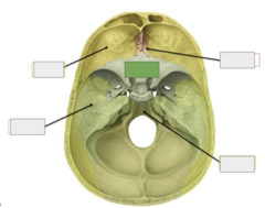
ethmoid
Superior view: this is the
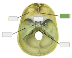
occipital bone
Superior view: this is the
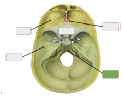
temporal bone
Superior view: this is the
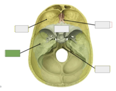
frontal
Superior view: this is the

the groove for middle meningeal a.
This is

the carotid sulcus
This is
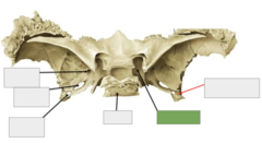
the clivus
This is
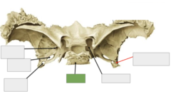
also the foramen spinosum
This is the

also the foramen ovale
This is the
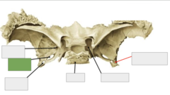
also the foramen rotundum
This is the
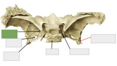
of the superior orbital fissure
This is a different view
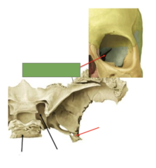
the superior orbital fissure
This is

carotid sulcus
This is the
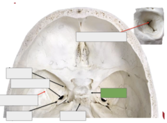
clivus
This is the
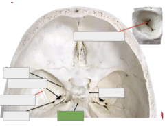
foramen spinosum
This is the
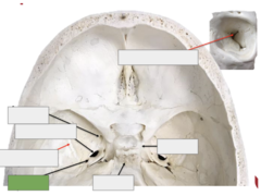
groove for middle meningeal a.
This is the
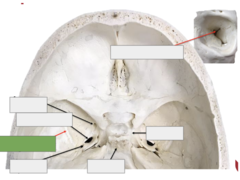
foramen ovale
This is the

the foramen rotundum
This is
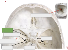
the jugum sphenoidale
Cranial fossa: this is
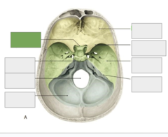
is the dorsum sellae
Cranial fossa: this

foramen magnum
Cranial fossa: this is the
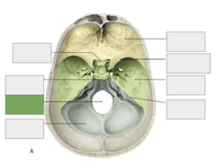
posterior cranial fossa
Cranial fossa: this is the

petrous ridge/crest
Cranial fossa: this is the

middle cranial fossa
Cranial fossa: this is the
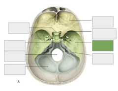
sphenoid bone, lesser wing
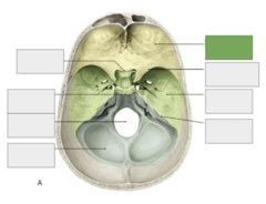
Cranial fossa: this is the

anterior cranial fossa
Cranial fossa: this is the
palatine
Inferior view: this is the
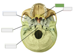
vomer
Inferior view: this is the
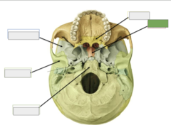
occipital
Inferior view: this is the
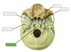
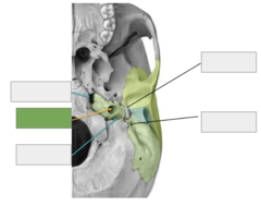
temporal
Inferior view: this is the

sphenoid
Inferior view: this is the
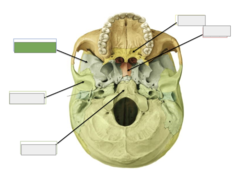
foramen ovale & spinosum
This is the

is the medial pterygoid plate
Pictured here
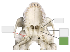
is the lateral pterygoid plate
Pictured here
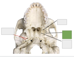
is the pterygoid hamulus
Pictured here
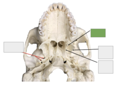
Pterion
Lateral view: this is the

lesser wing
This is the
greater wing
This is the
the body
This is

pterygoid process
This is the
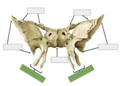
the lateral pterygoid plate
Anterior view: This is
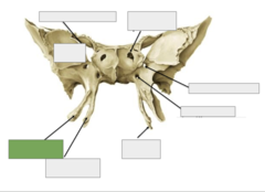
the medial pterygoid plate
Anterior view: This is
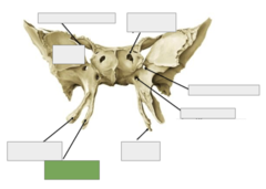
the pterygoid hamulus
Anterior view: This is
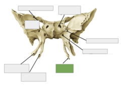
pterygoid canal
Anterior view: This is the
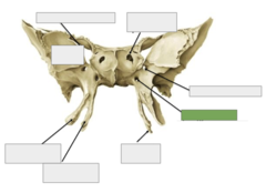
foramen rotundum
Anterior view: This is the

orbital surface
Anterior view: This is the
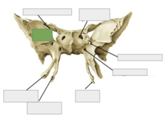
aperture of sphenoid sinus
Anterior view: This is the
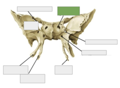
superior orbital fissure
Anterior view: This is the
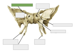
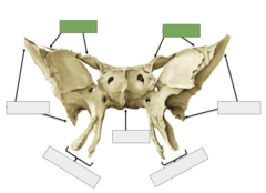
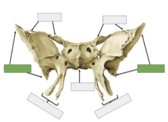
greater wings
Posterior view: These are the
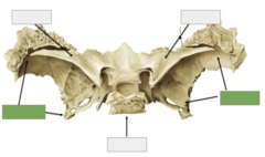
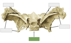
body
Posterior view: This is the
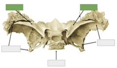
the lesser wings
Posterior view: These are
optic foramen
Posterior view: This is the
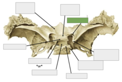
anterior clinoid process
Posterior view: This is the
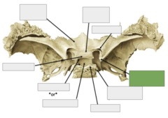
posterior clinoid process
Posterior view: This is the
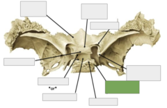
the dorsum sellae
Posterior view: This is
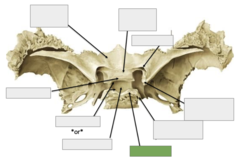
the hypophysial fossa
Posterior view: This is
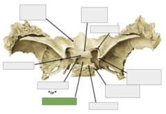
sella turcica
Posterior view: This is the
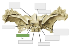
the tuberculum sellae
Posterior view: This is
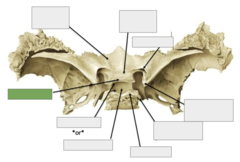
chiasmatic groove
Posterior view: This is the
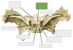
jugum sphenoidale
Posterior view: This is the
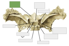
inferior orbital fissure
Inferolateral view: This is the

pterygopalatine fossa (space)
Inferolateral view: This is the
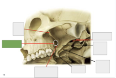
pterygomaxillary fissure (opening)
Inferolateral view: This is the
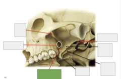
pterygoid hamulus
Inferolateral view: This is the
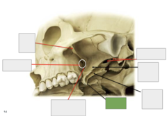
medial pterygoid plate
Inferolateral view: This is the

lateral pterygoid plate
Inferolateral view: This is the
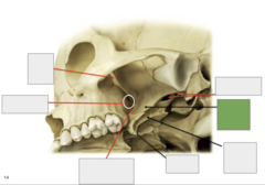
foramen ovale and spinosum
Inferolateral view: This is the
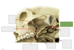
groove for middle meningeal a
5

tuberculum sellae
12
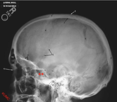
dorsum sellae
13
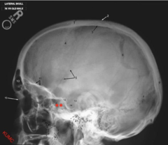
hypophysial fossa
14

is the sphenoid sinus
**
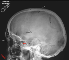
wings

sphenoid sinus

lateral pterygoid plate
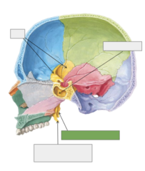
medial pterygoid plate (and hamulus)

- paired bones
- petrous part = endochondral
- squamous part = intramembranous
- mastoid part = intramembranous
- tympanic part = intramembranous
Temporal bones are
petrous part
the only part of the temporal bone that is formed via endochondral ossification is the
squamous, mastoid, tympanic
these parts of the temporal bone are formed via intramembranous ossification
squamous
This is the

petrous
This is the
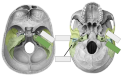
tympanic
This is the
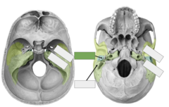
mastoid
This is the
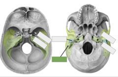
zygomatic process
This is the
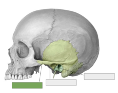
anterior tubercle
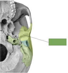
This is the

mandibular fossa
This is the

anterior eminence
This is the
mastoid process (air cells within)
Mastoid part: this is the
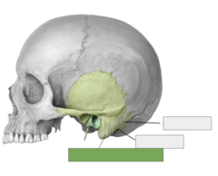
mastoid notch
Mastoid part: this is the
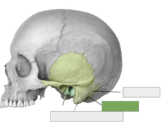
mastoid foramen
Mastoid part: this is the

jugular foramen
This is the
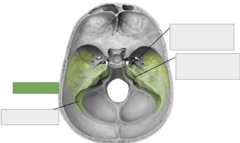
groove for sigmoid sinus
This is the
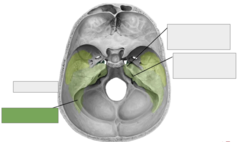
internal acoustic meatus (canal inside bone)
This is the
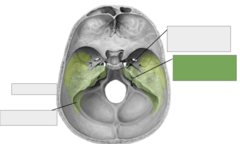
internal opening of carotid canal (deep to arrow)
This is the
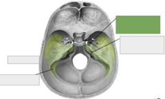
styloid process
This is the
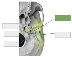
stylomastoid foramen
This is the

jugular foramen
This is the
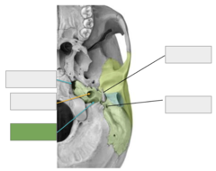
carotid foramen (to canal)
This is the