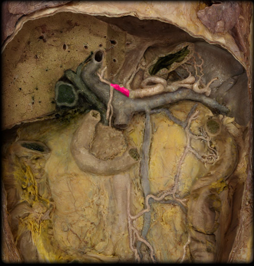
IU ANAT-A 225 Block 5
 Call Kai
 Learn
Learn Practice Test
 Spaced Repetition
 Match
Match Flashcards
 Knowt Play
1/273

There's no tags or description
Looks like no tags are added yet.
Name | Mastery | Learn | Test | Matching | Spaced | Call with Kai | Chat |
|---|
No analytics yet
Send a link to your students to track their progress
274 Terms
Gastrointestinal (GI) tract
tube that extends from mouth (oral cavity) to anal canal, includes: oral cavity, pharynx, esophagus, stomach, small intestine, large intestine, anal canal
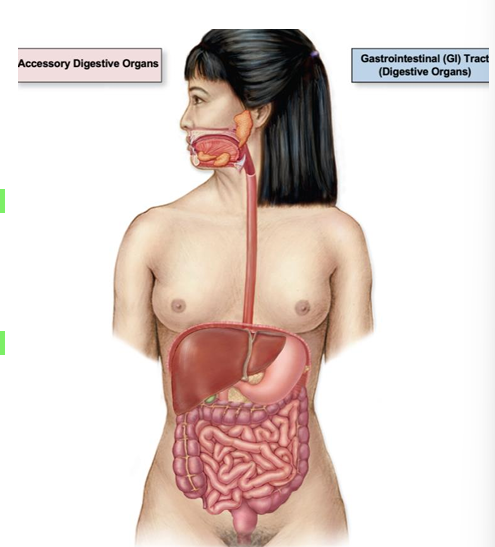
peristalsis
waves of contraction, pushes food further along
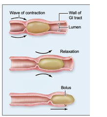
Accessory Digestive Organs
not part of GI tract, but assist with digestion, tongue, teeth, salivary glands, liver, gallbladder, pancreas
Mechanical digestion
physically breaking down food, teeth chewing, stomach muscle churning
Chemical digestion
salivary glands/saliva uses enzymes to break down food
Absorption
water and food molecules pass through the intestines and move into other structures
GI lymphatics (lacteals)
absorb dietary lipids and lipid-soluble vitamins
Blood vessels
absorb all other digested nutrients
parietal peritoneum
outer lining of abdominopelvic cavity
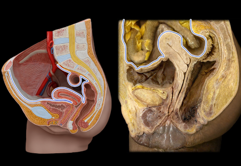
visceral peritoneum
inner lining of abdominopelvic cavity
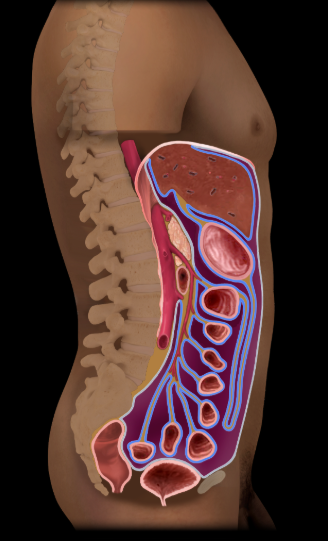
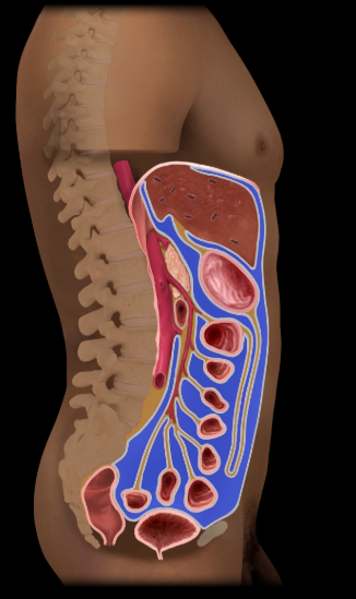
peritoneal cavity
spaces in abdominopelvic cavity
Oral cavity
continuous posteriorly with oropharynx, nonkeratinized stratified squamous epithelium, includes palate, teeth, tongue, salivary glands
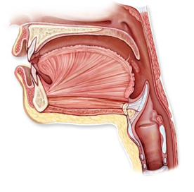
hard palate
anterior 2/3 of palate, bony, palatine bone and maxilla
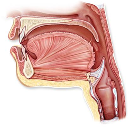
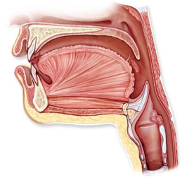
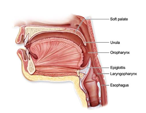
soft palate
posterior 1/3 of palate, formed from skeletal muscle, contains uvula
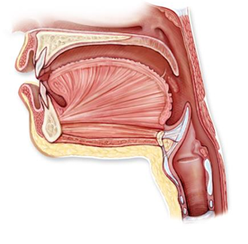
uvula
rises to close off the nasopharynx when we swallow
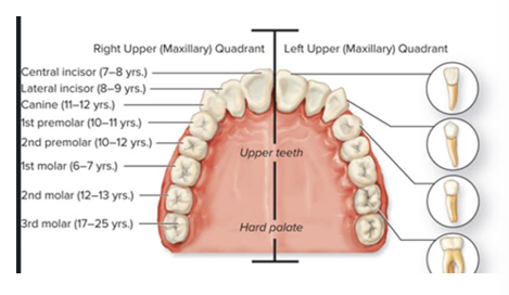
permanent teeth
32, 2 incisors, 1 canine, 2 premolars, 3 molars on each side
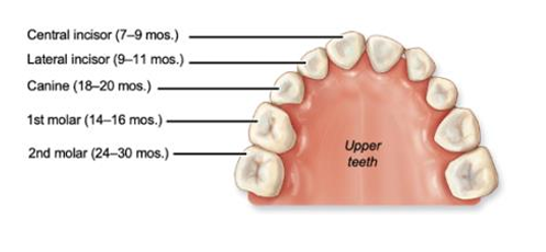
deciduous (baby) teeth
2 incisors, 1 canine, 2 molars on each side
Tongue
skeletal muscle covered with mostly nonkeratinized stratified squamous epithelium, perceives taste, mechanical digestion
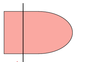
tongue innervation
motor: CN XII - Hypoglossal, taste: CN VII - facial and CN IX - glossopharyngeal, sensation: CN V - trigeminal, and CN IX - glossopharyngeal
salivary glands
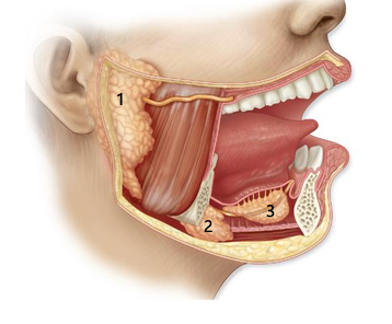
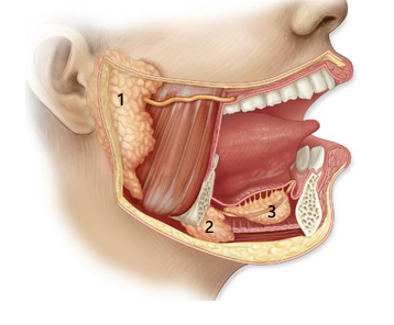
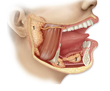
three pairs, parotid, submandibular, sublingual, secrete saliva
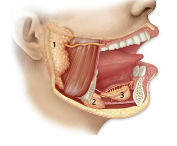
parotid gland
largest of the salivary glands and are located on each side of the head anterior and inferior to the ear, partially overlying the masseter muscle
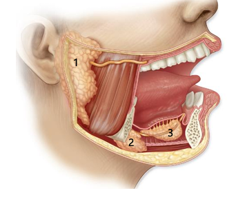
submandibular gland
2
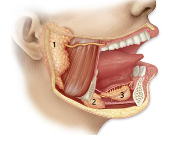
sublingual gland
3
functions of saliva
dissolves food molecules for taste, moistens food, cleanses mouth, chemical digestions of carbs, lysozyme and antibodies in saliva inhibit bacterial growth in mouth, innervated by CN IX - glossopharyngeal nerve
Pharynx
throat, skeletal muscle, skeletal muscle tube that connects nasal cavity and mouth to larynx and esophagus, skeletal muscle propels food and drink to esophagus or air to larynx, only oropharynx and laryngopharynx conduct both food and air
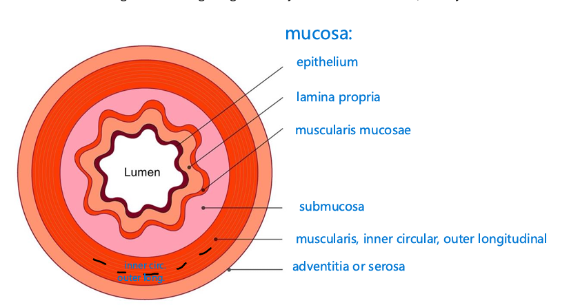
Mucosa
innermost tunic, esophagus: nonkeratinized stratified squamous, stomach to anal canal: simple columnar epithelium
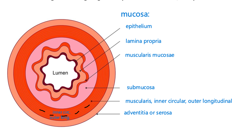
lamina propria
areolar connective tissue
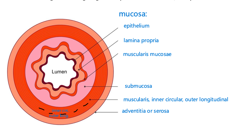
muscularis mucosae
thin layer of smooth muscle
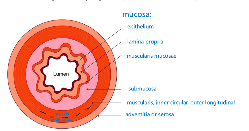
submucosa
connective tissue, contains major blood vessels
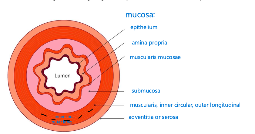
muscularis
2 layers of smooth muscle, inner circular and outer longitudinal, esophagus has two layers of skeletal and smooth muscle, and the stomach has 3 layers of smooth muscle, responsible for peristalsis
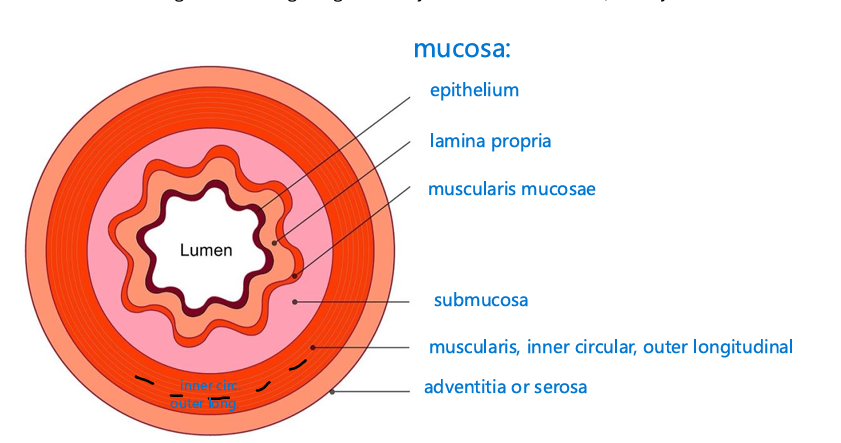
Serosa
outermost layer, visceral peritoneum + areolar connective tissue
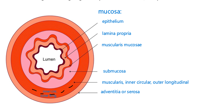
adventitia
outermost layer, areolar connective tissue only
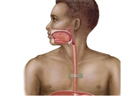
esophagus
propels food to the stomach, muscular tube that connects the pharynx to the stomach
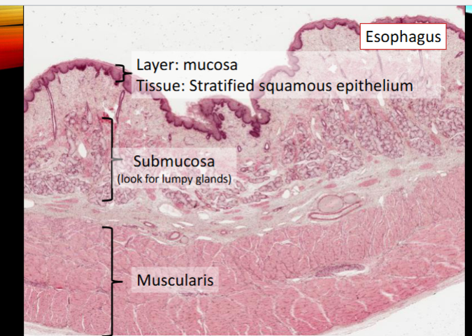
esophagus mucosa
nonkeratinized stratified squamous epithelium (histology)
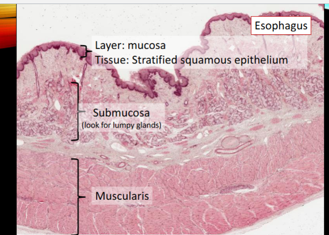
esophagus muscularis
2 layers of muscle, upper 1/3: skeletal muscle, middle 1/3: skeletal + smooth muscle, lower 1/3: smooth muscle only
reflux esophagitis (heartburn)
stomach contents reflux into esophagus, causing burning, risk factors: overweight, smoking, large meals before bedtime
vestibule
space between the teeth and the lips or cheeks
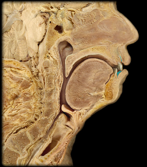
palatine tonsils
paired lateral masses of lymphatic nodules at the border between the oral cavity and the oropharynx
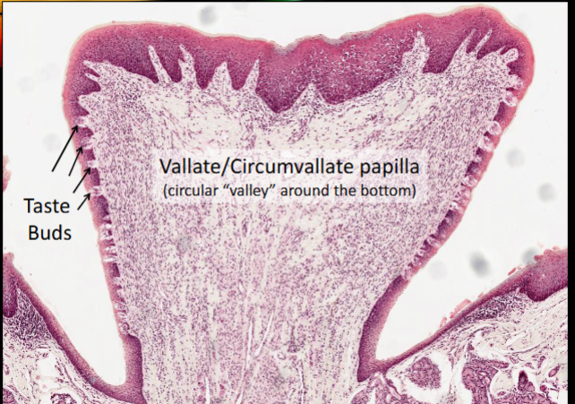
vallate papillae
form an inverted V a the boundary between the anterior two-thirds and posterior one-third of the surface of the tongue
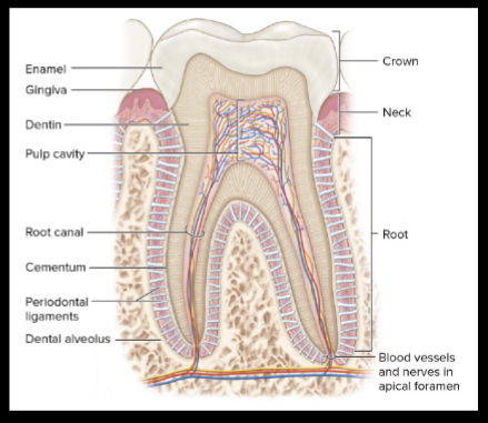
crown
extending into the oral cavity, covered by enamel
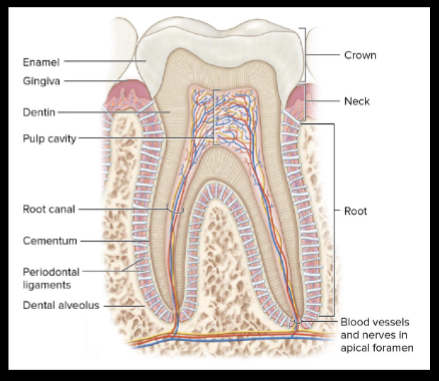
neck
narrow, slightly constricted region below the crown, in contact with the gums that cover the alveolar processes of the mandible and maxillae
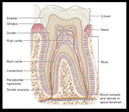
root
one or more to anchor the tooth into itsalveolus
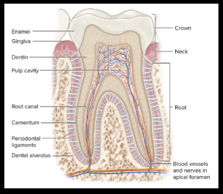
dentin
forms the bulk of the tooth; it is a hard, yellowish, calcified substance
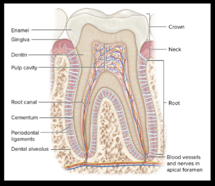
enamel
the hardest substance in the body and is composed largely of inorganic salts, the enamel covers the dentin of the crown
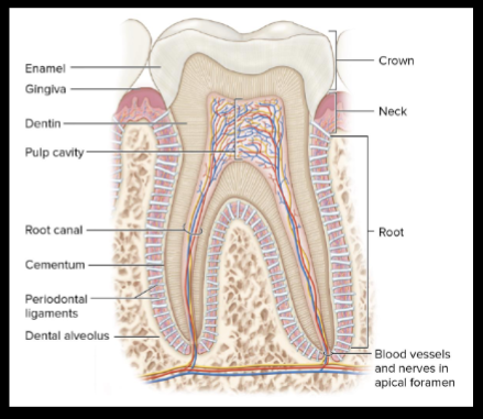
cementum
bonelike substance which surrounds the dentin of the root and helps anchor the tooth
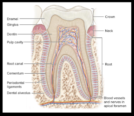
pulp
connective tissue with blood vessels and nerves found in pulp cavity
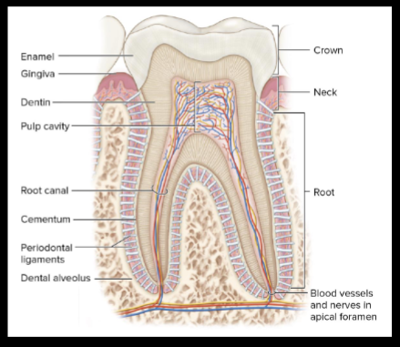
pulp cavity
holds pulp
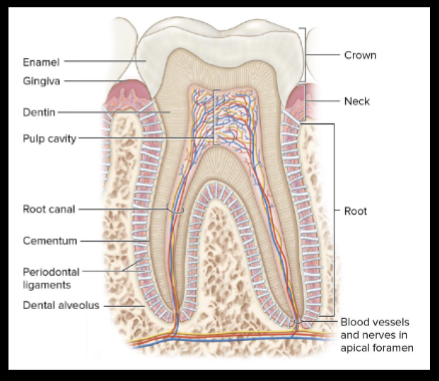
root canal
narrow core of each root
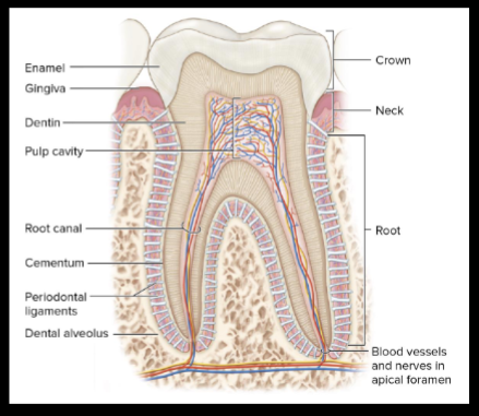
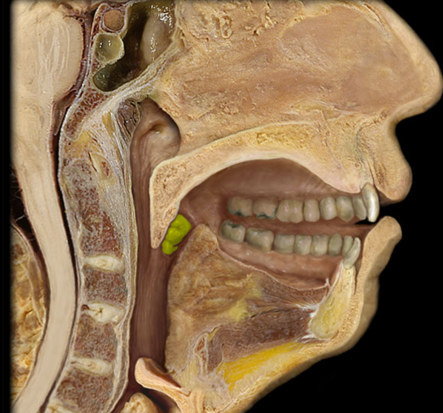
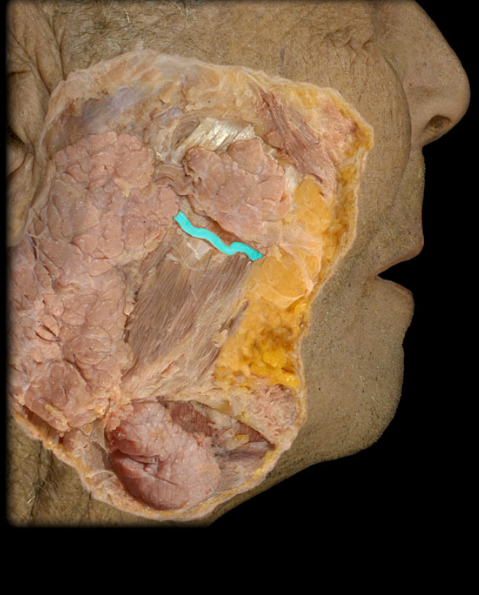
parotid duct
passes over the masseter muscle, pierces the buccinator muscle and enters into the vestibule of the oral cavity opposite the second upper molar tooth
periodontal ligaments
composed of dense regular connective tissue and run between the bone of the alveolus and the cementum to anchor the tooth
submandibular gland
located along the medial aspects of the mandible, each with superficial portion which is continuous posteriorly around the mylohyoid muscle with a larger deep portion
sublingual glands
on the anterior floor of the mouth, deep to the oral cavity mucosa
stomach
joins the esophagus and small intestine, shape and position are modified by its volume changes and surrounding structures
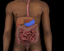
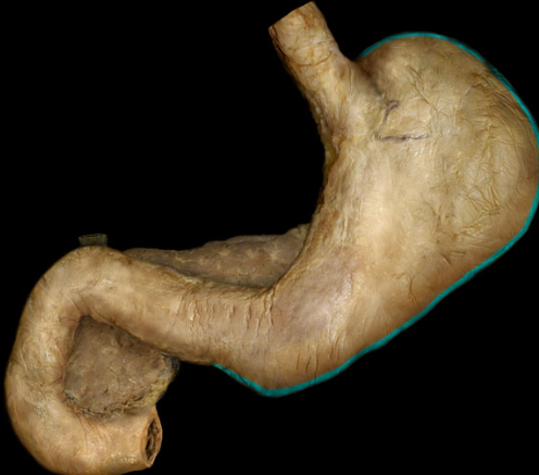
greater curvature
left and inferior border of stomach, attached to the greater omentum
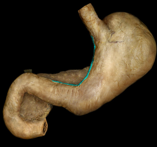
lesser curvature
left and inferior border of the stomach, attached to the lesser omentum
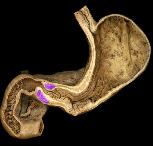
pyloric sphincter
thick ring of smooth muscle in the pyloric orifice
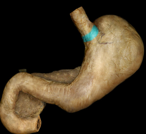
cardiac
area immediately surrounding the cardiac orifice
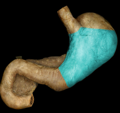
body
the main, central portion of the stomach
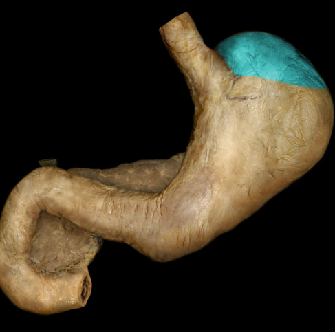
fundus
bulges superior to and to the left of the cardiac orifice
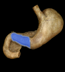
pylorus
tapers toward the pyloric sphincter at the distal end of the stomach
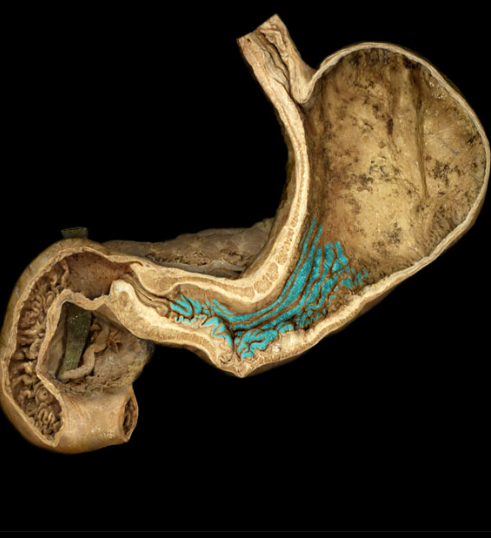
gastric folds
branching folds which run longitudinally, these stretch when stomach is full
stomach mucosa
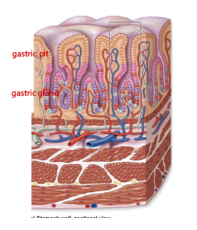
simple columnar epithelium, mucosa contains gastric pits, branching off gastric pits are gastric glands
stomach muscularis
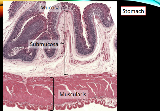
3 layers of smooth muscle, not 2, has innermost oblique layer, inner circular, and outer longitudinal
small intestine
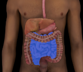
form pyloric sphincter to ileocecal valve to join the large intestine, duodenum, jejunum, and ileum. Finishes chemical digestion, absorbs 90% of nutrients
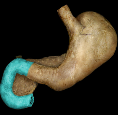
duodenum
first and shortest segment of small intestine, behind the parietal peritoneum, receives chyme from stomach, receives bile from liver and pancreatic enzymes through duodenal papilla
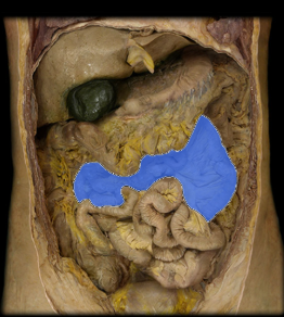
jejunum
second segment of small intestine, 2/5 of the small intestine, huge surface area for absorption
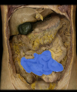
ileum
final 3/5 of the small intestine, loosely attached to the mesentery, connects cecum to large intestine, few folds but contains more MALT
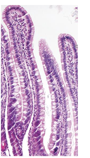
small intestine mucosa
simple columnar epithelium with goblet cells and microvilli, with lacteals
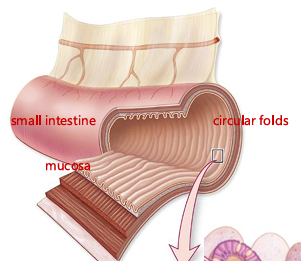
small intestine circular folds
folds of mucosa and submucosa, increase surface area for nutrient absorption, slow down chyme and allow it to mix with intestinal juices, allowing time for full nutrient absorption
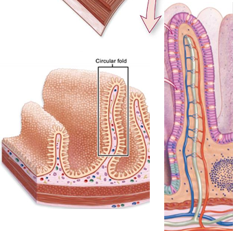
villi
folds on small intestine mucosa only, increase surface area, provide great distance for material to be ingested and absorbed, in the center of each villus are blood vessels and a lacteal
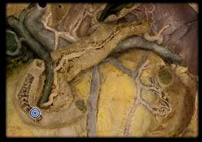
major duodenal papilla
helps bring bile and pancreatic enzymes into the duodenum
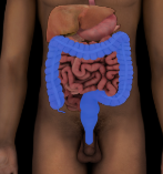
large intestine
extends form the end of the ileum to the anus, absorption of water and electrolytes, storage of undigested material until it can be expelled
cecum
connects to ileum, vermiform appendix attaches to this, lower right side of the abdominal cavity as, short wide pouch
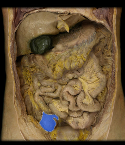
ascending colon
right side of body, ascends along the posterior abdominal wall from the cecum to near the liver
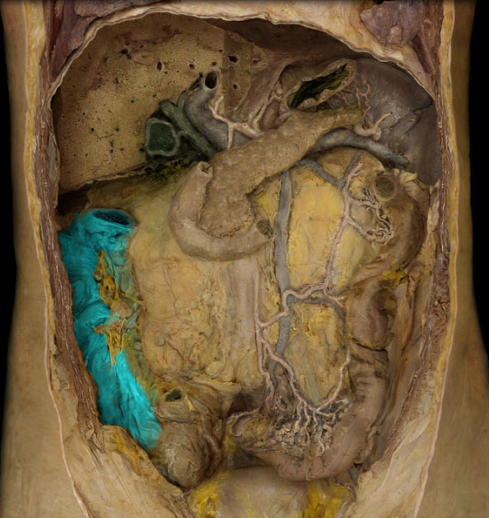
transverse colon
runs from right to left side of body
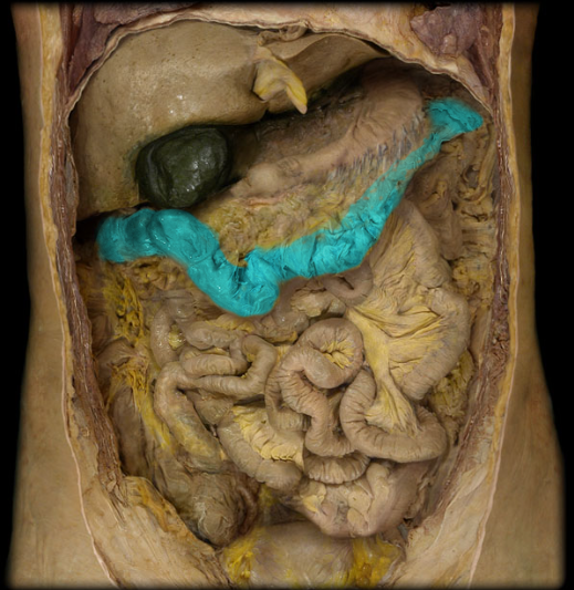
greater omentum
hangs inferiorly from the transverse colon in front of the small intestine
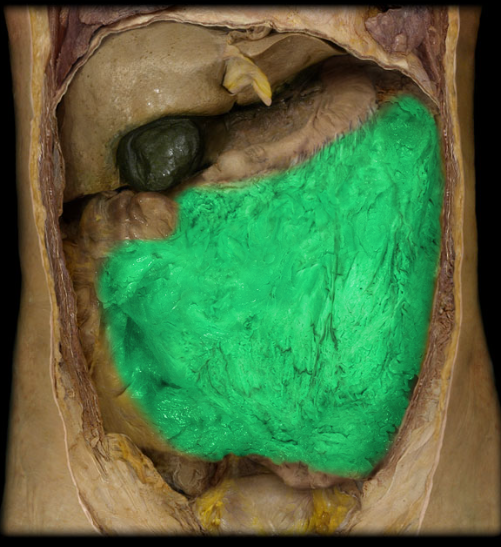
descending colon
descends along the posterior body wall from near the spleen to the pelvic region
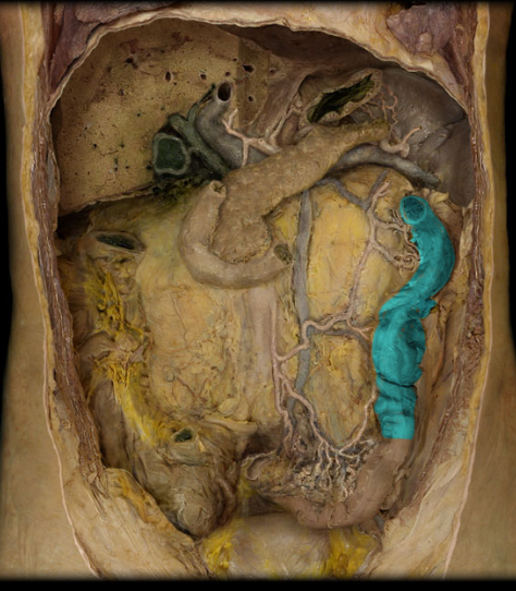
sigmoid colon
as the descending colon enters the pelvis, it curves toward the midline of the body in an S-shape
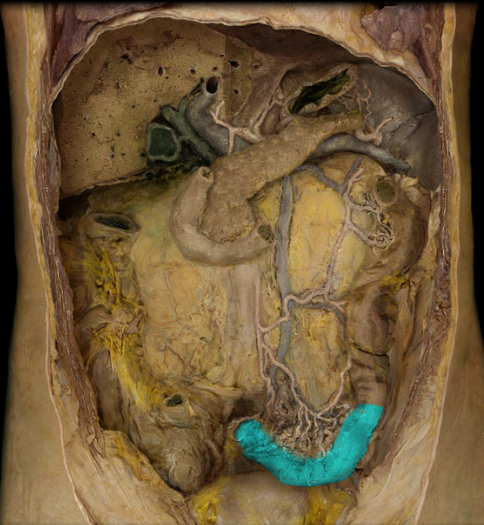
rectum
begins where the sigmoid colon passes beneath the parietal peritoneum and runs inferiorly along the sacrum toward the pelvic diaphragm

anal canal
last and shortest segment of large intestine, begins at the pelvic diaphragm and ends at the anus
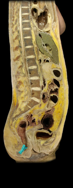
anal sphincter
anal canal surrounded by internal and external muscles
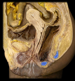
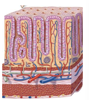
large intestine mucosa
simple columnar epithelium with goblet cells
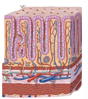
large intestine muscularis
2 layers of smooth muscle, outer longitudinal layer consists of teniae coli, and haustra
teniae coli
longitudinal bundles of smooth muscle fibers forming three distinct bands
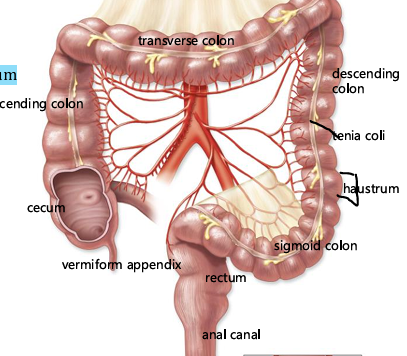
haustra
large intestine wall tends to form a series of sac-like structures
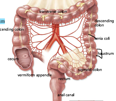
omental appendices
hanging from the haustra are folds of peritoneum filled with adipose tissue
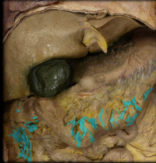
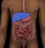
liver
produces and secretes bile, processes the products of digestion, stores glycogen, largest internal gland and is situated under the diaphragm in the upper right part of the abdominal cavity
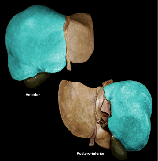
right lobe
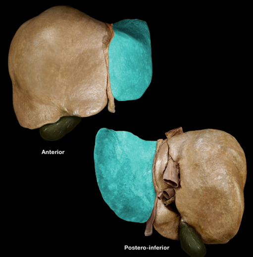
left lobe
quadrate lobe
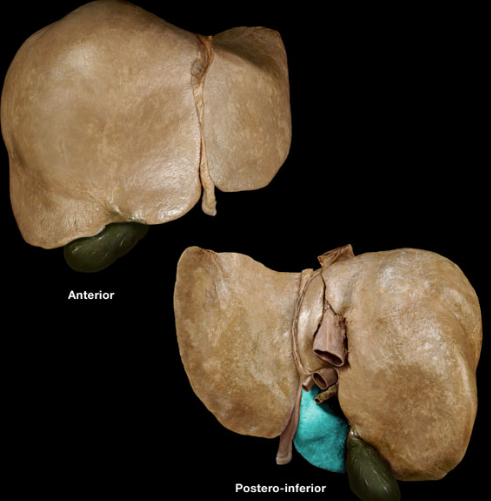
caudate lobe
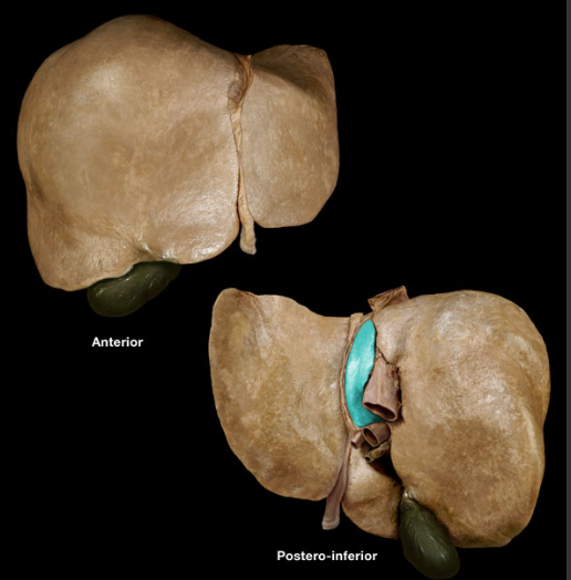
gallbladder
bile produced by the liver is carried by ducts to here where it is stored and concentrated until needed, by the inferior surface of the liver at the medial border of the right lobe
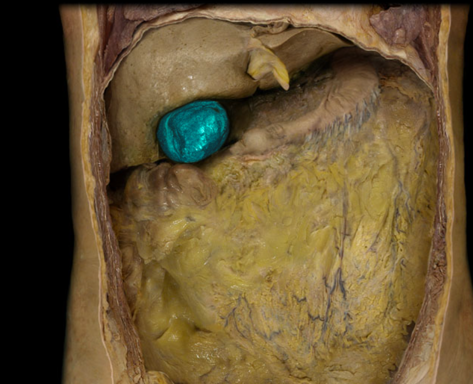
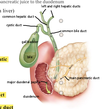
common hepatic duct
receives smaller ducts from the lobes of the liver and is joined by a duct from the gallbladder
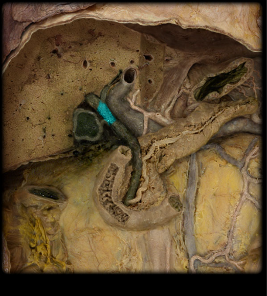
cystic duct
duct from the gallbladder, carries bile from the common hepatic duct to the gallbladder, or from the gallbladder to the common bile duct
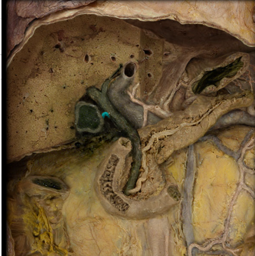
common bile duct
runs from the junction of the common hepatic duct and cystic duct to the duodenal wall, it is joined by the main pancreatic duct to enter the duodenum at the major duodenal papilla
pancreas
located behind the stomach and runs from the inner curve of the duodenum to the vicinity of the spleen, acinar cells secrete pancreatic juice
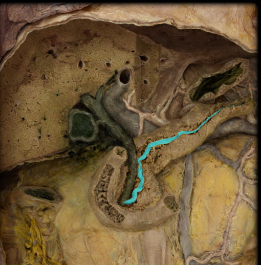
pancreatic duct
runs the length of the pancreas to join the common bile duct at the duodenum, pancreatic juice leaves via this
celiac trunk
supplies abdominal digestive organs
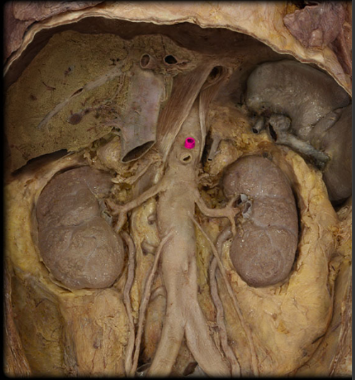
left gastric artery
branch of celiac trunk
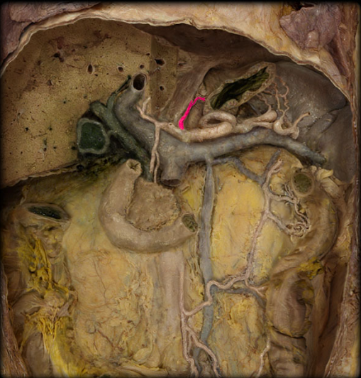
common hepatic artery
branch of celiac trunk