Module 1: Cells as the basis of life - biology
1/46
There's no tags or description
Looks like no tags are added yet.
Name | Mastery | Learn | Test | Matching | Spaced | Call with Kai | Chat |
|---|
No analytics yet
Send a link to your students to track their progress
47 Terms
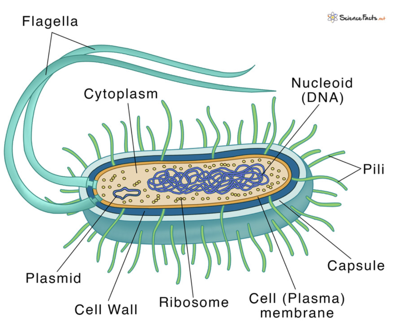
Prokaryotic cells
Prokaryotic cells are unicellular organisms that:
Do NOT have nucleus or any other membrane bound organelles
DNA floats around in the cytoplasm, is small and circular
Examples: archaea, bacteria
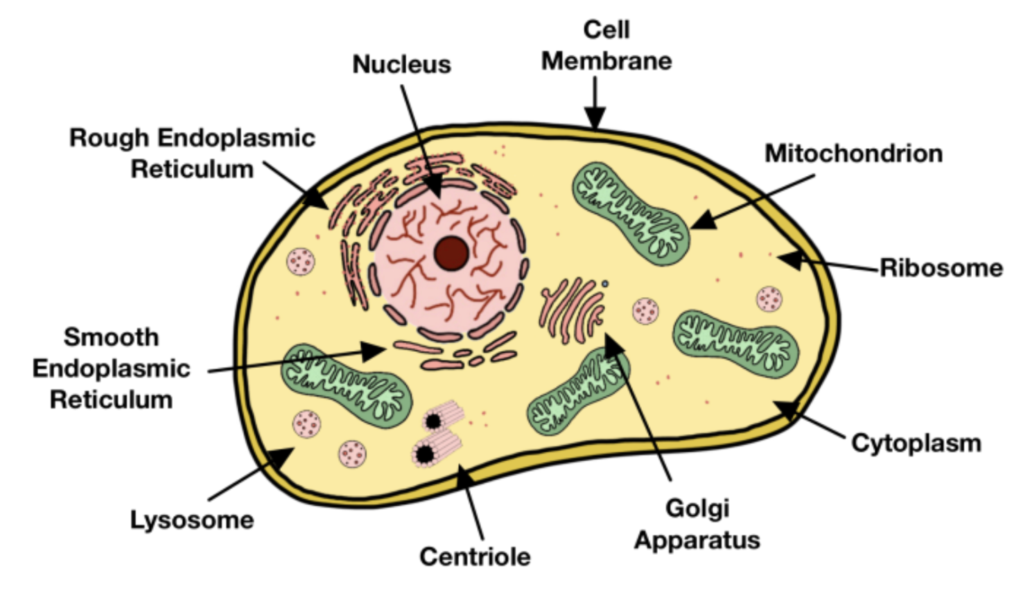
Eukaryotic cells
Eukaryotic cells are unicellular or multicellular organisms that:
Have a nucleus
Has membrane bound organelles
E.g. mitochondria, endoplasmic reticulum
Much more complex
DNA is contained within the nucleus, is straight and is a relatively large amount
Examples: animals, plants, protists, fungi
Light microscope - How does it work?
Most simple and commonly used
Uses visible light and two lenses to magnify a specimen
Light passes through the specimen
Light is refracted by the objective and ocular lenses
Light is refracted (bent) to form a magnified image
Light microscope - Specimen preparation
A specimen may be a whole organism, a smear of cells or a thin slice of tissue
Put specimen on glass microscope slide
Add dye to stain certain structures
Add a drop of fluid and a coverslip
Cell staining technqiue
Staining used to enhance appearance of cell structures
Involves adding a dye to the specimen
Which stains certain cell components e.g. cell wall
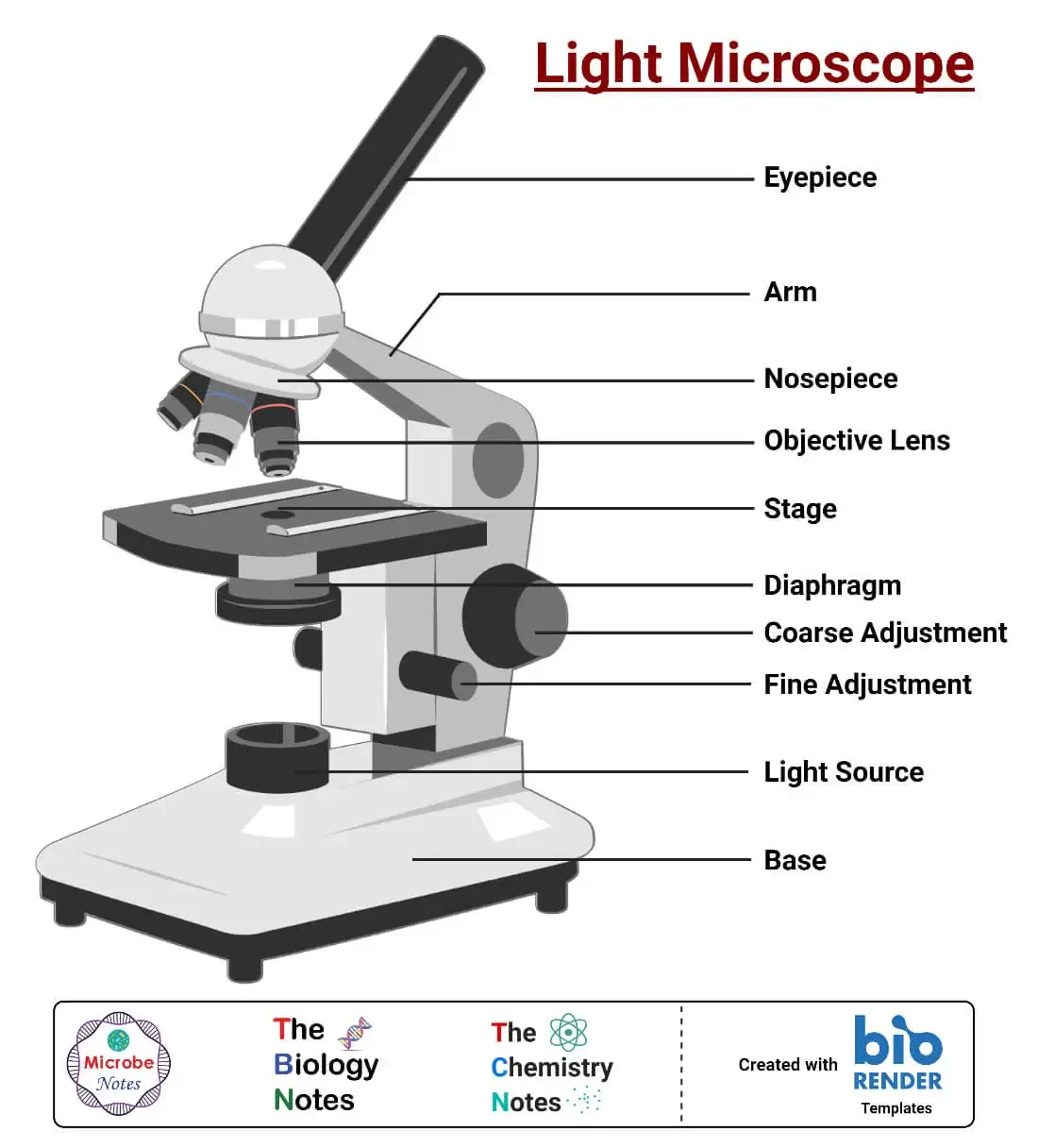
Light Microscopy set up
Place the microscope on a flat bench and adjust it to a comfortable height
Plug it in and turn the light on
Put the lowest power objective lens (4x) into position
Adjust the light using the diaphragm and focus carefully
Lower the objective lens using the coarse focus knob
Focus using the knobs to get a clear image
For a higher magnification switch to a higher objective and refocus
Magnification definition
The process of enlarging the apparent size of an object
Magnification formula
Image size / Actual size
Total magnification
The total magnification equals ocular lens magnification multiplied by objective lens magnification
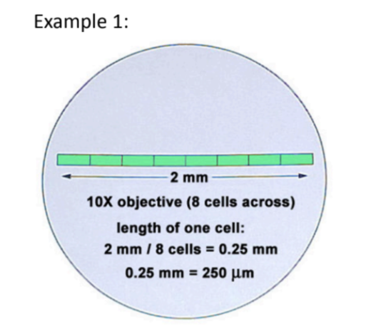
Magnified size
Magnified size is the diameter of the field of view divided by the number of cells that fit across it
Microscope Magnification Summary Table
Ocular lens | Objective lens | Total magnification | Diameter of F.O.V |
10x | 4x | 40x | 4.5 mm |
10x | 10x | 100x | 450 μm |
10x | 100x | 1000x | 150 μm |
Confocal microscope
An advanced light microscope
Works by passing a highly focused laser through the specimen
Creates a high quality image of a section of the specimen
Moving the laser slightly creates another image
A computer then enhances the images and stitches the sections all together to create a 3D image
Fluorescent microscope
An type of light microscope
The sample is labelled with a fluorescent substance that will attach to the structures that the scientist wants to observe.
The sample is illuminated with a high intensity source of light that causes the fluorescent substance to emit light.
This fluorescent light is directed through filters that separate it from surrounding light and the viewer is able to see only those areas of the sample that are fluorescing.
Parts of light microscope
Electron microscope - How does it work?
Uses a beam of electrons and electromagnets to make the specimen look bigger
Electrons are small and sensitive and will bounce off everything
The internal chamber of the electron microscope is under vacuum conditions (no air)
An electron gum shoots a beam of electrons at the specimen (in a vacuum)
The electron beam is controlled by electromagnets
When the electrons hit/interact with the specimen, the beam gets scattered
The way the electrons scatter depends on the structure of the specimen
This scattering is detected by different machinery and a computer turns the information into an image (called an electron micrograph)
Transmission electron microscope - How does it work?
Treat specimen with chemicals which give it increased structural strength as the electron beam is extremely hot
Water is removed from the specimen using alcohol
As water evaporates immediately in a vacuum - which could destroy the specimen
Specimen is embedded in a resin
Cut specimen into super thin slices
Then one broad laser beam is shot at specimen
The electron micrograph is a detailed image of the inside of the specimen
A TEM creates a 2D image of the ultrathin slice.
It can magnify up to 1,500,000x and has a resolution of about 2nm.
Scanning electron microscope - How does it work?
Treat specimen with chemicals which give it increased structural strength as the electron beam is extremely hot
Water is removed from the specimen using alcohol
As water evaporates immediately in a vacuum - which could destroy the specimen
Specimen is coated in a thin layer of gold
Electron micrograph is a detailed image of the outside of the specimen
Difference between SEM and TEM
TEM: one broad laser beam is shot at specimen
SEM: one super fine beam is systematically scanned across the whole specimen
The SEM has poorer resolution (about 10nm) than TEM but gives excellent 3D images of surfaces.
Comparison: Light microscope Vs Electron microscope
Feature | Light microscope | Electron microscope |
Purpose | Make a magnified image of a specimen | Make a magnified image of a specimen |
Radiation type | Light | Electrons |
Magnification | <20 000 X | <10 000 000 X |
Resolution | Lower (best resolution = 0.25μm) | Higher (best resolution = 0.0001μm) |
Price | Lower | Higher |
Preparation | Fast, cheap, low expertise | Slow, expensive, high expertise |
Specimen: alive? | Yes | No → need to use vacuum = no oxygen = death |
Colour? | Yes | No (black and white) |
Resolution definition
Shortest distance between 2 points on a specimen that can still be distinguished by the microscope as separate entities
Higher resolution = clearer image
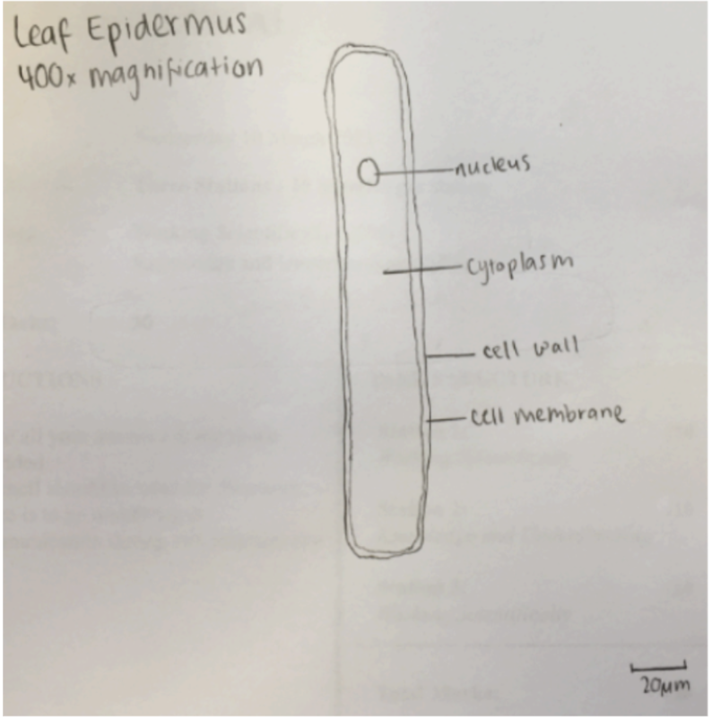
Rules for biological drawings
Always use pencil
Labels should be drawn using a ruler, should never cross each other and should not have arrowheads
Centre drawing in middle of page
Diagram should take up about ½ the page (about 10cm by 10cm)
Draw using simple clear lines
Do NOT sketch or shade
Only draw 2 or 3 cells
Only draw the structures that you see
Diagram should include title of the name of specimen which should be underlined
Record the magnification next to the diagram
OR
Requires a scale bar
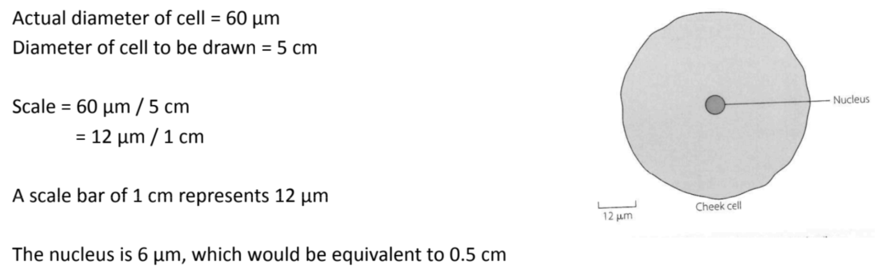
Cell scaled diagrams example
Scale - mm to μm
Cells are measured using micrometres
1mm = 1000μm
What is a scale bar?
A scale bar is a capped horizontal line that represents the ratio between the drawn size of the object and the actual size of the object
Calculating scale bar
Scale = Actual size of specimen / Size of drawing
Determine the actual size of the specimen (usually given in the question).
Choose a size that you will use to draw your diagram (if not specified)
Draw and label image
Determine scale bar using formula
Draw scale bar onto diagram
Size of one cell formula
Size = Diameter of field of view / Number of cells that fit across the diameter
What are organelles?
Organelle = membrane-bound compartments within the cell
Each organelle performs a different function that keeps the cell alive
Creates small, enclosed spaces for specific processes
Help keep the cell organised
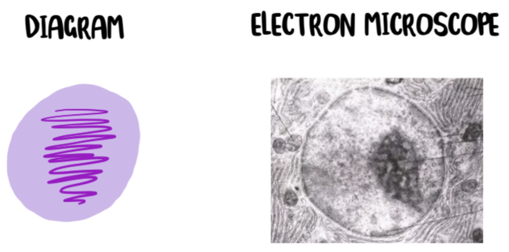
Nucleus - Structure and function of organelles
Large in size
Spherical in shape
Contains genetic information needed for growth, repair and proper functioning
Contains chromosomes made of DNA
Bound by a double membrane
Controls the cell by directing its activities
The nucleus has pores which control the movement of substances into and out of the nucleus.
Contains an organelle called the nucleolus
Nucleolus - Structure and function of organelles
Spherical in shape
NOT bound by a membrane
Made of protein and ribonucleic acid (RNA)
Ribosomes are made here
Which play a major role in protein synthesis