Microbiology Lab Practical 1
1/117
There's no tags or description
Looks like no tags are added yet.
Name | Mastery | Learn | Test | Matching | Spaced | Call with Kai | Chat |
|---|
No analytics yet
Send a link to your students to track their progress
118 Terms
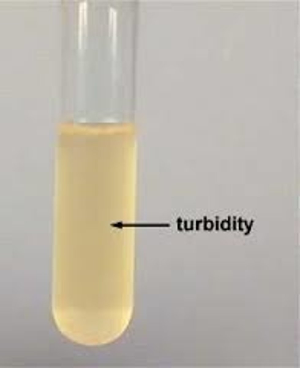
Turbidity Count: indirect method using _____ to measure absorption by bacterial _______ used.
light, culture
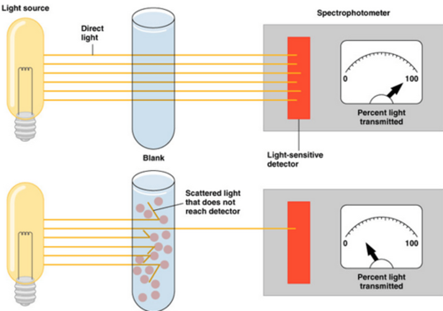
More turbidity = ______ growth (light absorption)
more
Less turbidity = _______ growth (light absorption)
less
For calculating Cellular Densities (CFU/ml) you NEED to know:
______ of colony forming units, ________ factor used to inoculate, and __________ used to inoculate.
number, dilution, volume
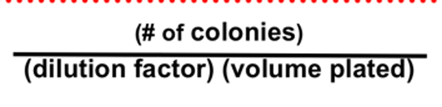
Units used to count microbes are: __________
(CFU/mL)
Growth + division = _____________
proliferation
Temperature • pH• Osmotic pressure• Presence of carbon, nitrogen, sulfur, phosphorous and trace elements• Presence or absence of oxygen• Organic growth factors are ALL factors that affect _________ growth
microbial
No _________ ___________ = No ability to use nutrients, no ability to obtain life building blocks, ability to generate proteins, lipids, nucleic acids and lipids, growth and proliferation
metabolic enzymes
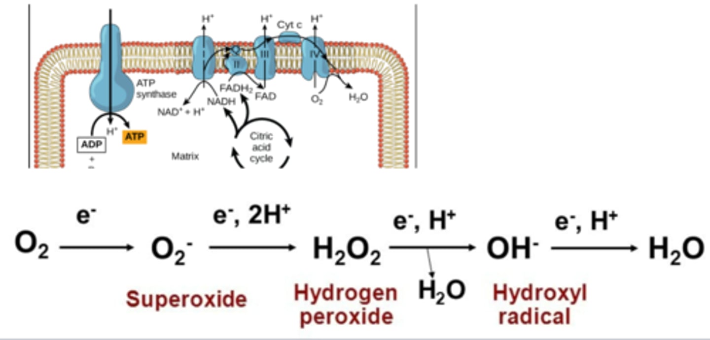
Oxygen can harm organisms in different ways known as _________ _______ ________ (____) (Generated during oxidative phosphorylation --> Partial reduction of oxygen). It attacks membranes, proteins, and DNA.Obligate aerobes • Must have oxygen to live
Reactive Oxygen Species(ROS)
Obligate aerobes must have _________ to live.
oxygen
Facultative anaerobes can use _________ when present but can get by ________ as well.
oxygen, without
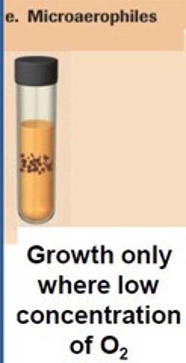
Microaerophiles are _________, but picky, picky.
• Must have just the right amount of _______, less than found in normal air
Aerobic, oxygen
Different Aerobes
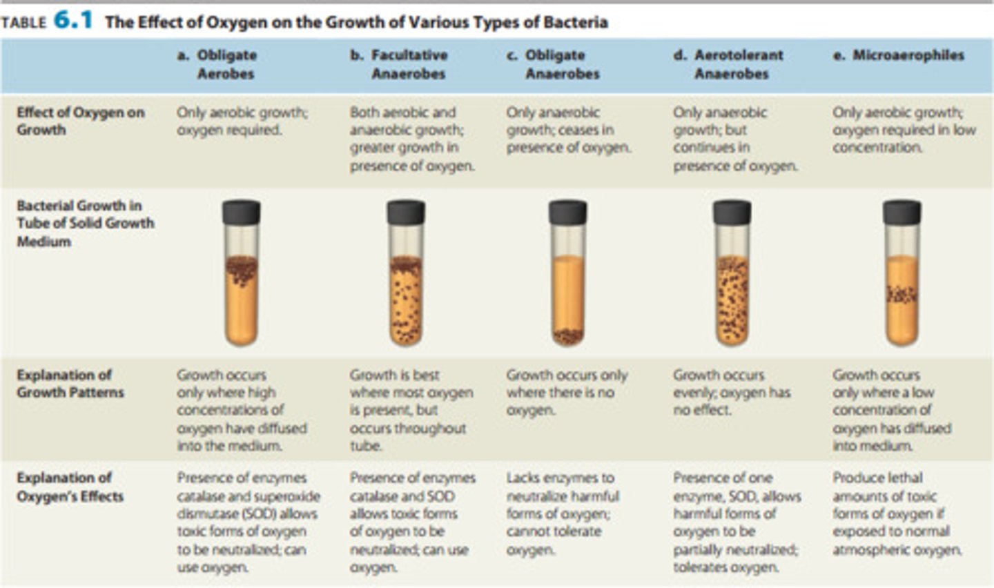
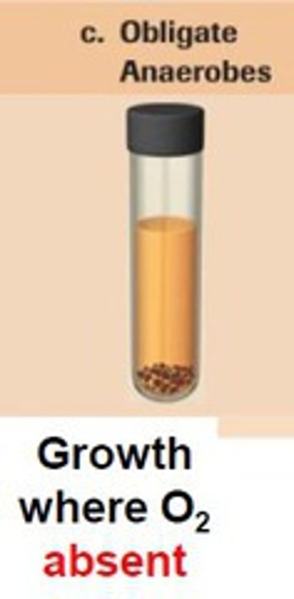
Obligate anaerobes can't use oxygen bc it is _________ to them
toxic
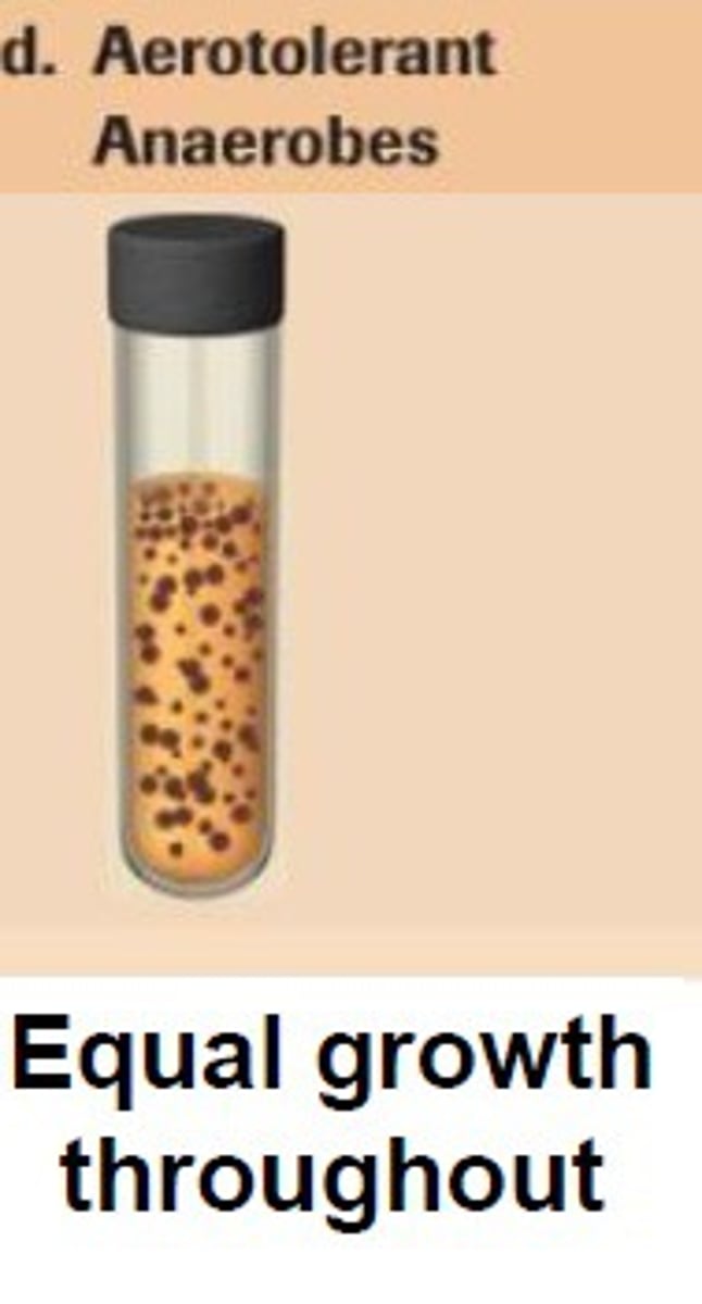
Aerotolerant anaerobes ______ use oxygen - but it isn't toxic to them
don't
How do we determine Oxygen requirement experimentally?
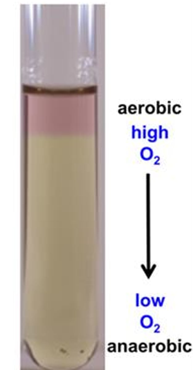
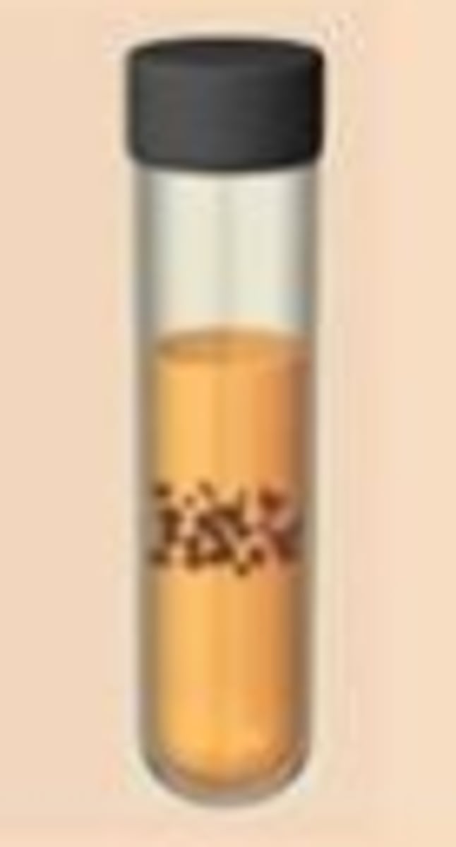
A) Fluid Thioglycolate Medium (FTM)•
Key reagents:•
-Sodium Thioglycollate (_________ oxidation/reduction potential)
•Resazurin (____ indicator: redox sensitive dye that turns pink in the presence of ____)
•Agar (___________ oxygen diffusion)
lowers, O2, O2, prevents
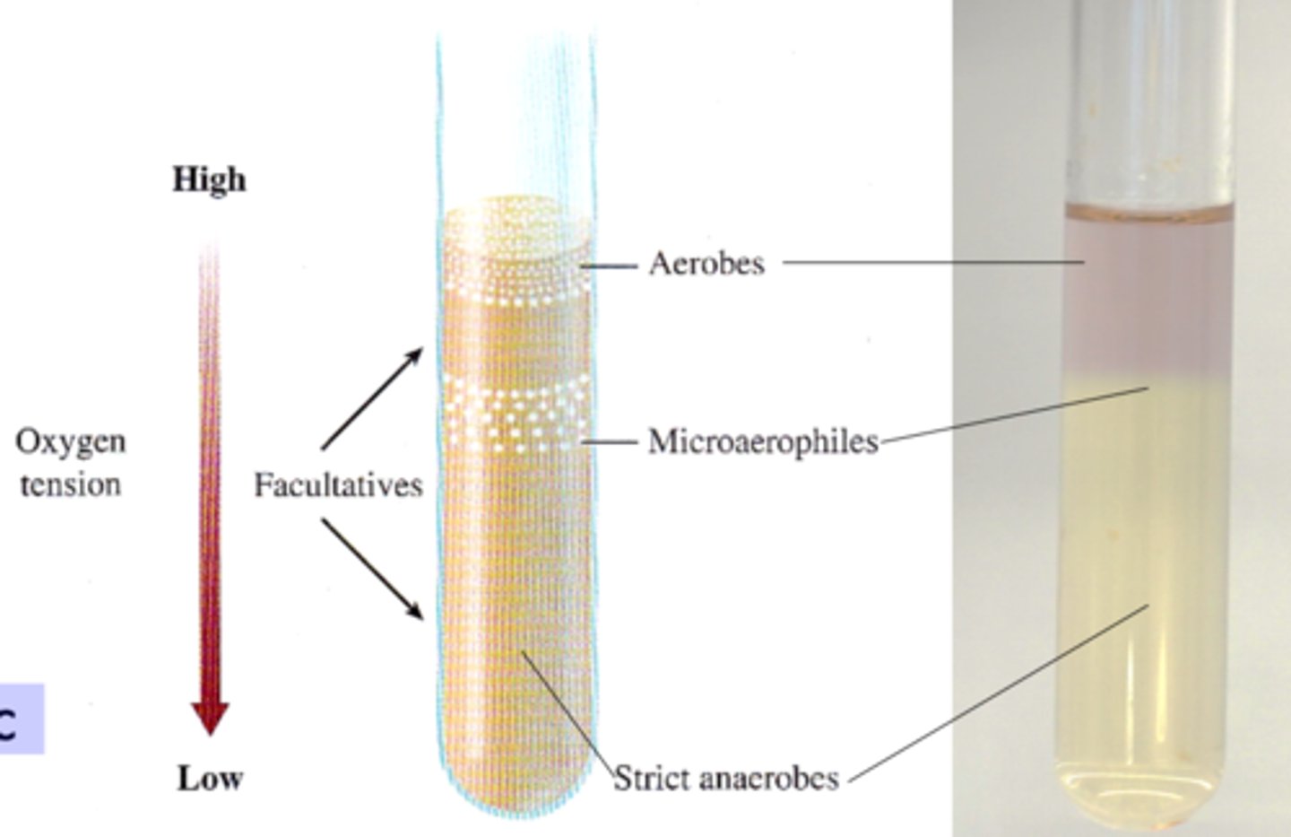
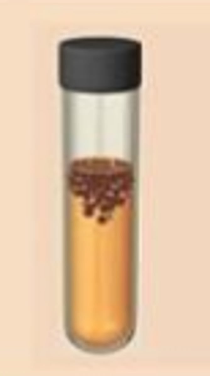
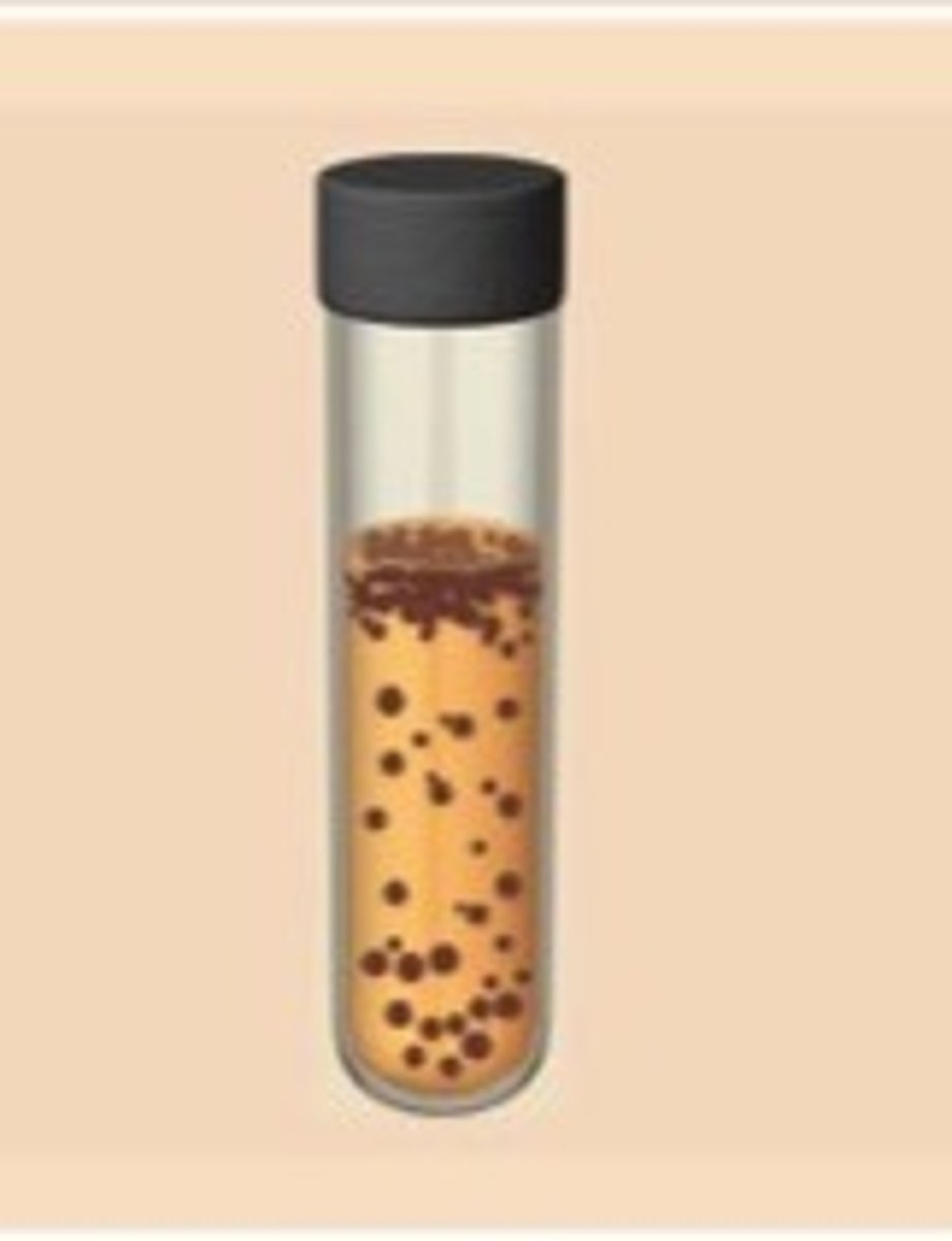
FTM (fluid thioglycollate medium) functions to create an oxygen gradient that differentiates ____________
aerotolerance

w/i FTM aerobes are the ____, microaerophiles are ________, & strict anaerobes are the _______
top, middle, bottom
______________ require oxygen concentration lower than air. i.e., w/i a candle jar (high CO2, low O2)
Microaerophiles
Bacteria with the ability to capture DNA from environment are known as ___________
competent
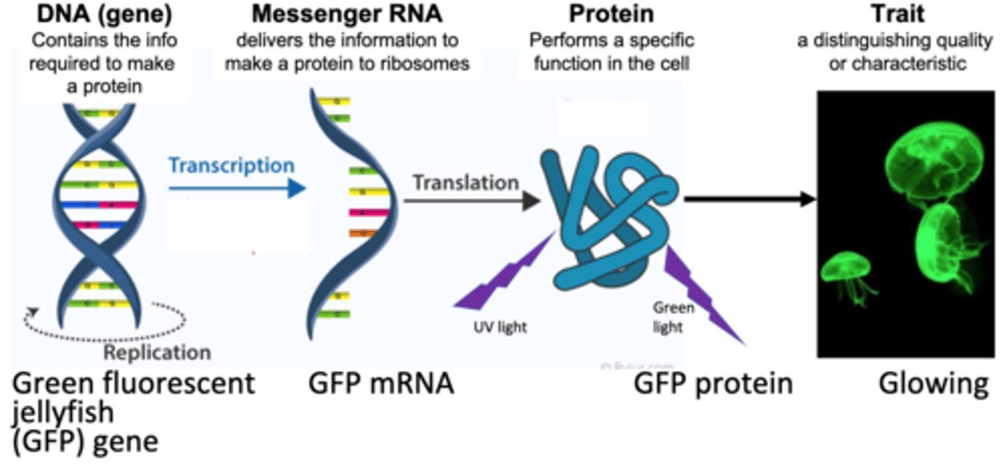
Central Dogma:
______ (gene) --> _______ --> _____ --> ____
DNA (gene) -(transcription)-> mRNA -(translation)-> protein --> trait
Linear DNA is degraded inside the cell, therefore, ________ must be used to to transfer DNA from one cell to another
plasmids
__________ ________ is an engineered plasmid used in biotechnology as a vector for creating genetically modified organisms. The plasmid contains several reporter genes, most notably for the green fluorescent protein (GFP) and the ampicillin resistance gene.
pGLO plasmid
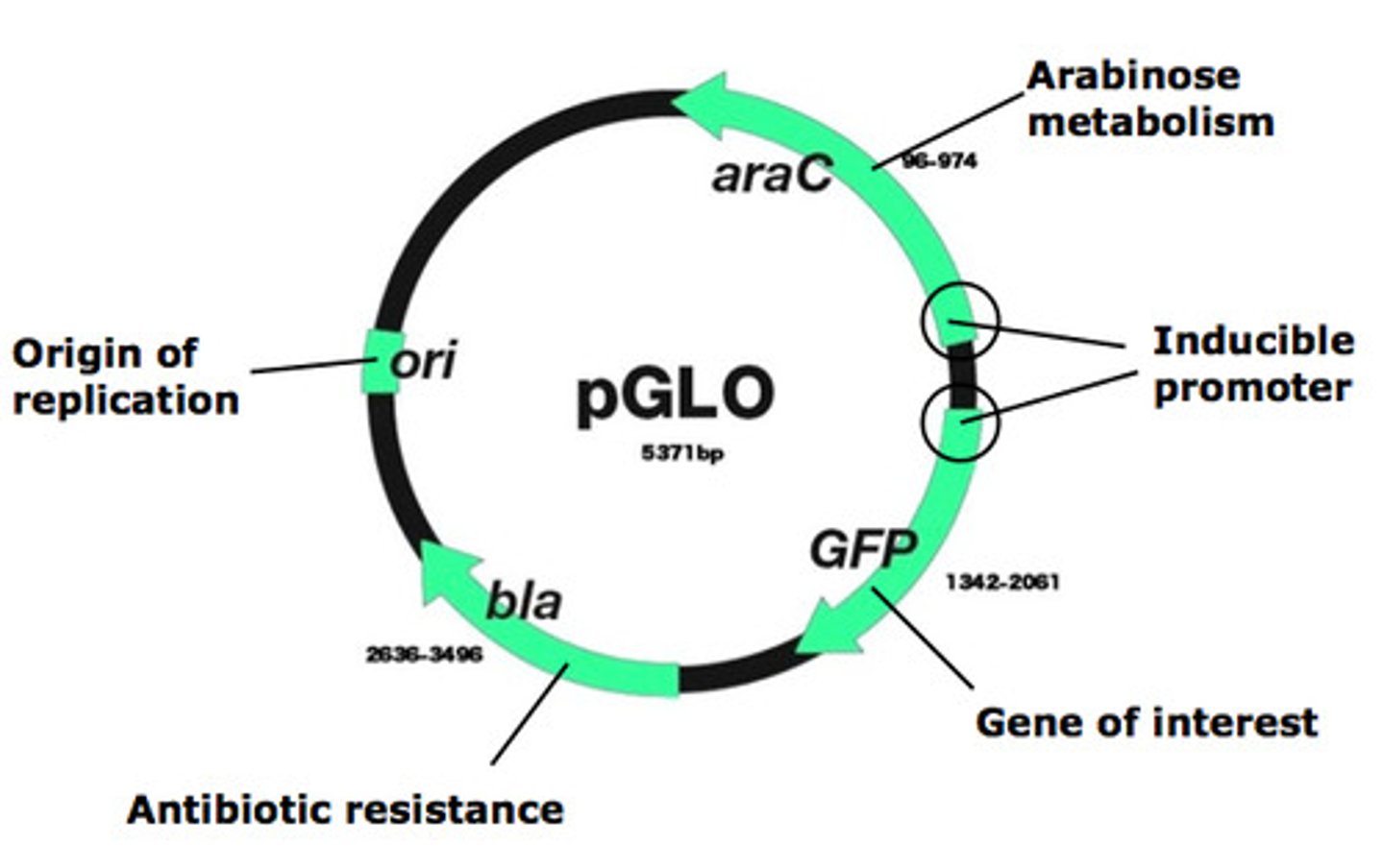
Ori = origin of _________
replication
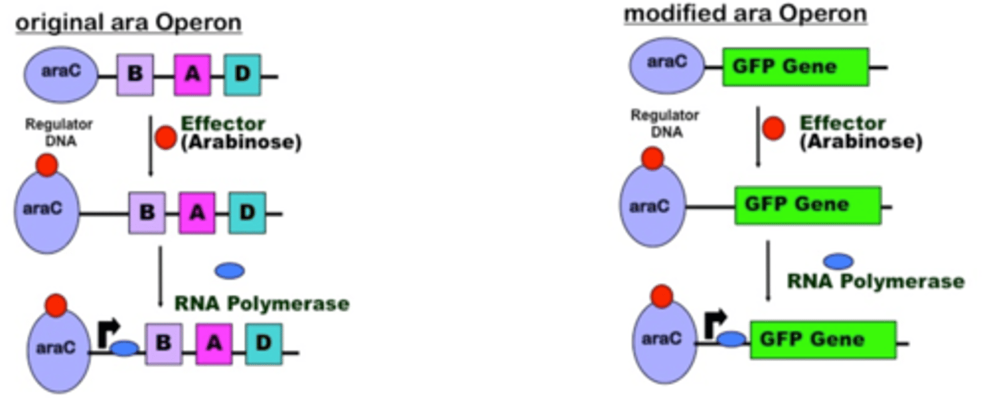
araC= arabinose detection gene that regulates production of ____
GFP
Bla= beta- lactamase enzyme gene (_________ reistence)
antibiotic
GFP= green fluorescent protein gene inserted into plasmids via ________ __________
restriction enzymes
We use ___________(sugar) to regulate the expression of GFP.
No ________ = No GFP
arabinose
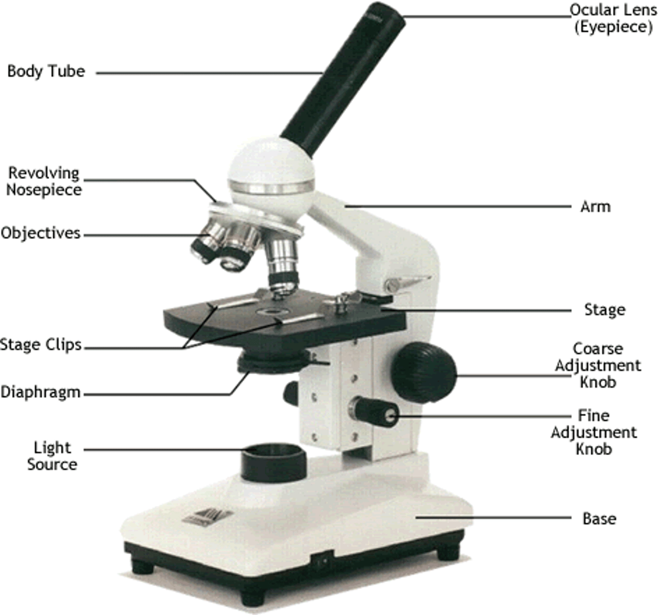
Lenses change the path of light rays known as ________
refraction
Total magnification depends upon the magnification of both the _____ lens and the ______ lens in use
ocular, objective
Maximum magnification of compound light microscope is about _________ due to limit of resolution of visible light
2000X
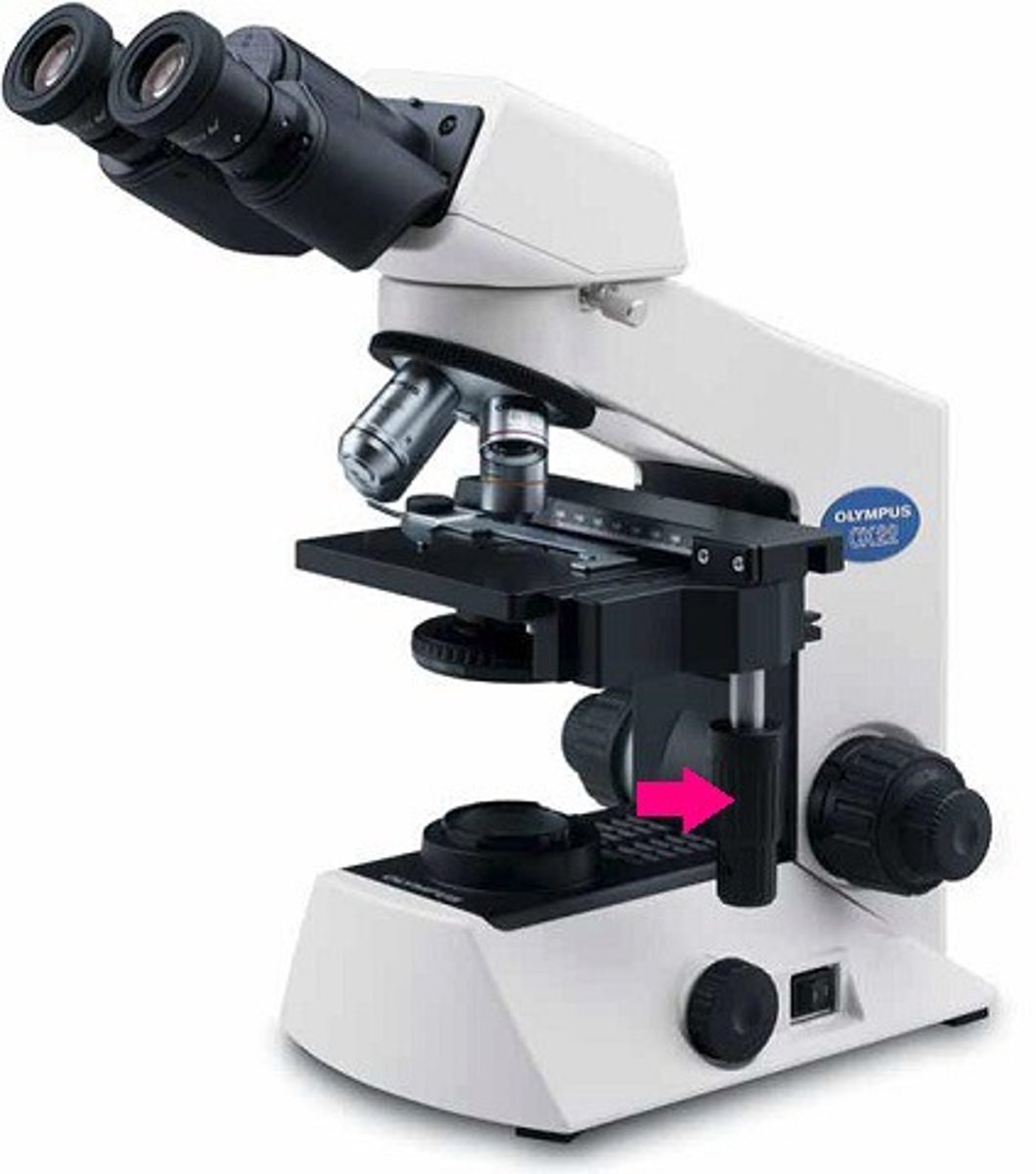
Fine Adjustment knob: ________ focusing
detailed
Coarse adjustment Knob: _____ focusing (scanning lens only)
Initial
X-Y Axis/Stage Adjustment Knob: move ______ right or left and backwards or forwards
stage
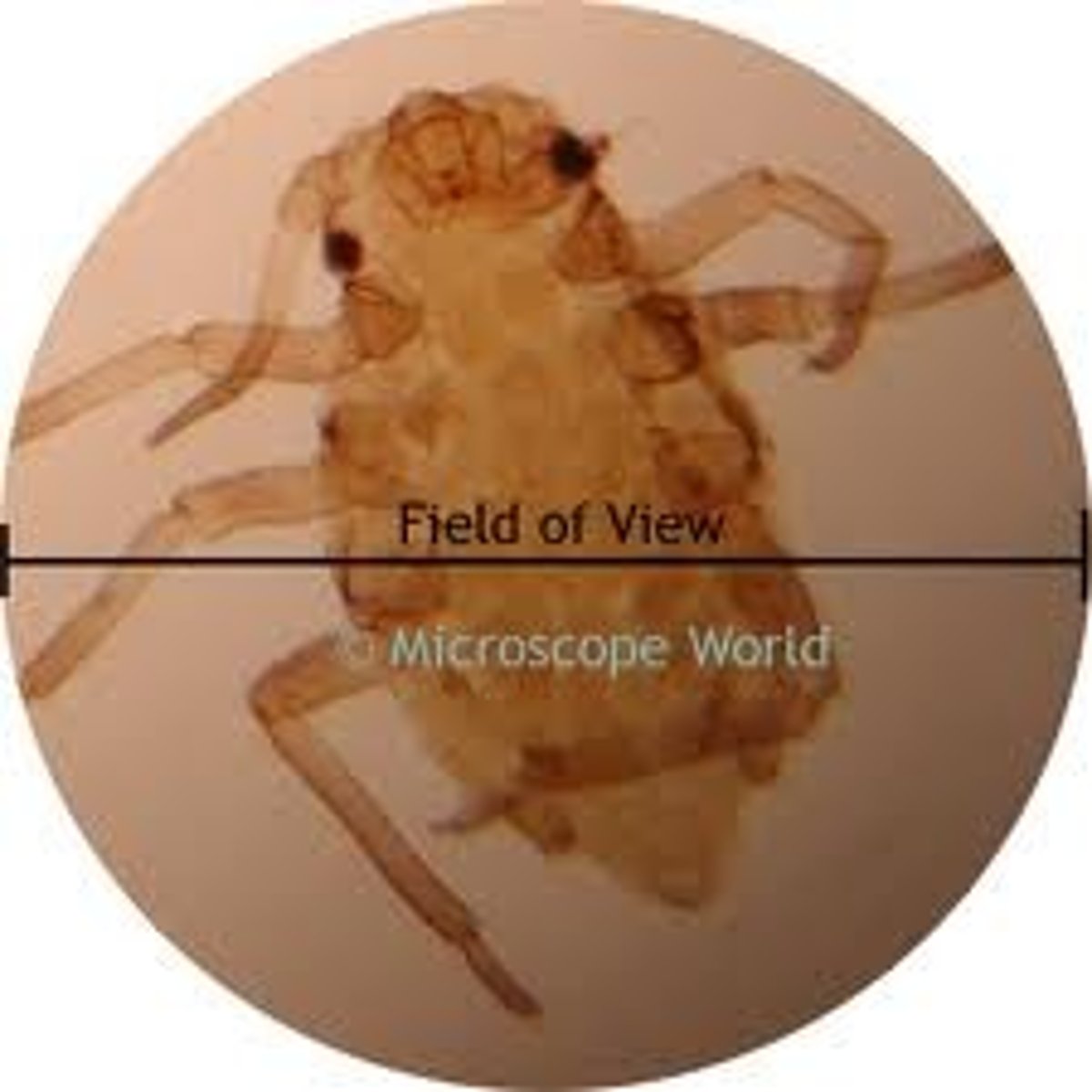
The area visible through the microscope eyepiece is known as the _________ of ______
field of view
microscope ___________ is the
ability of a lens to separate or distinguish small objects that are close together
resolution
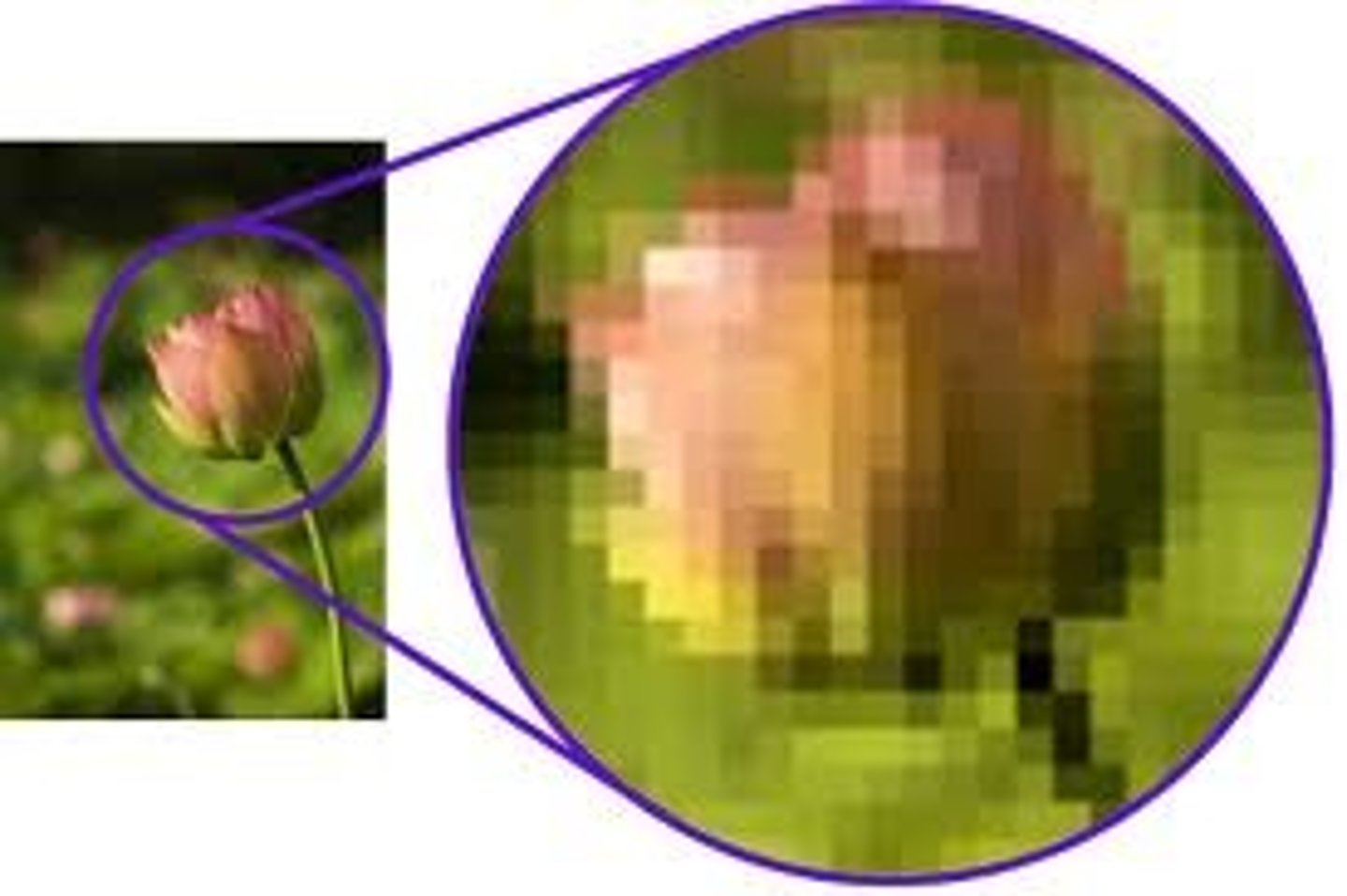
_________ resolution objects appear closer together & can be distinguished as independent entities.
Higher
The _______ the wavelength used the greater (better) the resolution
shorter
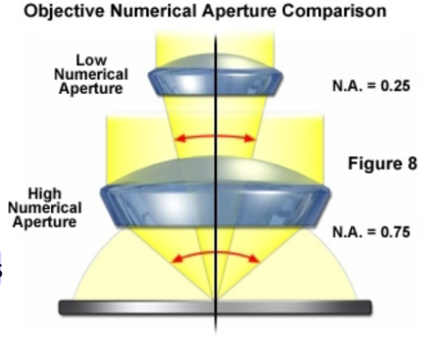
__________ power of a lens depends on its ability to gather light (numerical aperture)
Resolving
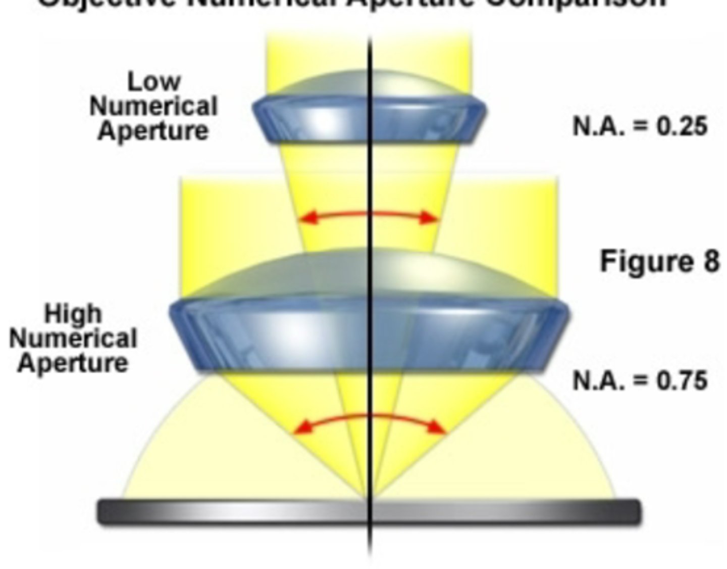
The ________ the numerical aperture (NA), the greater the resolving power of the lens
higher
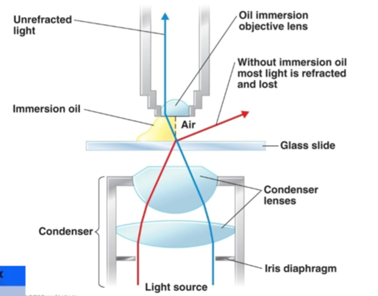
At ______ magnifications, light rays will refract from a straight path by bending away from the specimen (why we need immersion oil)
high (100x)
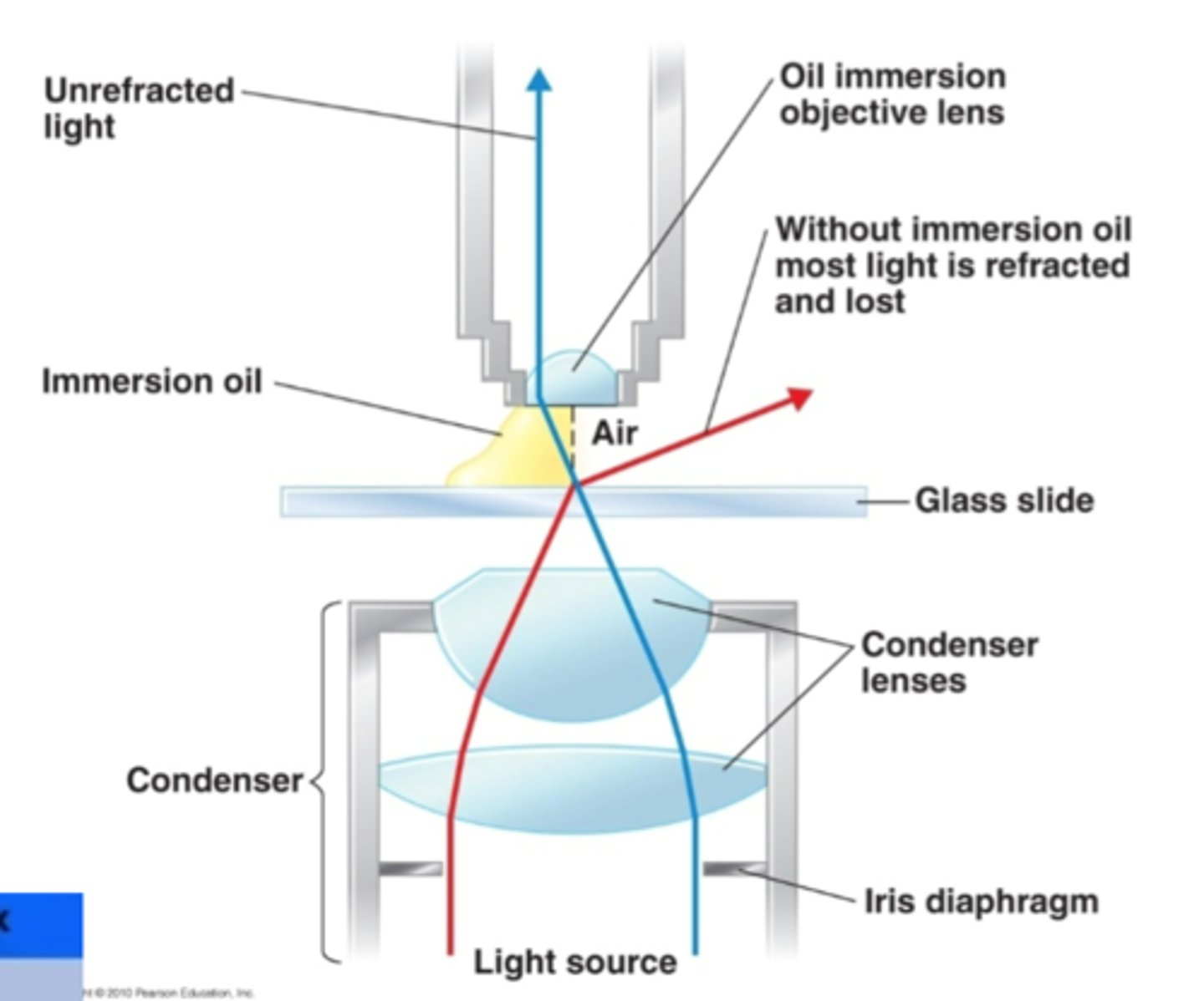
To preserve the direction of light rays at the highest magnification (100x) __________ oil is added between the lens and slide; it has the same refractive index as glass, and so the oil becomes part of the optics of the glass of the microscope.
immersion
staining allows for the visualization of bacterial cells because it increases _________ during microscopic imaging
contrast
Basic dyes: color is carried on _______ ion
positive
Acidic dyes: color is carried on _______ ion
negative
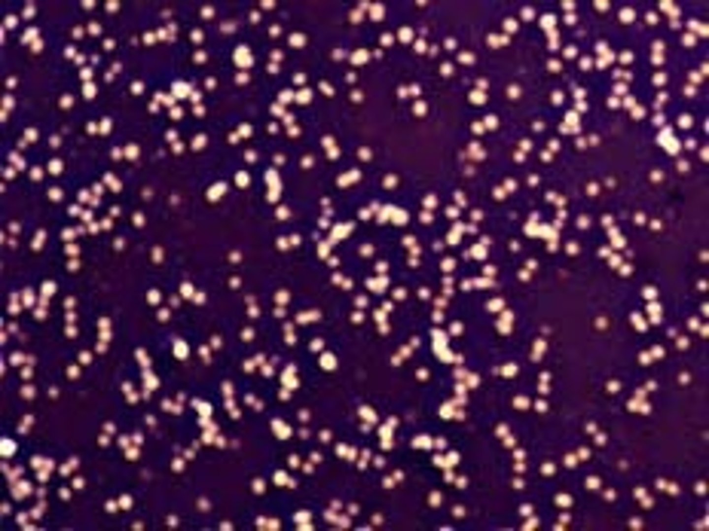
Negatively charged bacteria will attract _________ charged (basic) dyes
e.g., crystal violet, methylene blue, malachite green, safranin
positively
Simple Stain: Aqueous or ______ solution of a single, basic dye.
Applied to "fixed" smear then washed off.
Determines ____________.
alcohol, morphology
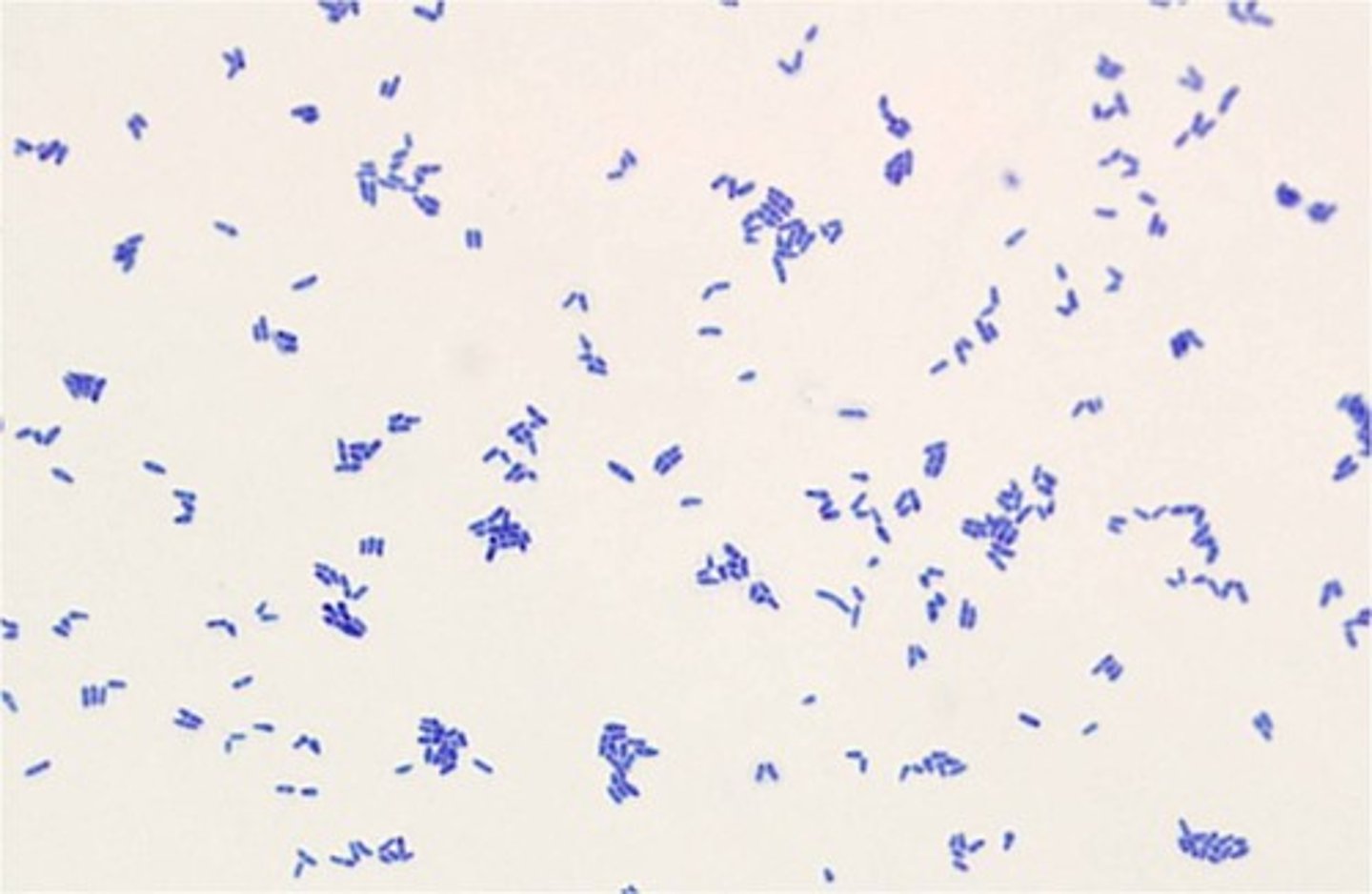
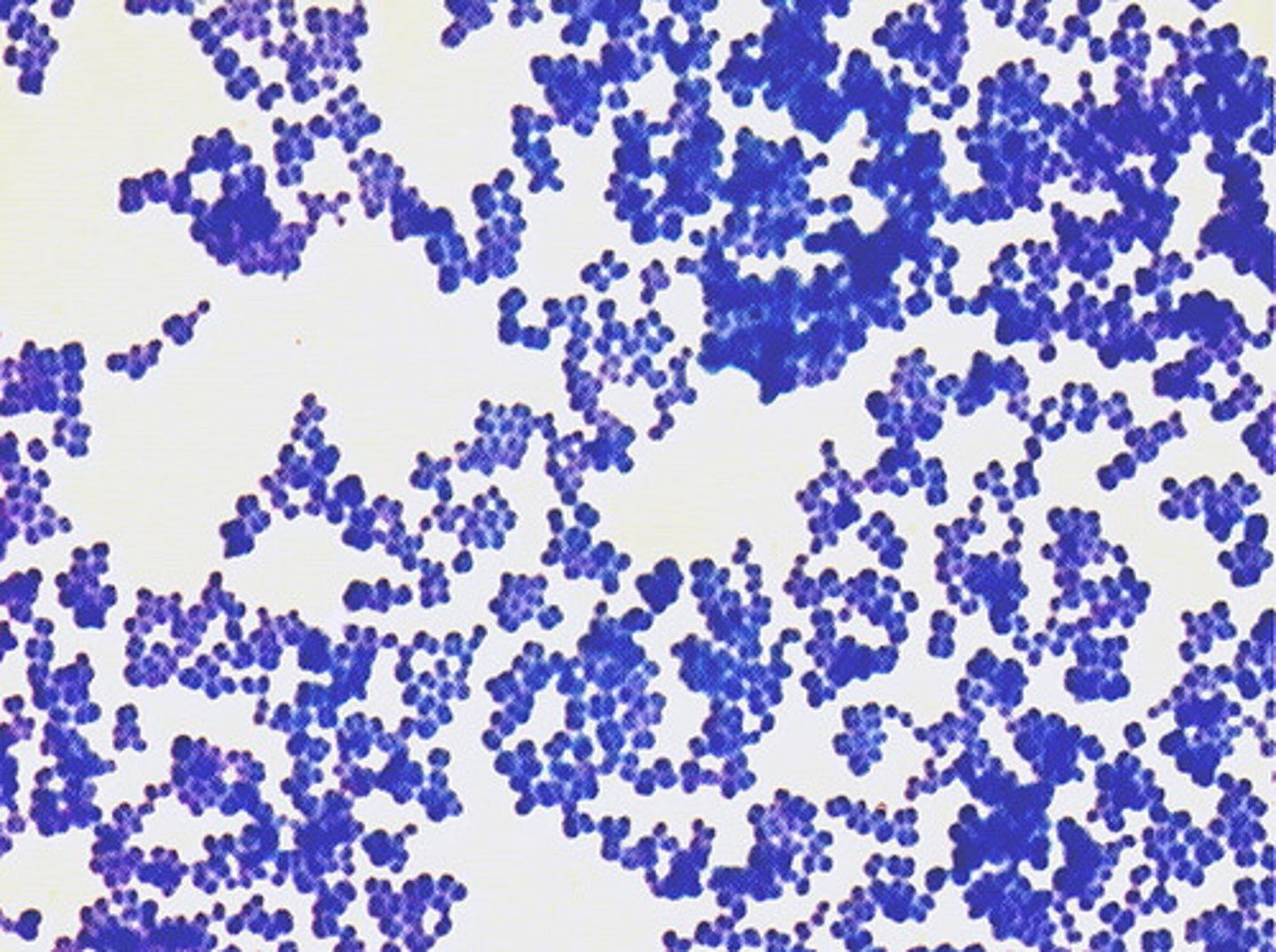
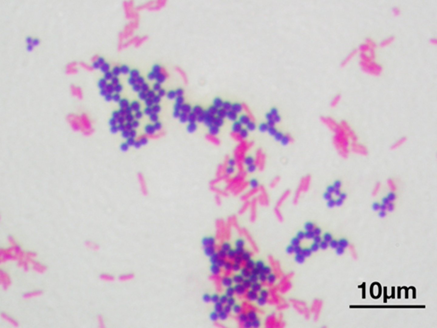
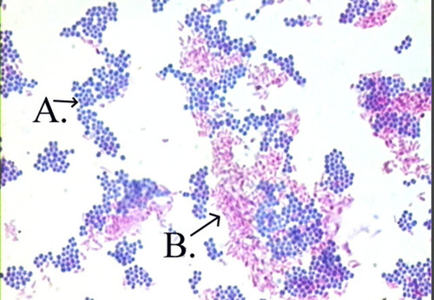
Gram stain is most frequently used differential stain (based on ___ ____ composition)
cell wall (gram +), (gram -)
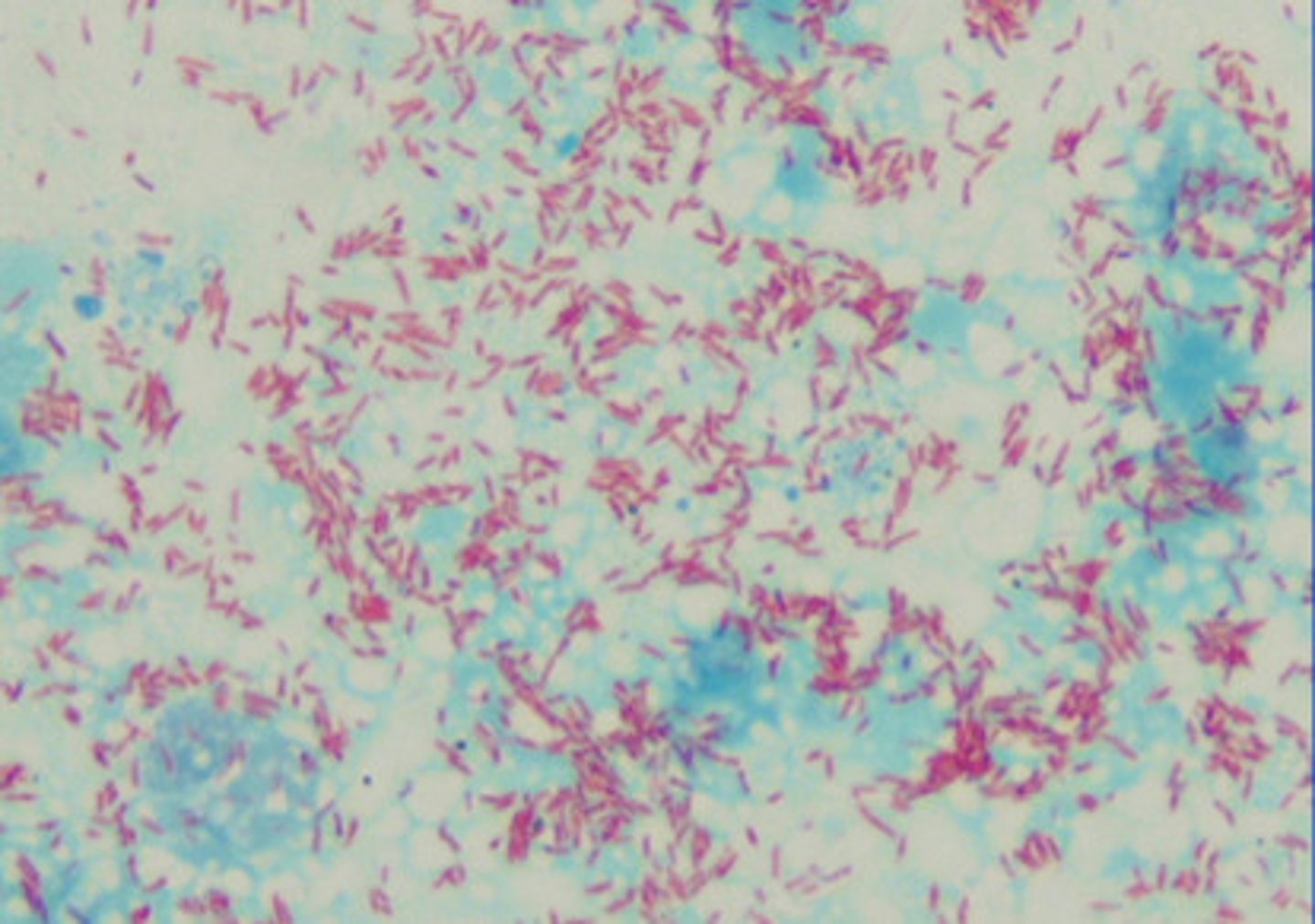
Acid fast stain is based on presence/absence of ________ acids
mycolic
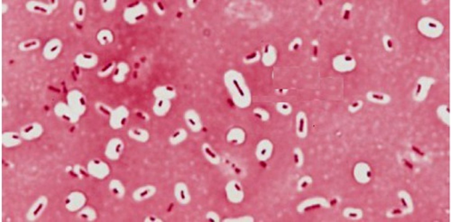
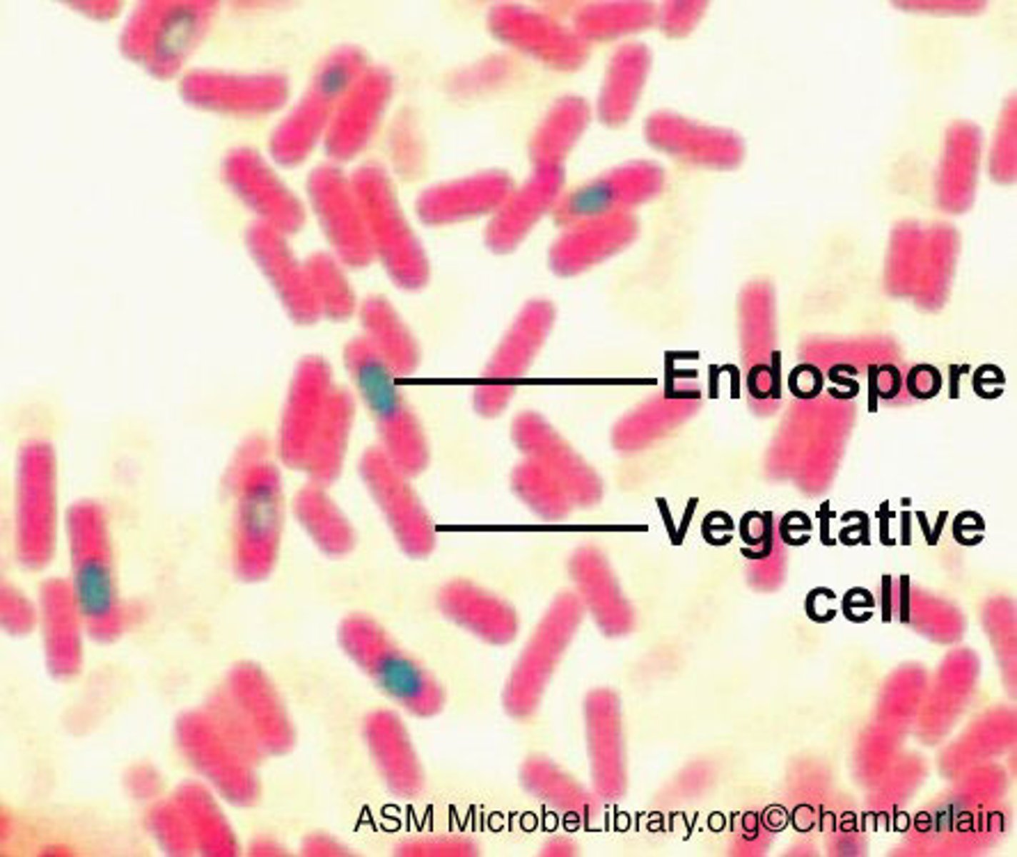
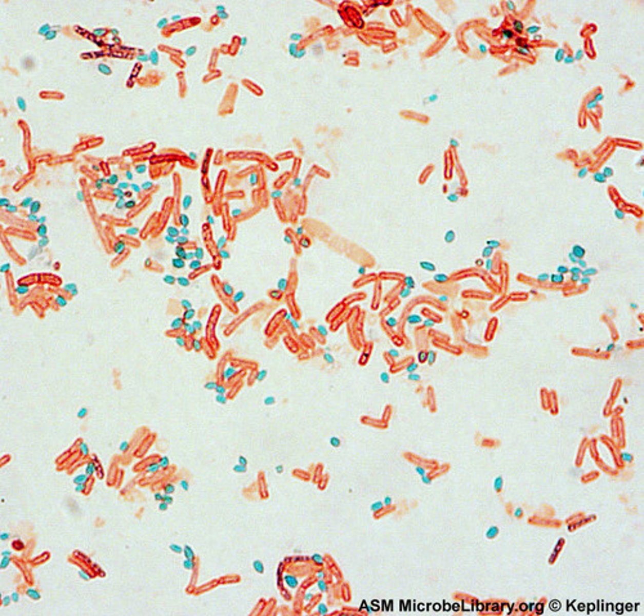
Endospore stain is based on presence/absence of _________.
endospores
Negative staining is
staining the __________ instead of the cell
background
Negative staining typically used to trouble shoot when ________ is a problem (shrinking, morphology, arrangement, heat sensitive bacteria)
fixation
Acid Fast staining AIM: Differential stain used to identify acid fast positive organisms (organisms with cell walls rich on ________ _____).
mycolic acids (red stain)
Reagents used for Acid Fast Staining include:
1. __________________ - organic solvent, primary dye
2. ____ __________- wash
3. ___________ ___- counterstain
Carbolfuchsin
Acid-alcohol
Methylene Blue
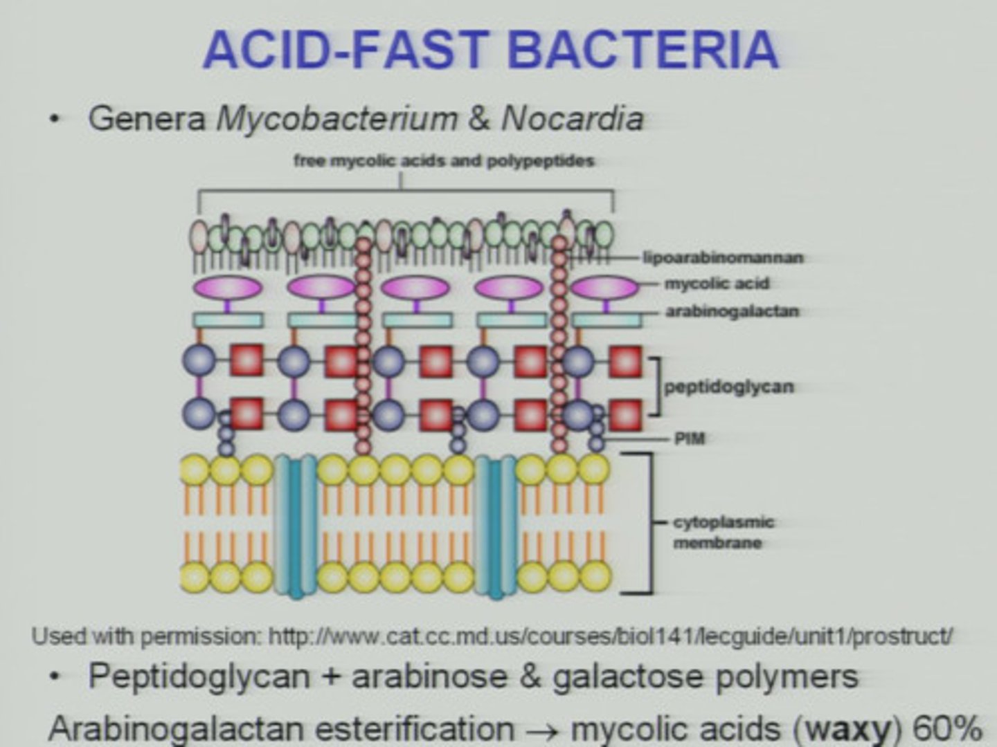
Mycobacteria and Nocardia contain high concentrations of a hydrophobic waxy lipid called ________ ______
mycolic acid
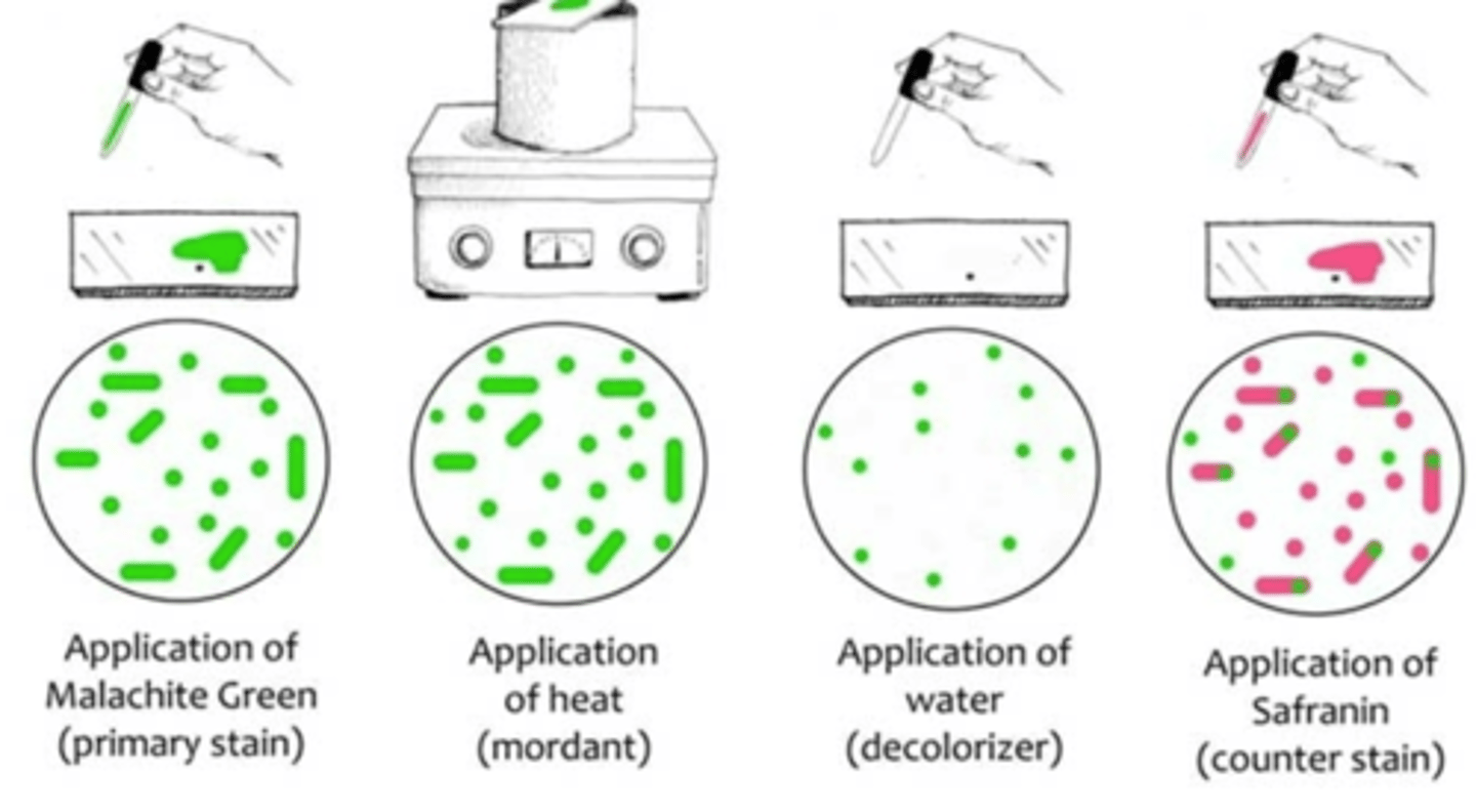
Endospore staining reagents:
1. _________ _________ - primary dye. ONLY retained by endospores
2. _________
3. __________- counter stain
1. Malachite green
2. water
3. Safranin

Endospore Staining:
Green structures are ______ (malachite green), pink are _________ cells (safranin)
spores, vegetative
Endospore Stain Procedure:
1. Prepare heat-fixed smear of ___-______ former on clean slide
2. Apply __________ ________ to bibulous paper placed over smear while steaming, 7 min (don't let paper dry)
3. Remove bibulous paper w/ forceps and gently rinse slides w/ ______ water
4. Counterstain smear w/ _________ for __ min, rinse w/ distilled water.
5. Gently blot w/ _____ paper
6. Observe under ___ _______
1. non-spore
2. malachite green, 7 min
3. distilled
4. safranin 3 min
5. bibulous
6. oil immersion
pGLO Process of Transformation in Living Organisms:
1. GFP gene (__________)
2. GFP mRNA (_______)
3. GFP _______ made
4. __________ organism (phenotype)
transcription, translation, protein, Glowing
GFP Gene Regulation in pGLO
Genes normally regulated by _____ (operaon) are replaced by _____ gene resulting in the expression of the GFP gene upon ________ presence.
araC, GFP, arabinose
No DNA should be placed in pGLO medium with or w/o ___________. Only ________ bacteria should be!
ampicillin, competent
What happens to a ray of light when it passes through a lens?
it increases the image make it appear larger
If the light rays from an object enter the eye at a small angle, then the object will cover a _____ portion of the back of the eye
larger
The closer you move an object to your eye, the ____ the angle of light from that object entering your eye. Therefore, the object appears to be _____.
smaller, larger
The magnifying lens _____ the angle of light coming from the object viewed. This results in _____ spread of the image at the back of the eye.
decreases. increased
Define resolution
The ability to distinguish between two points
Is resolution the same as magnification?
no
What is the total magnification if the objective lens is 4x and the ocular lens is 10x?
40x
What is the field of view?
entire area of view and the surrounding distance-proportional to magnification
What happens to the size of the field of view as you rotate the nosepiece and change magnification?
less magnification, bigger view-more magnification, smaller view
Which one of the objective lenses give the greatest magnification?
100x
Which one of the objective lenses gives the greatest field of view?
4x
When searching for a specimen, which lens should you use first?
4x
What is the depth of focus?
the zone of clear focus in the microscope-proportional to magnification
How does depth of focus change with magnification?
the greater the magnification, the less depth of focus
What quality of your vision is being lost up close to your eye?
resolution
What quality of your vision has been improved by the pinhole paper?
depth of focus
What has happened to the orientation of the image as it passes through the lenses of the microscope?
it flips to the opposite side
When you move the slide forward on the stage, in what direction does the "e" appear to move when viewed through the microscope?
when you move the slide forward, it moves upwards
When you move the slide in one direction, in what direction does the "e" appear to move?
it moves in the opposite direction
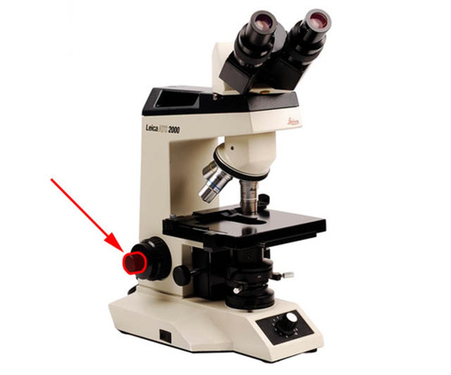
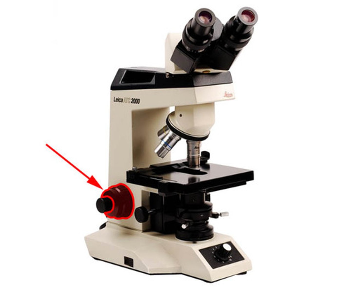
What microscope part is used to change the light intensity?
iris
What happens to the light intensity as you increase or decrease magnification of the objective lens?
increase-gets darker, decrease-gets lighter
When you switch to higher magnification, what should you do to the light intensity?
increase light intensity
Describe the proper focusing technique in terms of moving the lens
moving lens upwards focuses on subject
Why is this focusing technique so important?
an accurate field of view
Steps to Start Microscope
Microscope Set up:
• Quickly clean lenses with lens paper.
• Illumination: maximum power, iris closed.
• ALWAYS start on 4X power. Take stage all the way up.
• Place object into the light path.
• Use coarse focus (stage down) until object is in focus (around 45° angle rotation).
-Center object in field of view.
• Move to 10X power. Fine focus and center.
• Move to 40X power. Fine focus and center.
• Move 40X objective away, add oil drop to slide, move to 100X objective (careful not to touch stage at ALL).
• Fine focus and center.Note: Open iris (more light) as needed bright background
Steps to Put Away Microscope
Remove any slide.
• Clean lenses, specially the 40X and 100X:
-Lens paper, rotate until no oil comes of.
-Apply lens cleaner to paper and rotate on lens.
-Clean stage (no oil left)
-Lower stage all the way
-Point 4X lens at stage
-Turn dimmer all the way down
-Wrap cord
-Put it back in drawer.
Stains allow visualization by light ______________
microscopy
________ are salts composed of a positive and a negative ion, one of which is colored (chromophore)
Stains
______ dyes: color is in the positive ion
Basic
______ dyes: color is in the negative ion
Acidic
_____________ charged bacteria will attract positively charged (basic) dyes (Cystal Violet, methylene blue, malachite green, safranin)
Negatively
General Stain Steps
1) appropriately label slide
2) apply ________ to the slide using aseptic technique
3) ___ drying
4) ____ fixation
5) apply ________ method (gram, acid-fast, endospore)
1. label
2. bacteria
3. air
4. heat
5. staining
Purpose of simple stain
• Determine the _____ of the cells (i.e., cocci, bacilli, spirilla)
• Look at the ___________ of the cells e.g., Singles, pairs, tetrads, etc.
• Estimate ___ of cells
• Can use ocular micrometer if available to estimate size based on ______ __ ______ diameter
shape, arrangement, size, field of view
Smear (Methanol & Heat fixation)
1. Wash slide with _____ and ___ water. Clean w/ _____ & dry w/ paper
*don't touch slide w/ hand (oils!)*
2. ______ (stain, microbe, name date)
3. Smear & ____ fix (3x over flame)
1. soap, hot, alcohol
2. label
3. heat
______ Stain
1. Start w/ heat fixed emulsion
2. Cover smear w/ stain (tray to catch excess dye)
3. Tilt slide, rinse w/ water
4. Gently blot dry w/ bibulous paper
Simple
_____ stain is most frequently used differential stain (based on cell wall composition) (negative/positve)
Gram
_____ ____ stain (based on presence/absence of mycolic acids)
Acid fast
________ stain (based on presence/absence of endospores)
Endospore
Gram Stain (rinse w/ water between steps)
1. Apply _____ _____ (purple) for __ minute (already heat fixed)
2. Apply ______ (mordant) for __ minute
3. ________ wash ____ seconds!!
4. Apply _________ (counterstain) __ minute
1. crystal violet (1 min)
2. iodine (1 min)
3. alcohol, (15-25 sec)
4. safranin (1 min)