Principles of Parasite Diagnosis
 Call Kai
 Learn
Learn Practice Test
 Spaced Repetition
 Match
Match Flashcards
 Knowt Play
1/69

There's no tags or description
Looks like no tags are added yet.
Name | Mastery | Learn | Test | Matching | Spaced | Call with Kai | Chat |
|---|
No analytics yet
Send a link to your students to track their progress
70 Terms
The feces is the sample how do we see it
Direct smear · flotation
Sedimentation · Baermann
Egg counts · coproculture
THe sample feces what is the immunoligcal and molecular
coproantigen test
Copro-PCR
The sample is blood how do we see it
Thick and thin smears
Modified Knott's
The sample is feces what is immunoligcal and molecular
heartworm anitgen
anitbody ELISA IFAT and CFT
PCR
The sample is skin and hair what does it look like
Direct collection
Scraping · KOH · tape
The sample if skin and hair what is immunoligcal and molecular
no immunoligcal
PCR
The sample is tissue and organs what is do we see
Pepsin digestion · histology
Imaging · necropsy
The sample is tissue and organs what is the immunoligcal and molecular
immunohistochemistry
PCR
Three questions before you order a test
1 · Where does it live? (Gut · lung · blood skin · tissue)
2 · What stage does it shed? (Egg · oocyst · larva microfilaria · antigen · DNA)
3 · Is it time to look yet? Is the animal past the prepatent period?
Skip the third question why
and a perfectly good test will mislead you. During the prepatent period the animal is genuinely infected — and genuinely negative.
What is the Direct fecal smear test
Feces is the most common sample in practice — cheap, non-invasive, and it samples the gut without opening the animal.
What is the Principle for Direct fecal smear
§ About 2 mg of feces — a match-head — in a drop of saline, coverslip, read at 100–400×.
§ The saline keeps protozoa alive and moving.
can also add legel’s iodine on the slide if we don’t see any motile
What are the strengths for Direct fecal smear
Quick and simple
A quick first look in urgent cases
The only routine method that shows motile trophozoites — Giardia, Tritrichomonas
What are the limitations for Direct fecal smear
A tiny sample — light infections are missed routinely
Debris obscures the field
A positive is as good as any positive. A negative cannot rule out infection.
Two things decide whether a flotation works:
1 the solution
2 the procedure.
A solution denser than the eggs but lighter than the debris: what will happen
the eggs rise, the debris sinks, and a coverslip on the meniscus collects what floated.
Are fluke eggs heavier ?
are heavier and don’t float so we go through sedimentaition which simply using water/letting the eggs sediment to the bottom
If we choose Sodium nitrate, commercial what its for
The routine choice — common eggs and cysts
If we choose zinc sulfate, commercial what its for
Giardia cysts; some lungworm larvae
If we choose Sheather’s sugar, commercial what its for
Cryptosporidium; least distortion
If we choose Ssaturated sodium chloride , commercial what its for
Cheap — but crystallizes and distorts
What is the trade off for Fecal flotation
trade offs that if you’re using high specific gravity solution the chances of getting the distored morpholgy of the egss is to increase because of the increase osmotic pressure
Passive / standing flotation what are the characteristics
1. Feces + flotation solution in a cup
2. Strain into a tube or fecalizer
3. Coverslip on; stand 10–20 min
4. Lift the coverslip and read it
Simple and fast · commercial kits · lower sensitivity
Centrifugal flotation (Wisconsin) what are the characterisitcs
1. Feces + flotation solution in a cup
2. Strain into a centrifuge tube
3. Coverslip on; spin ~1,500 rpm × 5 min
4. Lift the coverslip and read it
5. Can be used as a quantitative float — eggs per gram of feces
Several times more eggs · higher sensitivity ·
needs a centrifuge
Why is centrifugation important
Centrifugation is the single biggest improvement available to you
Centrifugation increases the sensitivity of the test!
What is the principle for Quantitative fecal float — McMaster, FLOTAC, Wisconsin
Flotation concentrates parasite
eggs/oocysts; count stages within the McMaster
chamber grids and convert the count to EPG/OPG.
What is the key output for Quantitative fecal float — McMaster, FLOTAC, Wisconsin
Eggs or oocysts per gram of feces
(EPG/OPG): an estimate of shedding intensity,
not a direct adult-worm count.
What are the strengths for Quantitative fecal float — McMaster, FLOTAC, Wisconsin
Rapid, inexpensive, standardized, and
useful for comparing animals or tracking group-
level shedding over time.
What are the limitations for Quantitative fecal float — McMaster, FLOTAC, Wisconsin
Lower sensitivity for low egg burdens;
results vary with sample quality, fecal consistency,
parasite biology, and the method’s detection limit.
What are the sensitivity for Quantitative fecal float — McMaster, FLOTAC, Wisconsin
McMaster 25–50 EPG; FLOTAC 1–5
EPG; Wisconsin flotation 1 EPG
What is the key use of Quantitative fecal float — McMaster, FLOTAC, Wisconsin
Herd/flock parasite surveillance, identifying high
shedders, targeted selective treatment, and fecal egg
count reduction testing (FECRT) for anthelmintic efficacy;
What is the McMaster method

What is FLOTAC method

What is the purpose of Sedimentation
§ Heavy eggs settle in water instead of floating
§ For fluke and acanthocephalan eggs
What is the limit for Sedimentation
the debris settles too — slow to
read
Why do we use Baermann
§ Live larvae migrate out of the feces into
water
§ For lungworm L1 and Strongyloides
What is the limit for Baermann
Limit: fresh feces only, and overnight
What is Coproculture
§ Eggs hatch to L3 in vermiculite over 7–14 days
§ Names the genus when all strongyle eggs look alike
What are the limits for Coproculture
Limit: slow; now yielding to sequencing
Blood tests for parasites — the modified Knott's test what is the principle
• Concentrates microfilariae from blood by lysing red blood cells and centrifugation.
• Sediment is examined microscopically for microfilarial detection and identification.
What are the strengths for Blood tests for parasites — the modified Knott's test
• More sensitive than a direct blood smear.
• Helps identify microfilariae based on size and morphology.
What are the limitations for Blood tests for parasites — the modified Knott's test
• Detects only circulatingvmicrofilariae, not adult worms.
• Negative in occult infections (adult worms present, no microfilariae) and when microfilariae are scarce.
When do we use skin - scraping, KOH, and tape
Ticks, lice, nits and fleas you collect and identify — intact, because a crushed mite or a tick without its mouthparts loses the characters you need. Flea dirt on damp paper leaves a red halo. Mites you cannot see, so you go into the skin.
What is superficial scraping
§ Broad and shallow, over a large area and several sites
§ Sarcoptes, Notoedres, Cheyletiella, Otodectes
§ Scrape the crusted places — ear margins, elbows, and hocks
A negative scrape does not rule out what
mange — burrowing mites are few and deep.
What is KOH digest
1 volume of scraping in 10 volumes of 5–10% KOH dissolves hair and crust; mites, mite eggs and lice survive it intact
What is Tape preparation
surface mites and lice; perianal tape can be used for pinworms such as Oxyuris equi, and it collects tapeworm segments in dogs and cats.
What are the characteristics of Deep scraping
§ Squeeze the fold to express mites from the follicle
§ Scrape until there is capillary ooze — that is the endpoint
§ For follicular mites — Demodex
Tissue and organ methods is utilized for
For a parasite that never leaves the tissues, or has no stage in the circulating blood (e.g. Trichinella), no fecal or
blood method will find it. You have to go to where it lives.
What is Pepsin digestion —
muscle in acid-pepsin at 37 °C for 60–90 min, sedimented and examined: the standard recovery of Trichinella larvae
Direct observation and necropsy what do we mean by that
total worm counts and organ washing (e.g. eyeworms)
What are the limitations for Tissue and organ methods
Invasive, slow, and needs a parasitologist or other specialist
What is the thin smear test
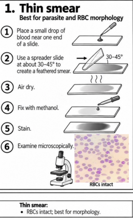
What is thick smear

What is wet mount

Antigen = parasite material test are for?
§ Detects parasite antigen/protein — mostly current infection
§ Heartworm antigen test — from adult female Dirofilaria
§ Coproantigen tests — Giardia in a SNAP device
§ SNAP 4Dx Plus: D. immitis antigen + five tick-borne antibodies, 8 minutes
Antibody = host response test for what
§ Detects the host's response — past or present
§ ELISA, IFAT, CFT, agglutination, immunodiffusion
§ Where antigen tests are insensitive — feline heartworm,
Toxoplasma, Neospora
§ A positive does not by itself mean current infection
What are the strengths for Immunological tests
finds infections microscopy cannot — prepatent, intermittently
shedding, or occult. Fast, in-house, and needs no eye for morphology.
What are the limitations for immunological test
cross-reactivity between related species and maternal antibody in the young; antibody
persists long after the infection has gone; and a test exists for only a handful of parasites.
What is Molecular tests — PCR, sequencing
Principle. Extract parasite DNA from feces, blood, tissue or environmental samples → amplify a marker gene → read
the product, or sequence it and name the species.
What are the strengths for Molecular tests — PCR, sequencing
Sensitivity — copro-PCR finds what microscopy cannot
Species and genotype, where every egg looks the same
What are the limitations for Molecular tests — PCR, sequencing
Expensive, and mostly not available in the clinic
Turnaround of days, when you need an answer today
A positive detects DNA — not necessarily a living, clinically
relevant worm
When the result does not fit the clinical picture WHat do we do
Recheck the biology and the specimen before you change the diagnosis.
How do we recheck the sample
§ Check the sample — right site, enough of
it, fresh? A frozen sample kills the larvae a
Baermann needs.
§ Check the target — does this method see
the stage? A flotation cannot find L1 larvae.
§ Check the timing — still prepatent, or
shedding on and off? A two-week-old
puppy sheds nothing yet.
§ Check the meaning — could a positive be
incidental? Giardia DNA in a well dog with
normal feces.
§ Then act — repeat, resample, add another
method, or rethink the differential.
How to approach any parasite diagnosis! Six steps, always in this order.
1 · SUSPECT signs, signalment, exposure
2 · LOCATE where does it live, general lifecycle?
3 · TARGET what stage does it shed?
4 · SAMPLE feces · blood · skin · tissue
5 · TEST the method that sees that stage
6 · INTERPRET does it fit the patient?
Errors arise at three stages : Pre-analytical
wrong patient or sample, poor collection, timing, transport, storage, contamination
Errors arise at three stages : analytical
wrong test, too insensitive, cross-reaction, inhibition, equipment or reader error
Errors arise at three stages : post-analytical
misidentification, wrong interpretation, transcription, host biology ignored
What are examples of False positive — parasite absent, test positive
Pseudoparasite – parasite look alike
Contaminated sample, or a specimen mix-up
Spurious parasitism – Coprophagy, prey
Cross-reaction in an antigen or antibody test
Antibody from past exposure, not current infection
DNA carry-over contaminating a PCR
What are exampls of errors in False negative — parasite present, test negative
Prepatent period – no shedding yet
Wrong sample, wrong site, wrong time
Spurious parasitism – Intermittent, Sporadic shedding or parasitemia
Occult infection, Single sex nematode infection
PCR inhibitors, or poor DNA extraction
Parasite sequestered in tissue, or recently treated