RADT Ch. 2: Chest Anatomy, Positioning, & Conditions
 Call Kai
 Learn
Learn Practice Test
 Spaced Repetition
 Match
Match Flashcards
 Knowt Play
1/101

There's no tags or description
Looks like no tags are added yet.
Name | Mastery | Learn | Test | Matching | Spaced | Call with Kai | Chat |
|---|
No analytics yet
Send a link to your students to track their progress
102 Terms
3 sections of the chest/thorax:
1. bony thorax
2. respiratory system proper
3. mediastinum
bony thorax
sternum, ribs, and thoracic vertebrae
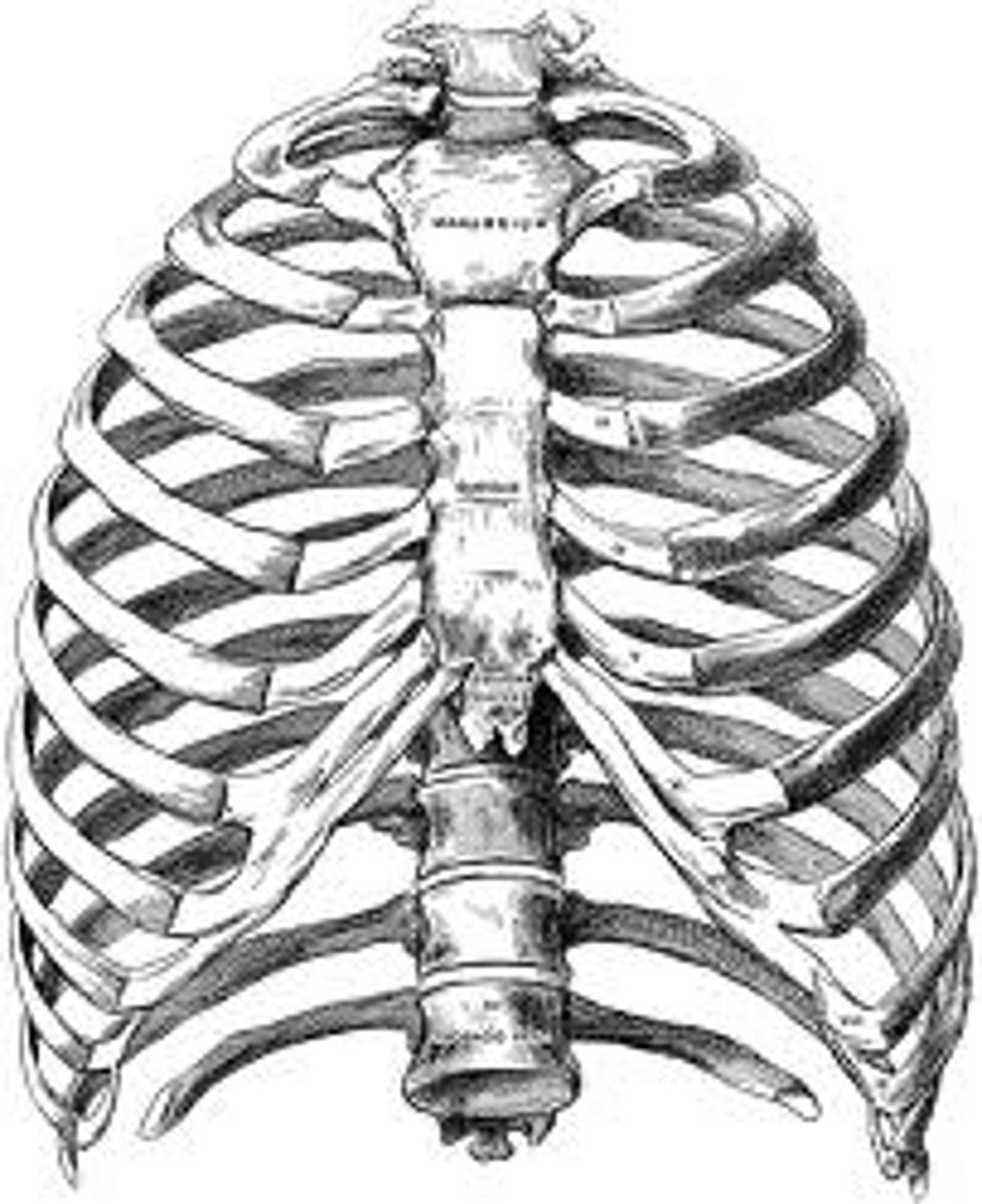
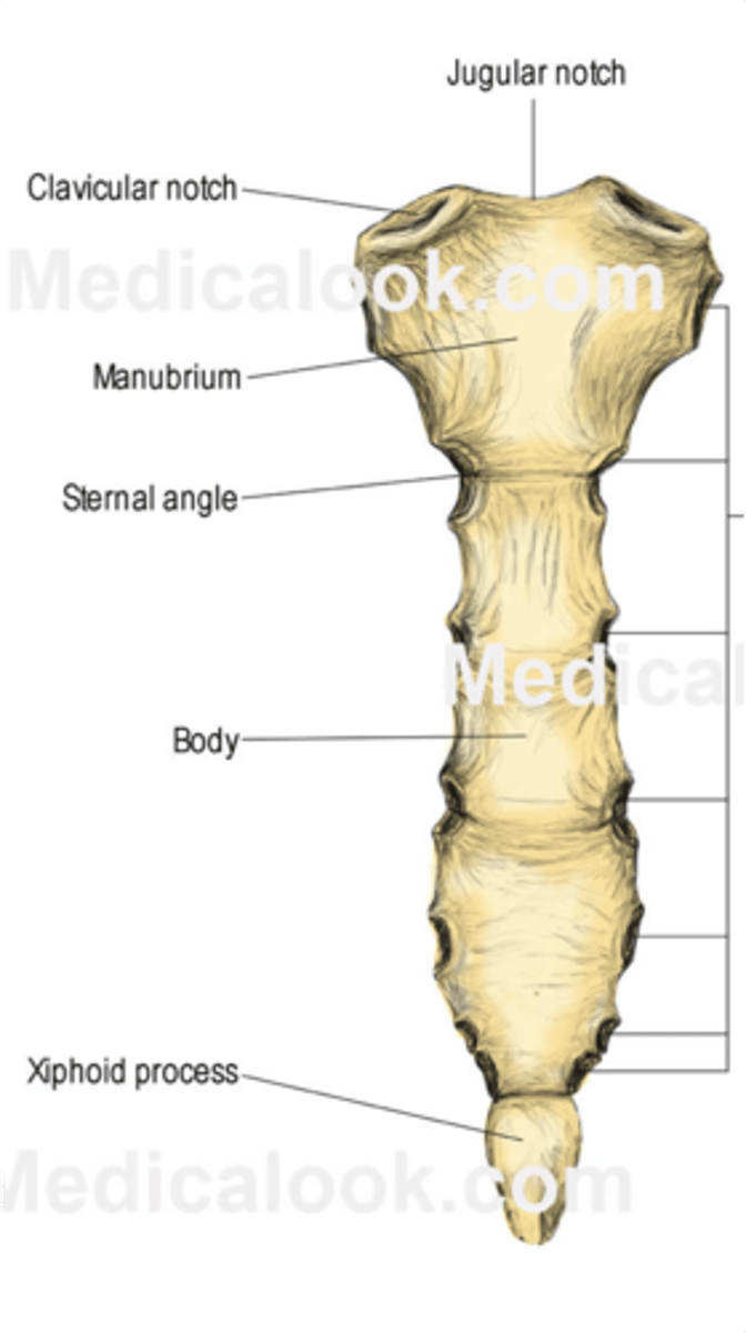
sternum's 3 sections:
manubrium, body, xiphoid process
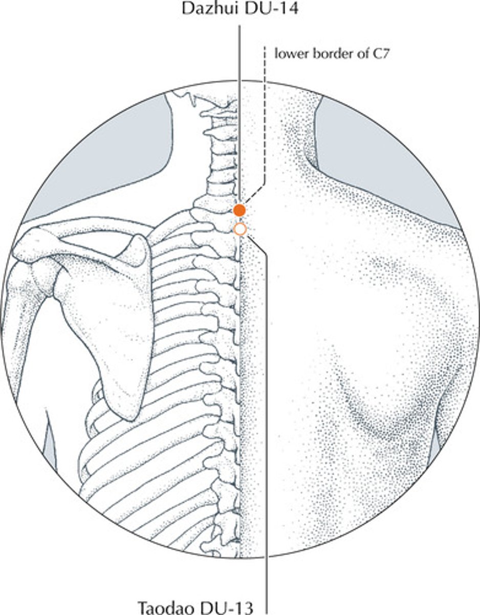
vertical prominens
-C7
-used to help determine the CR on a chest PA.
-palpitate base of neck
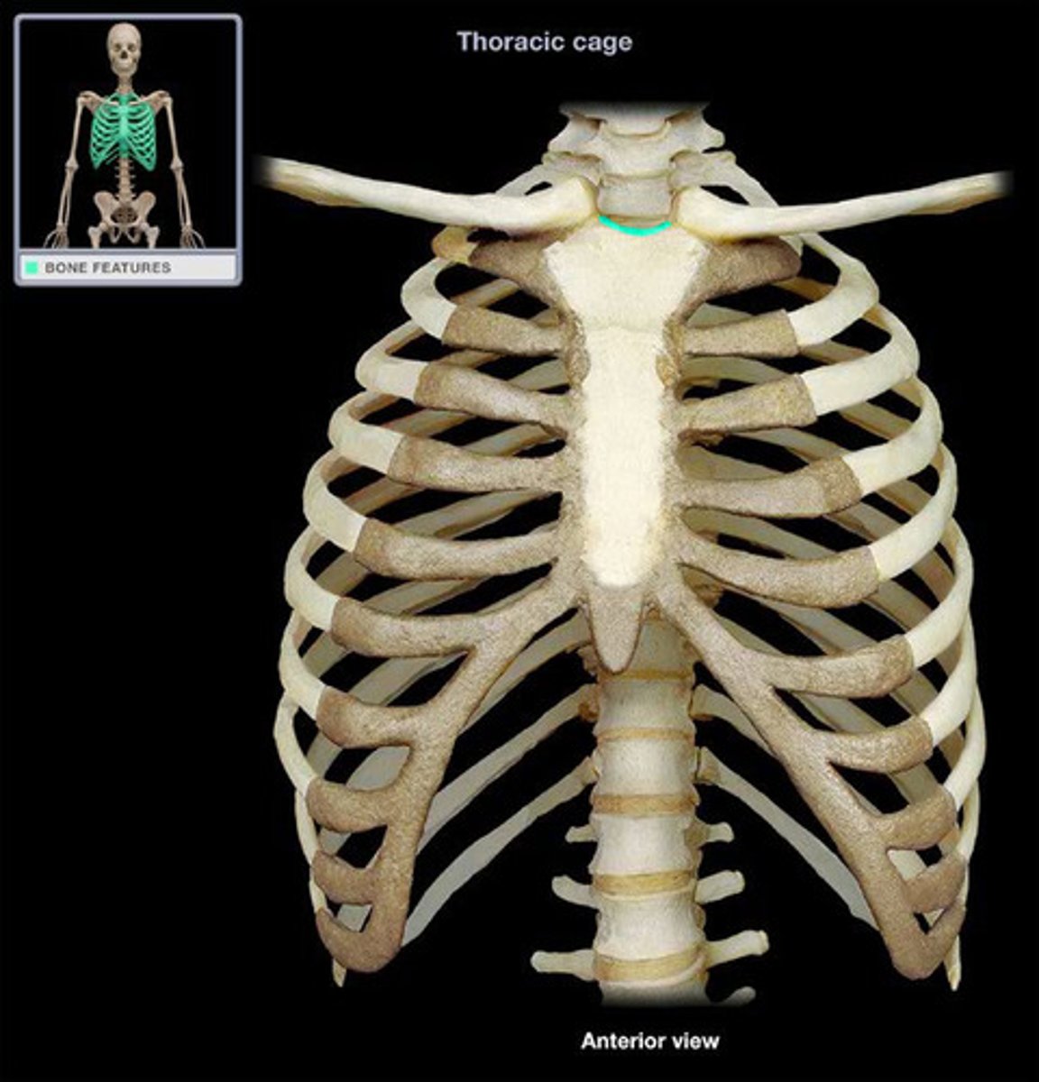
jugular notch
-felt as deep depression superior to sternum
-used to determine the CR on a chest AP
diaphragm
primary muscle of inspiration
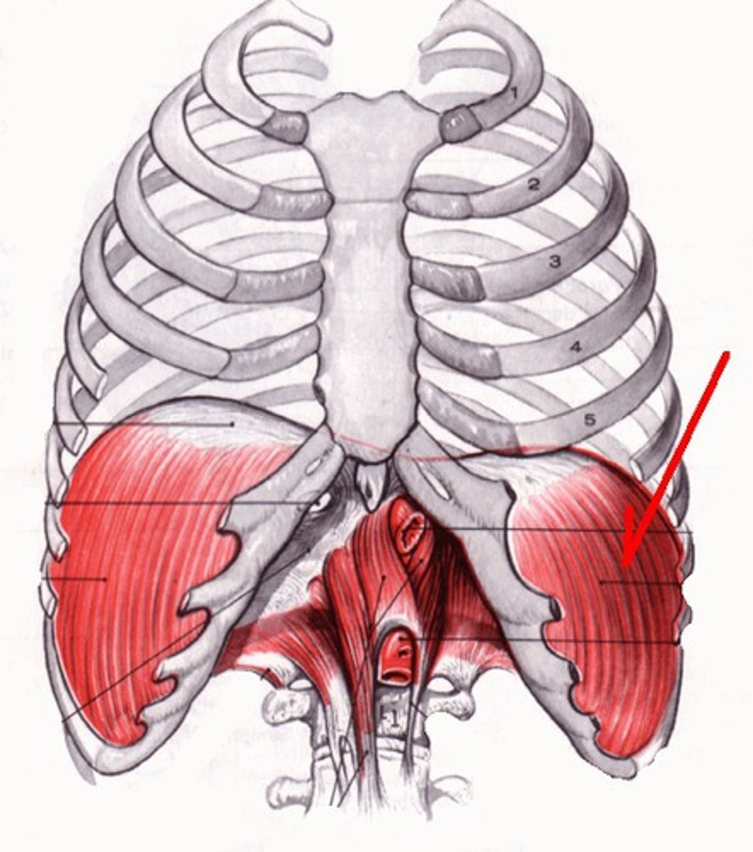
Hemidiaphragm
each half of the diaphragm
how does the diaphragm move during inspiration?
Contracts and pulls down, decreasing pressure in thoracic cavity
pharynx
-throat; passage for food, fluids, air
-3 divisions: nasopharynx, oropharynx, laryngopharynx
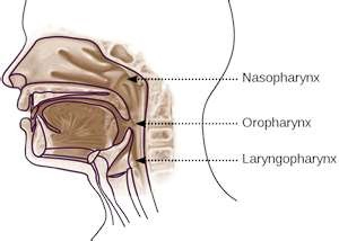
nasopharynx
-the area directly posterior to the nose
-oral cavity; contains the hard and soft palates
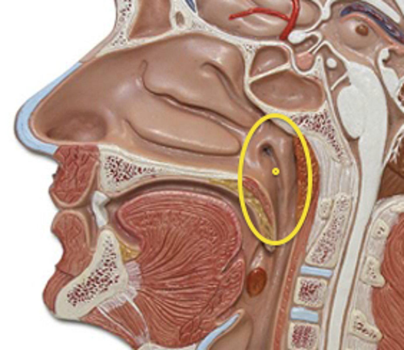
uvula
-small projection hanging from the back middle edge of the soft palate
-marks boundary between the nasopharynx and oropharynx
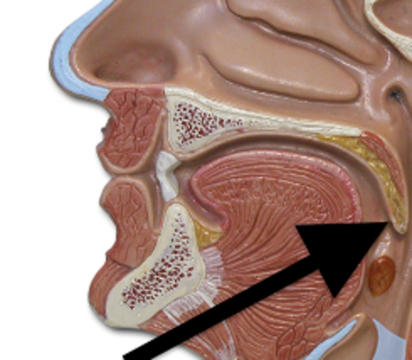
oropharynx
-posterior to mouth
-tongue is the anterior wall
-palatine and lingual tonsils
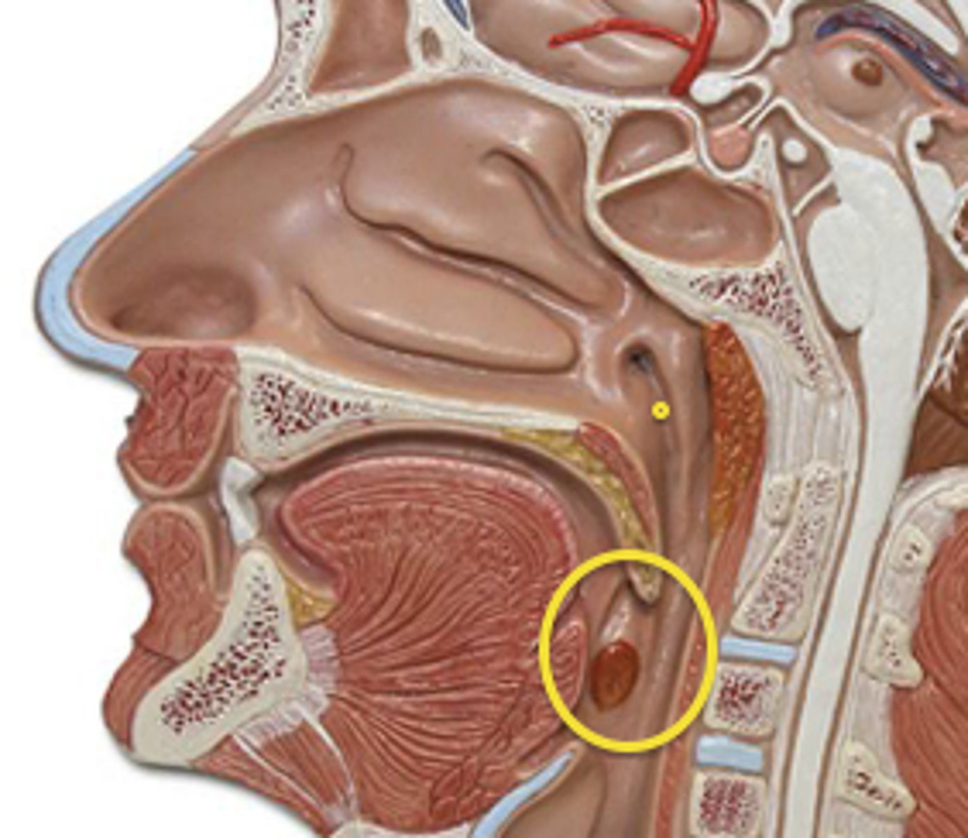
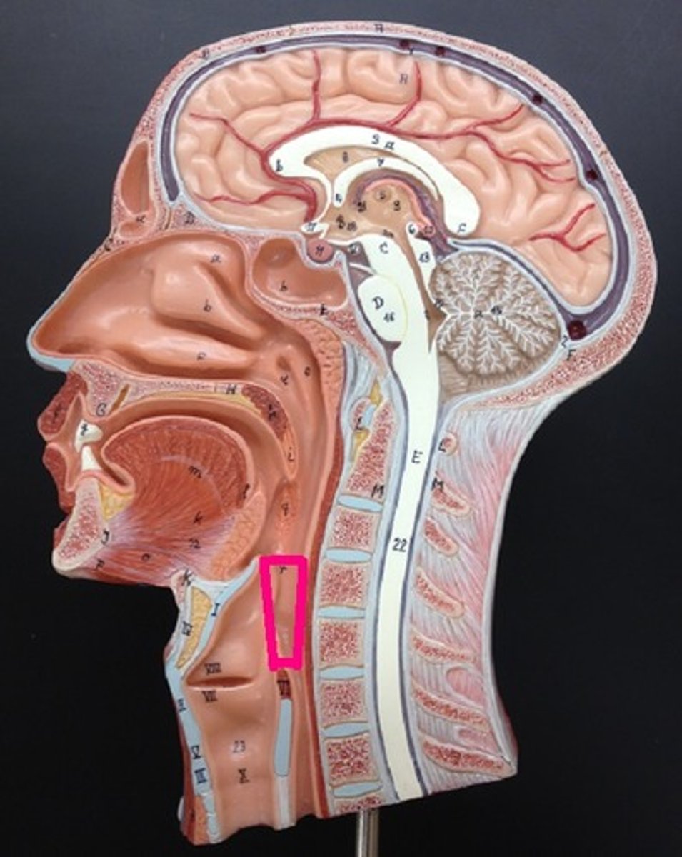
laryngopharynx
above and posterior to the larynx (C3-C6)
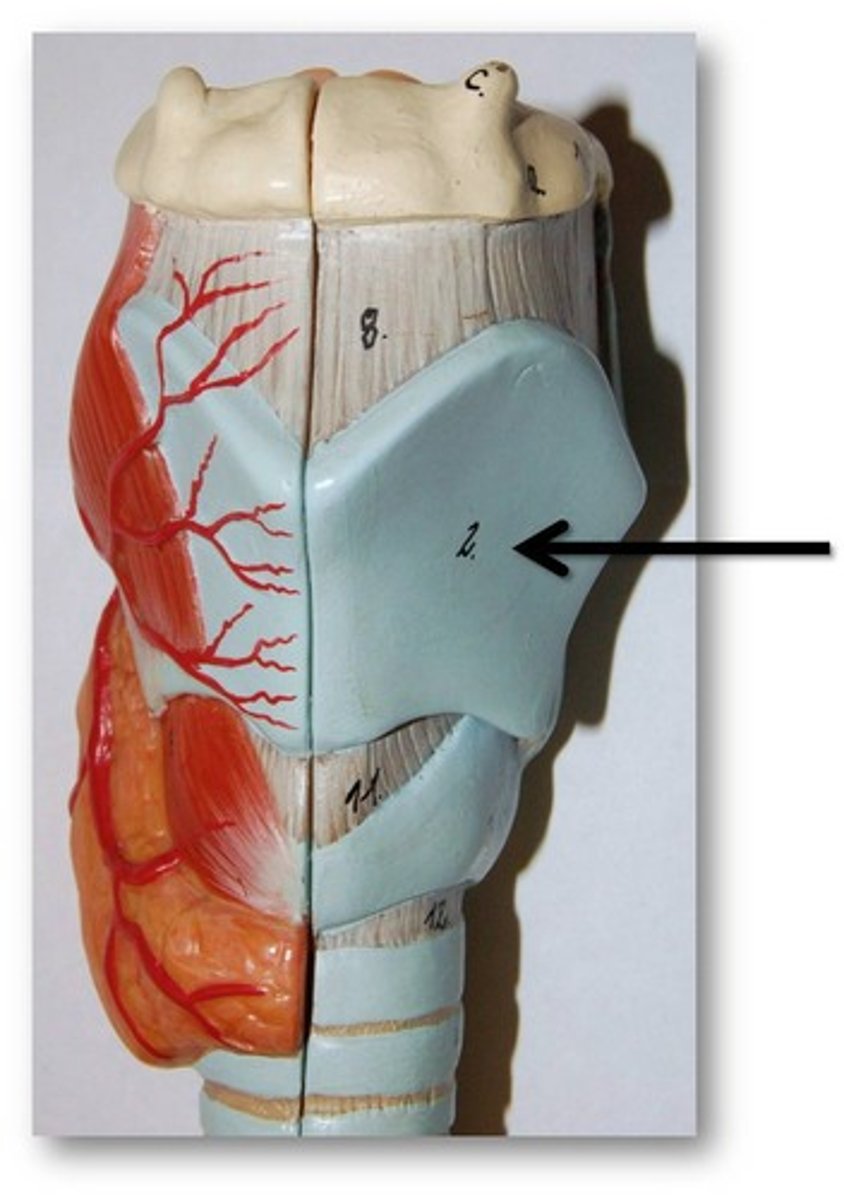
thyroid cartilage
anterior cartilage that covers the larynx
laryngeal prominence
-Adam's apple (outer projection of thyroid cartilage)
-C4-C5
cricoid cartilage
-the ring-shaped structure that forms the lower portion of the larynx
-connects to 1st ring of trachea
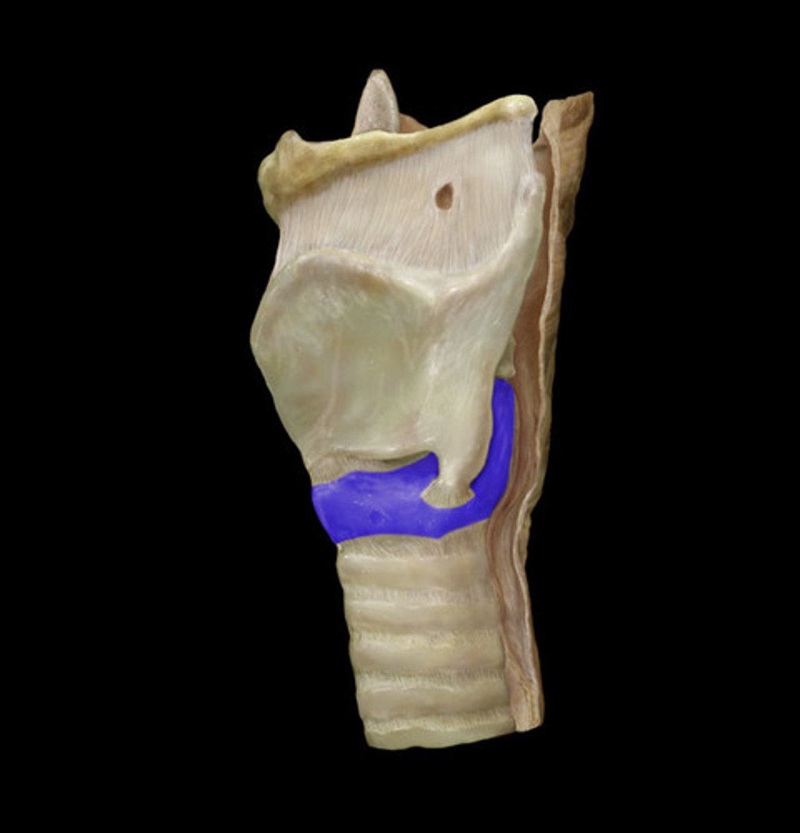
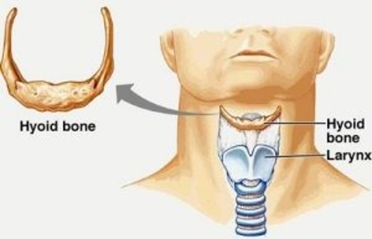
the larynx is suspended by the _______.
hyoid bone
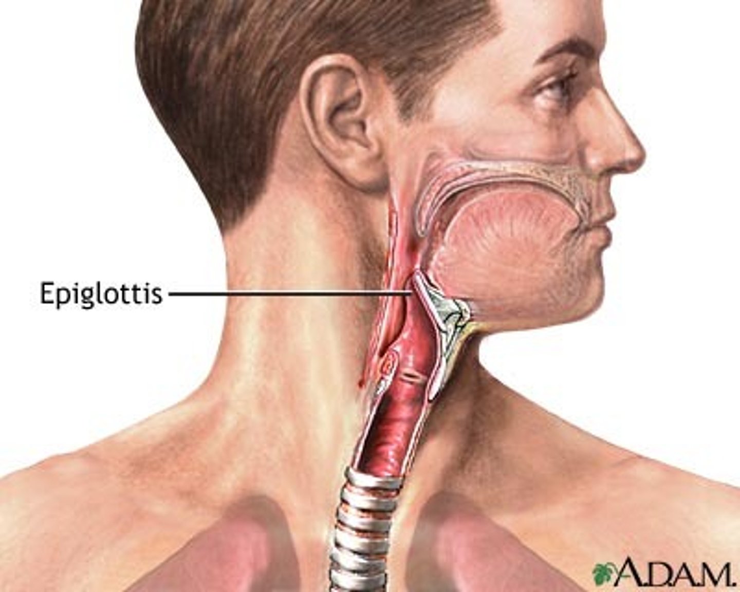
Epiglottis
-A flap of tissue that seals off the windpipe and prevents food from entering the bronchi
-posterior to larynx and trachea
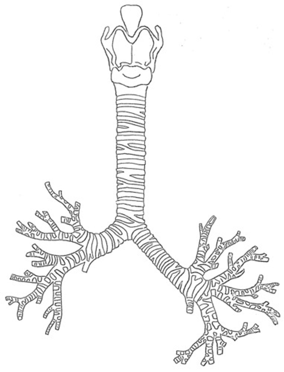
trachea
-windpipe
-16-20 cartilage rings to prevent collapse
-C6-T4/T5
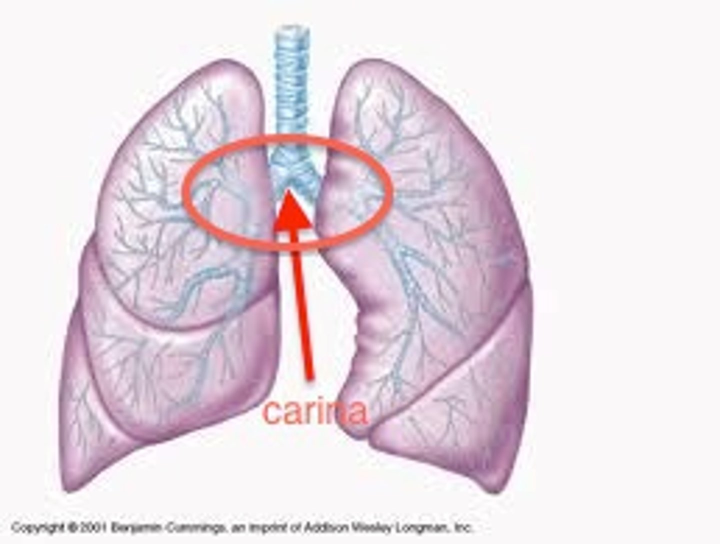
carina
Point at which the trachea divides into the left and right mainstem bronchi.
3 major endocrine glands associated with the trachea
1. thyroid gland
2. parathyroid gland
3. thymus gland
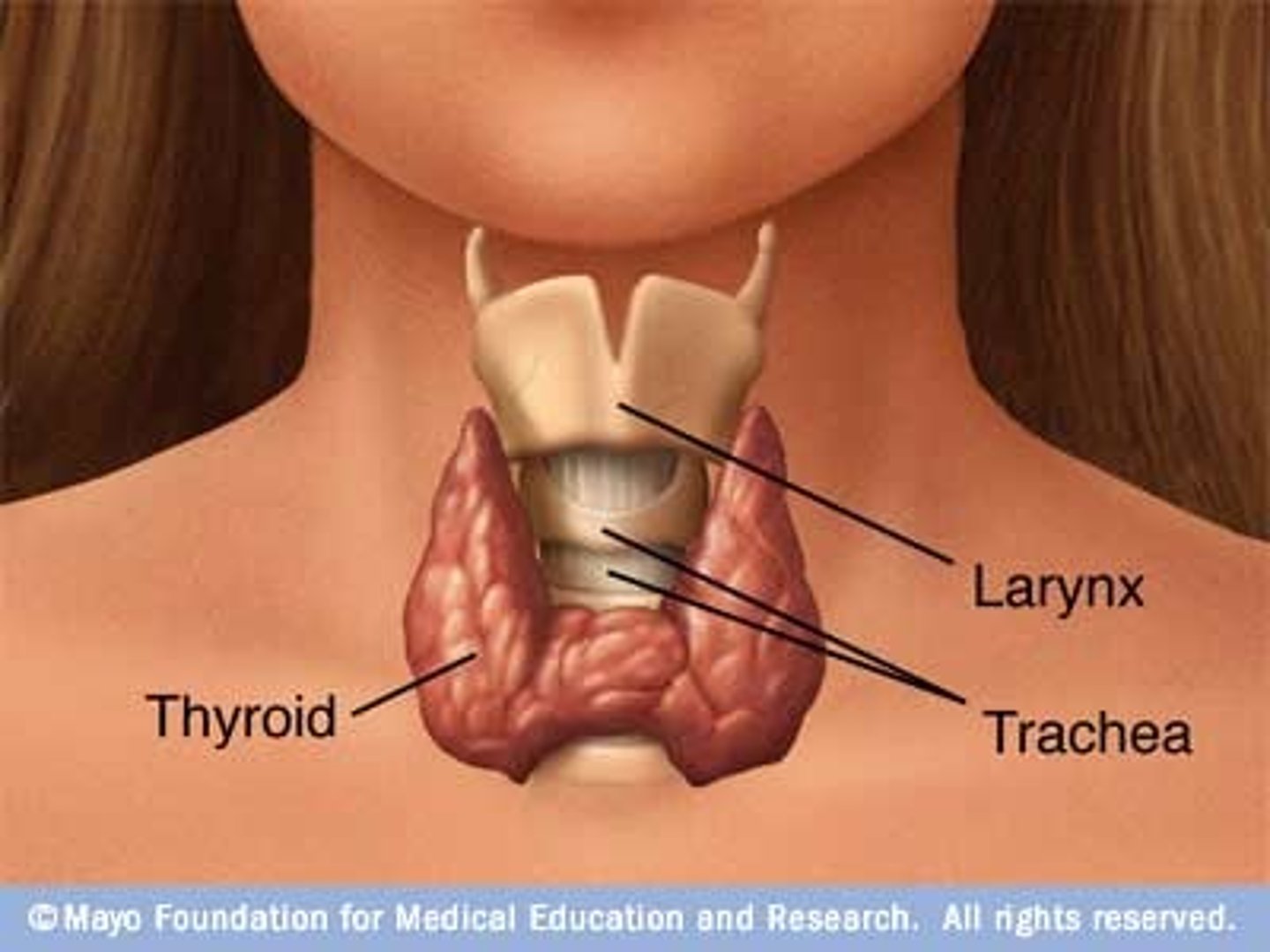
thyroid gland
-left and right lobes positioned on trachea
-regulate metabolic hormones
-IMPORTANT to shield from radiation
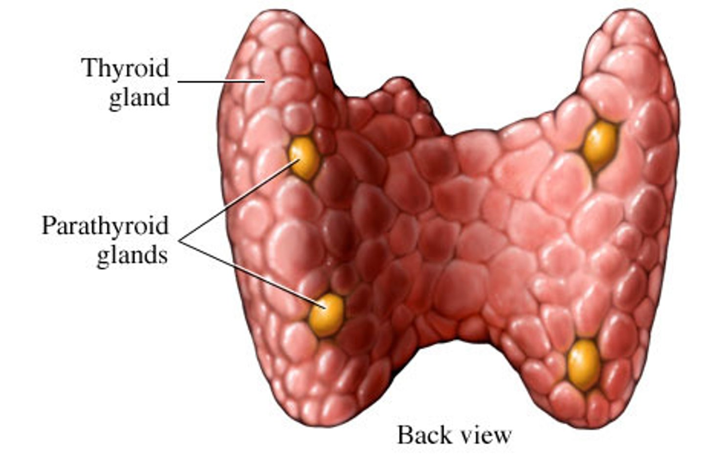
parathyroid glands
-four small glands on the posterior of the thyroid gland
-regulate blood hormones
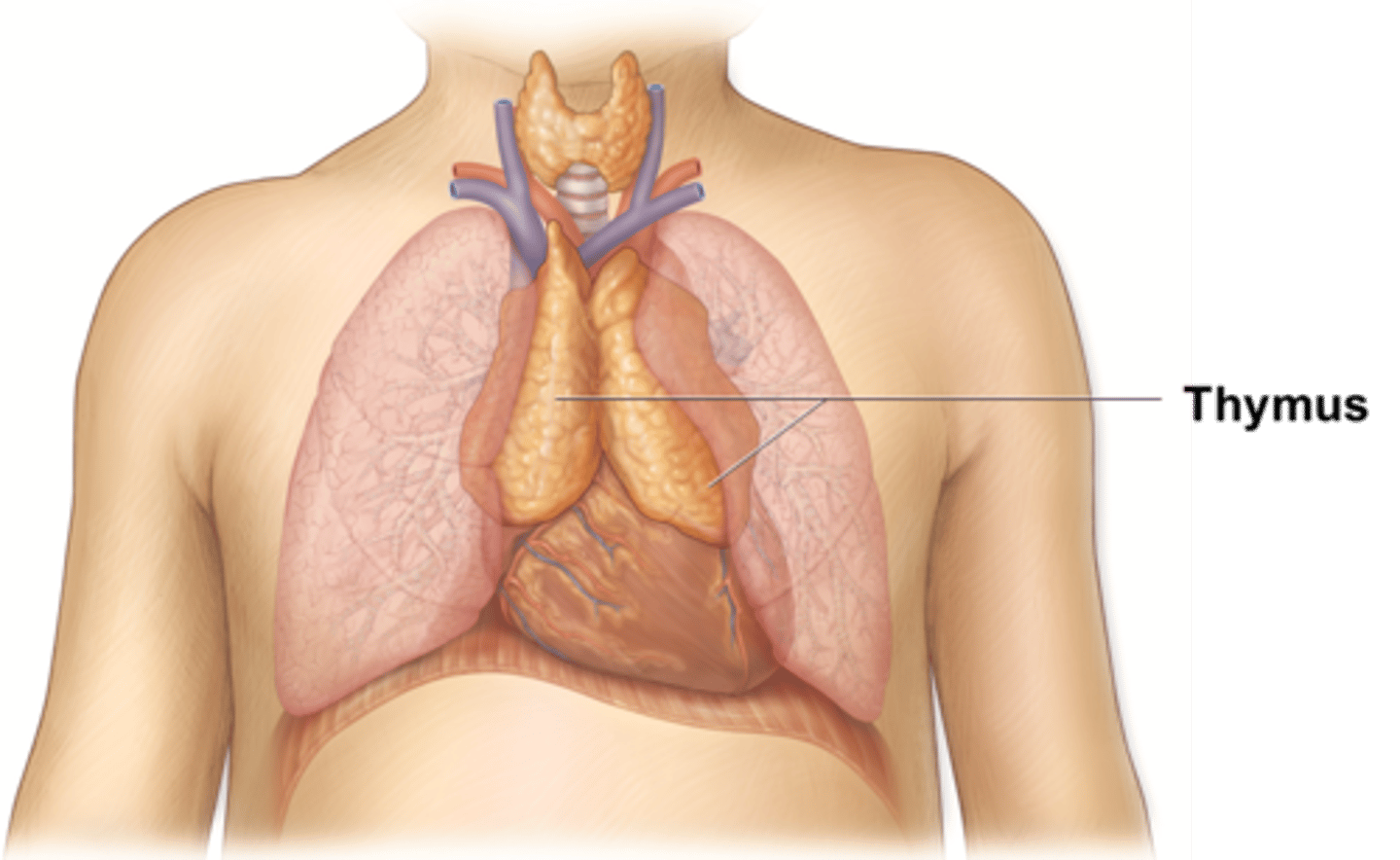
thymus gland
-anterior and superior to heart
-prominent in infants to build immune system; disappears completely into adulthood.
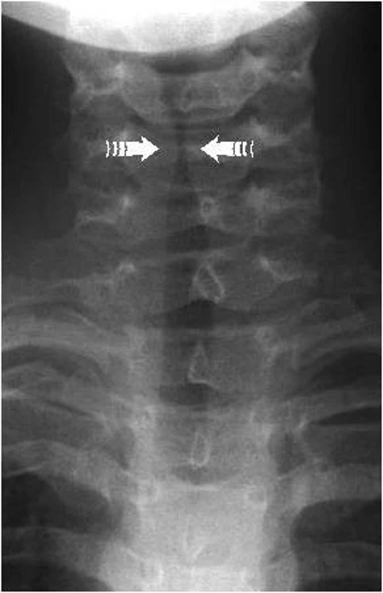
what does an AP trachea show?
-possbile abnormalities in thymus/thyroid glands
-air-filled trachea
what does a lateral trachea x-ray show?
esophagus and its relation to the trachea/larynx
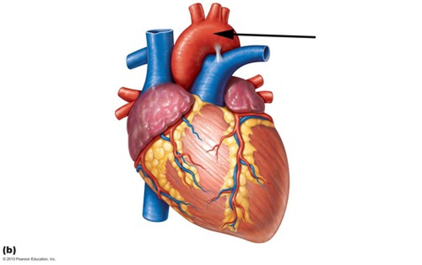
aortic arch
a curved blood vessel from which arteries branch to the head and neck.
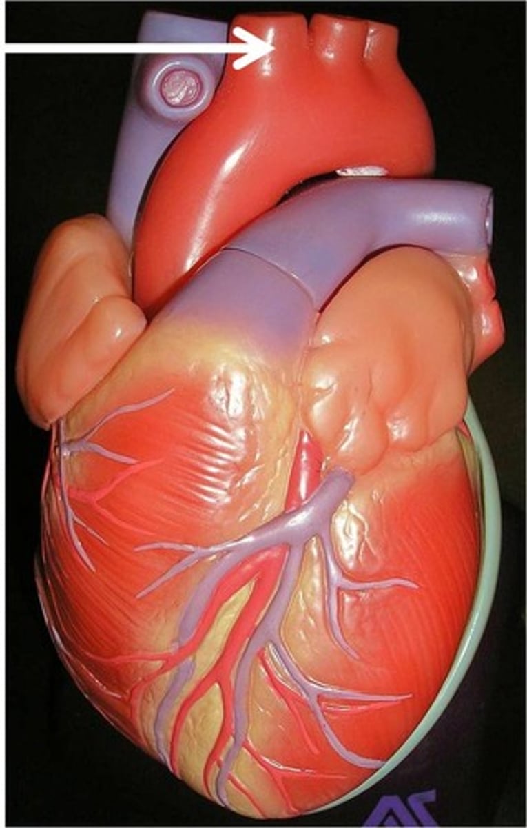
brachiocephalic artery
The first major branch off of the aorta and the major artery to the forelimbs and head.
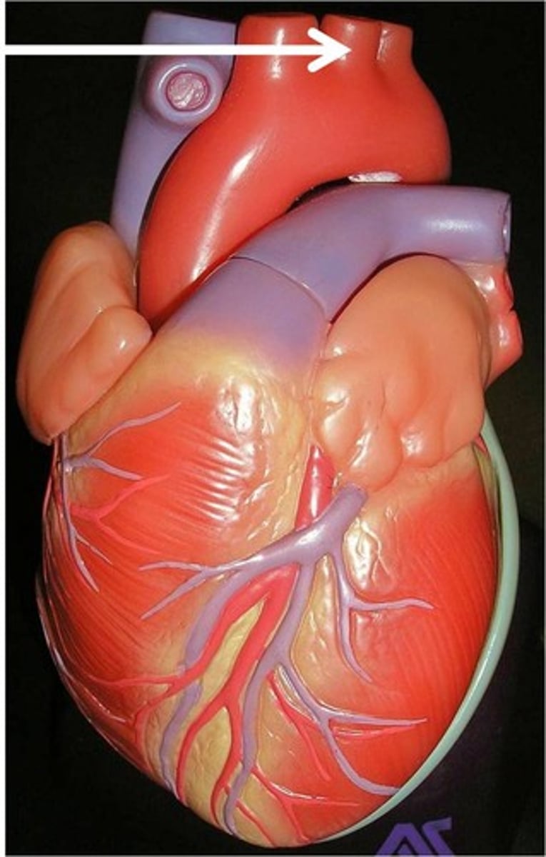
left common carotid artery
supplies left side of head and neck
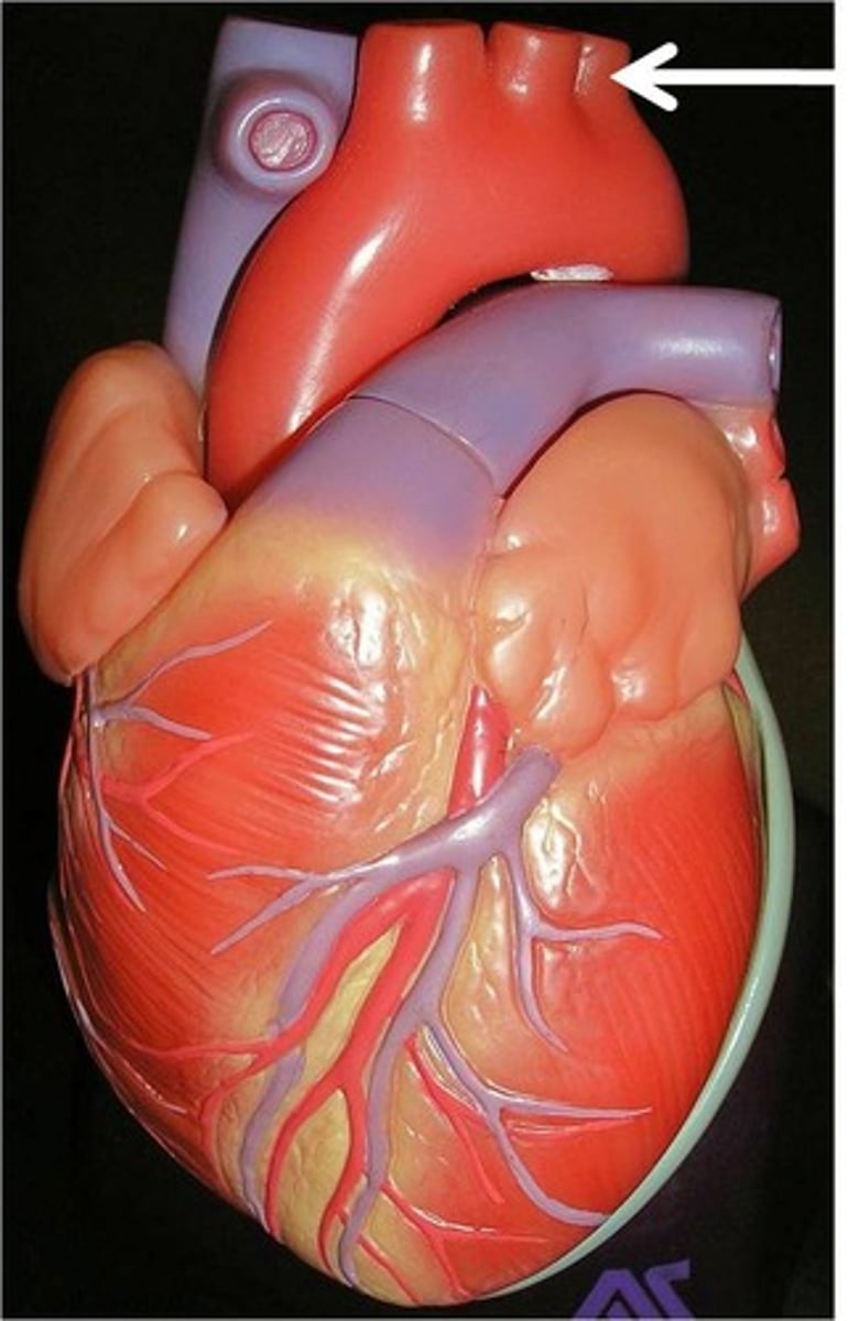
left subclavian artery
Third branch of the aortic arch that distributes blood to the left arm
right primary bronchus
-wider, shorter, more open, and more vertical than the left primary bronchus
-easier for foriegn material to enter
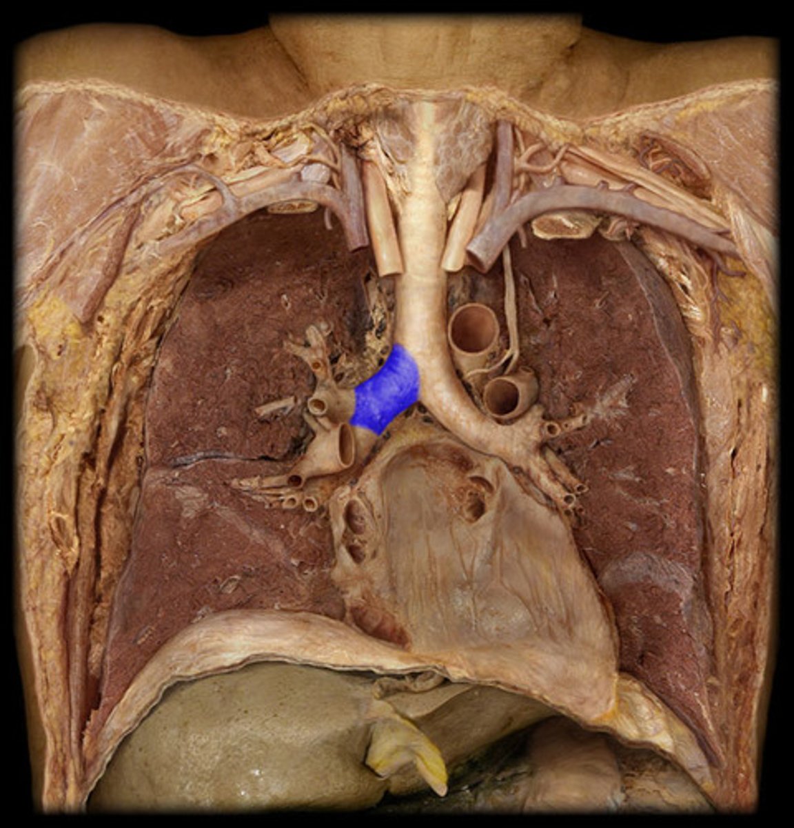
how many lobes in the left lung?
2 (superior and inferior) lobes\
2 primary bronchi
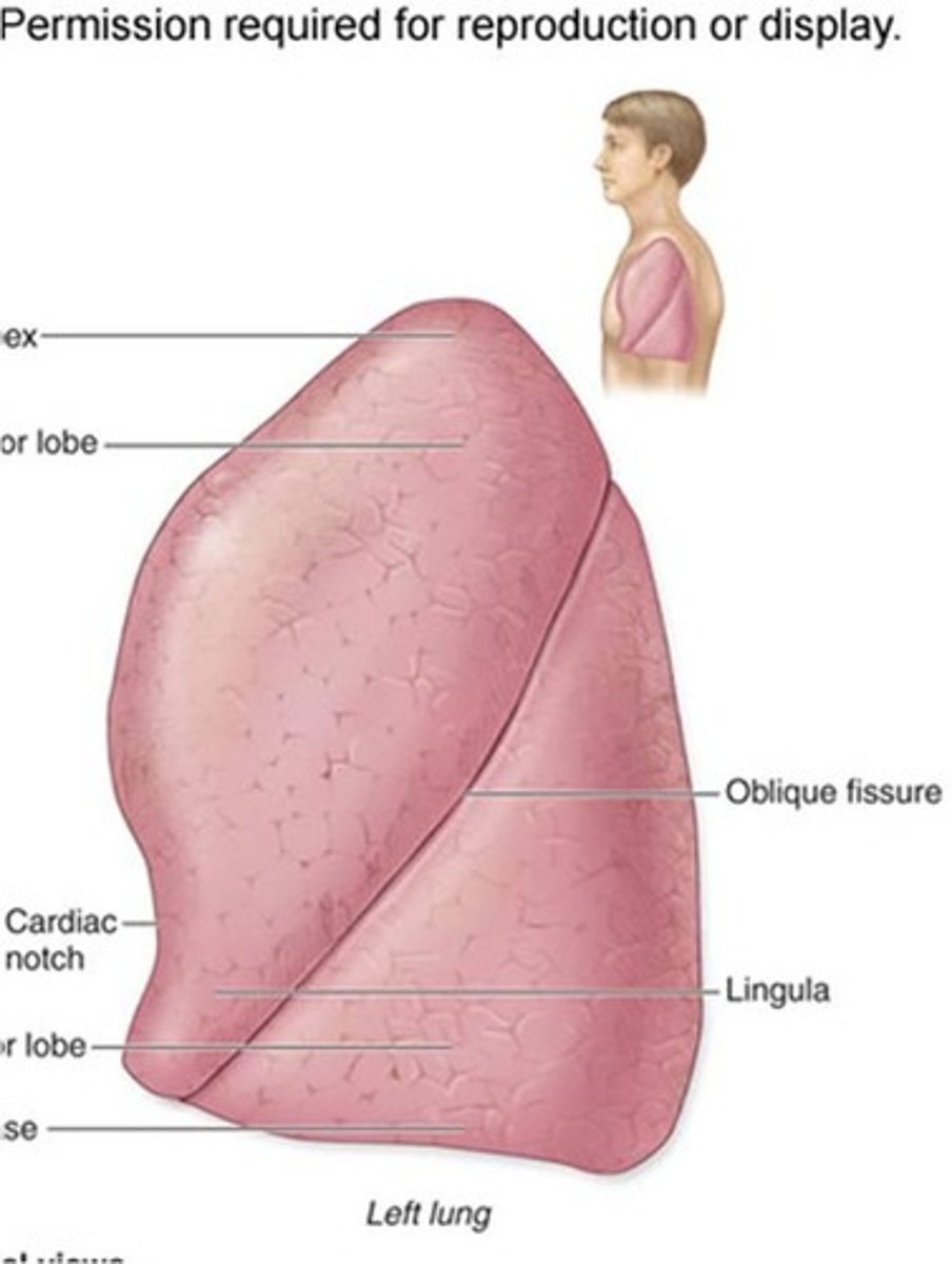
oblique fissure
-separates superior and inferior lobes of left lung
-separates middle and inferior lobes of the right lung
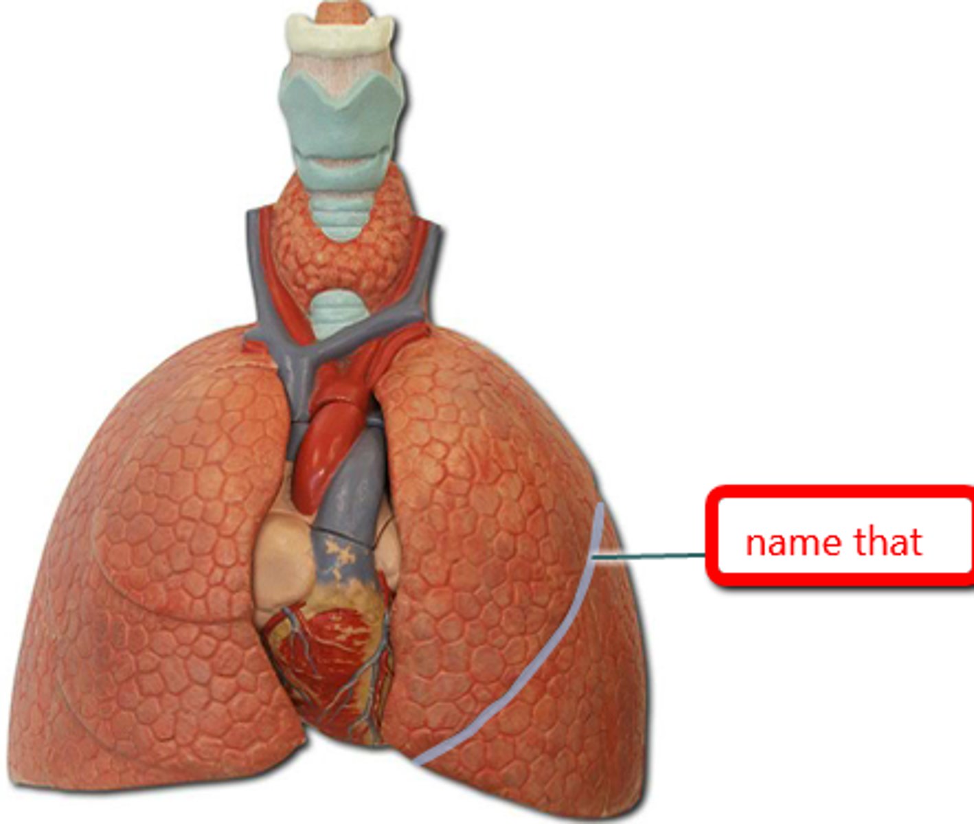
How many lobes in the right lung?
3 (superior, middle, inferior)
3 primary bronchi
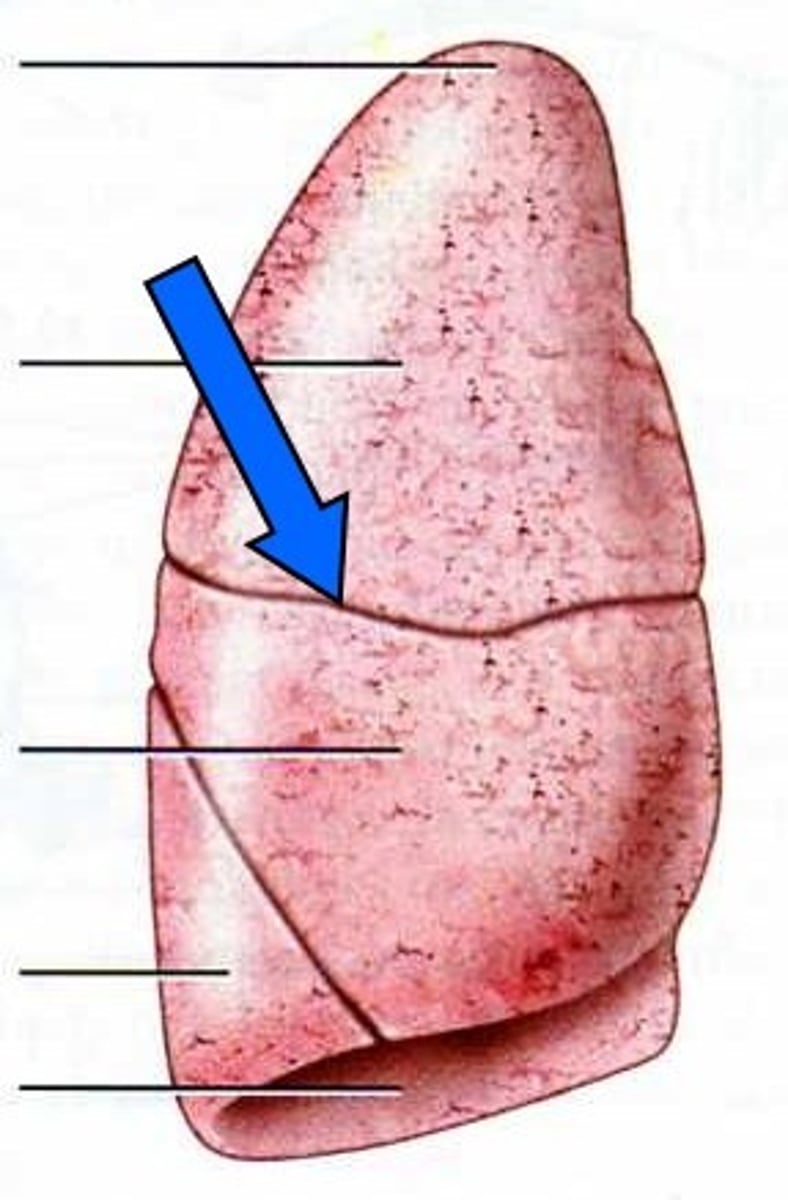
horizontal fissure
separates the superior and middle lobes of the right lung
lingula
flat, tongue-shaped process on the left lung
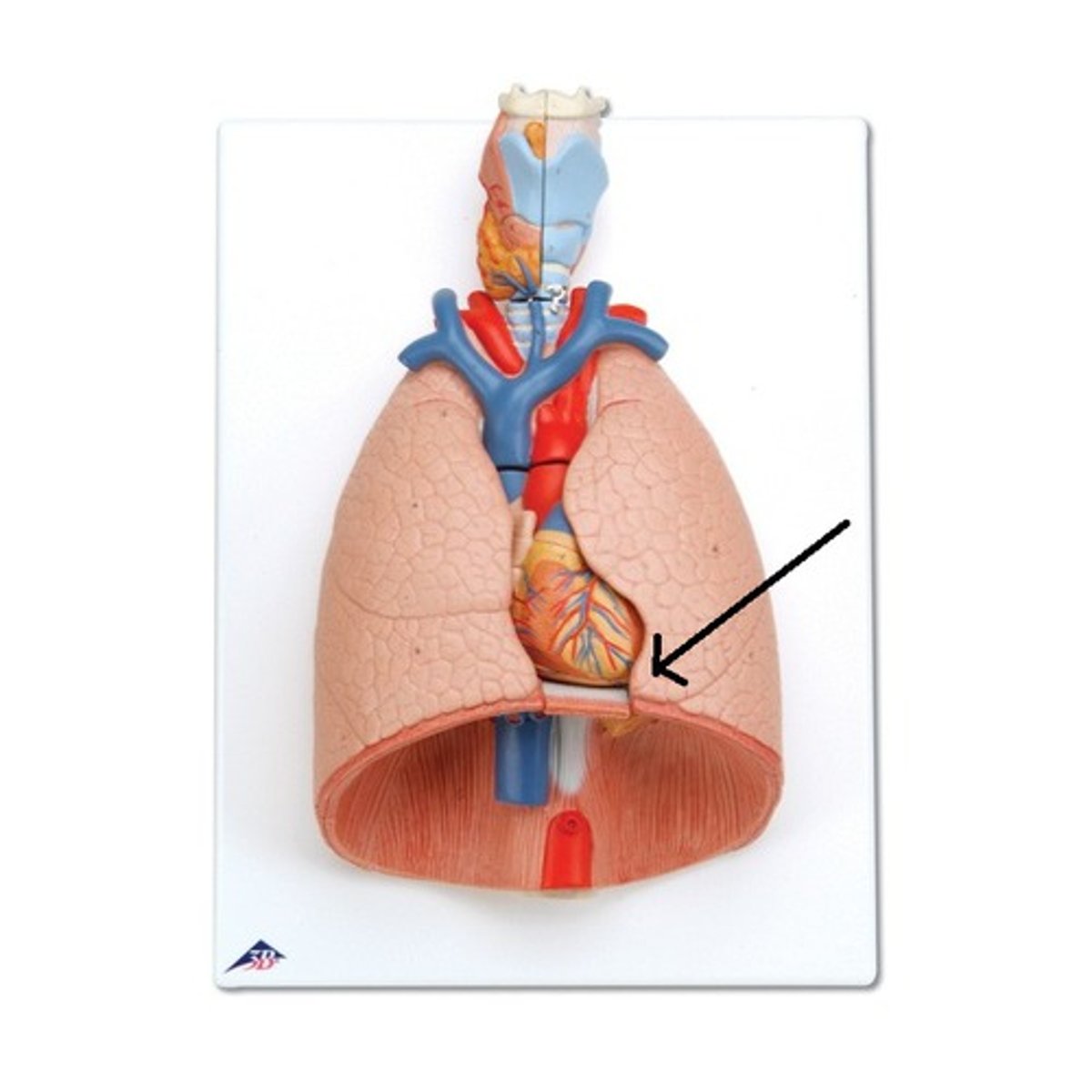
cardiac notch
a concave space on the left lung in which the heart lies
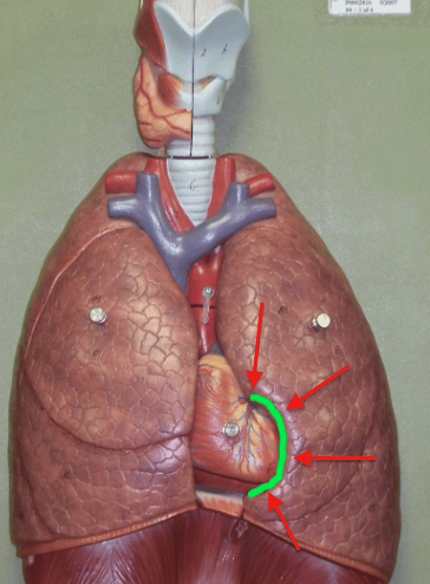
the lung tissue is composed of _______
parenchyma; provides elasticity
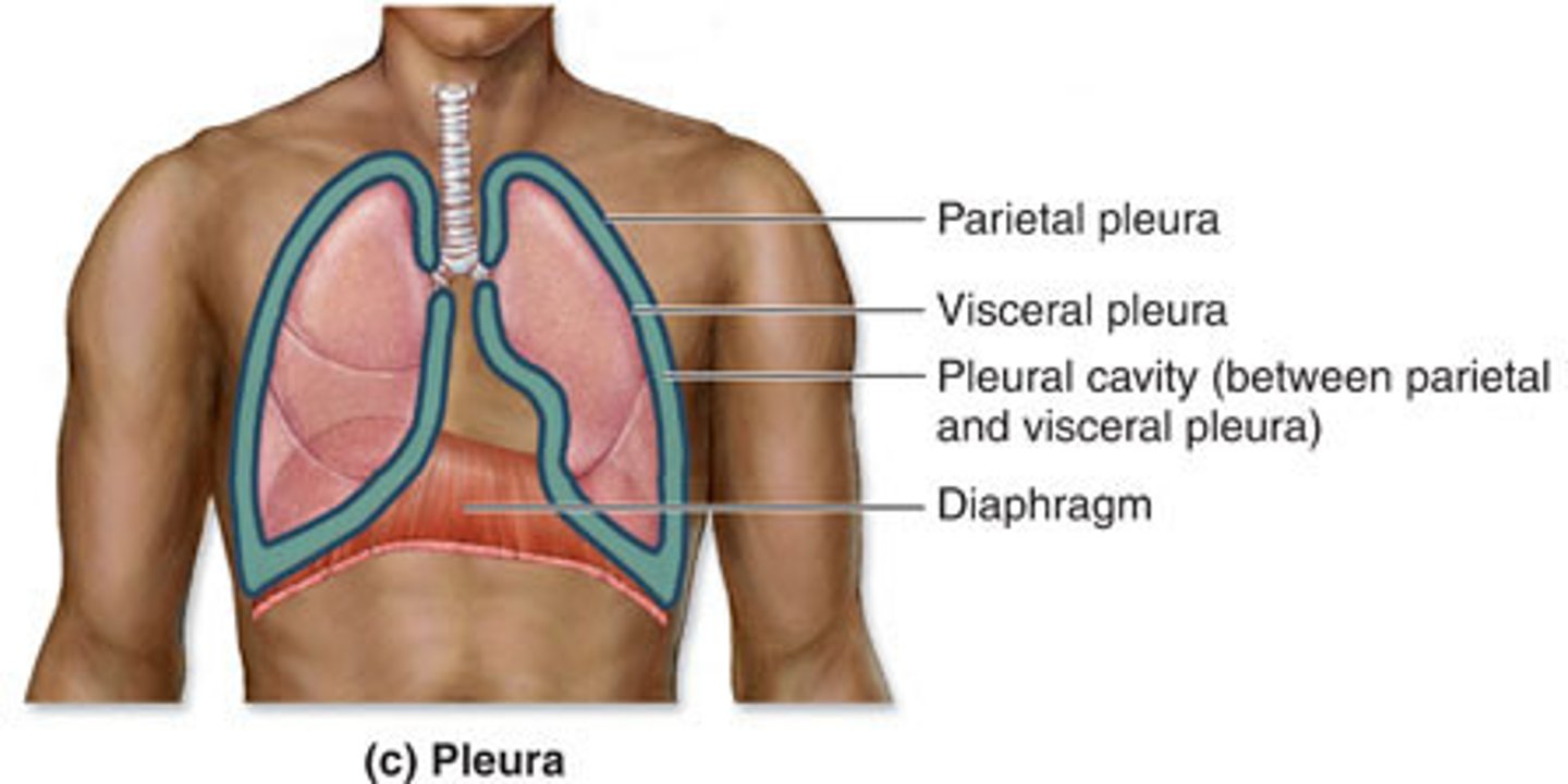
pleura
double-layered membrane surrounding each lung
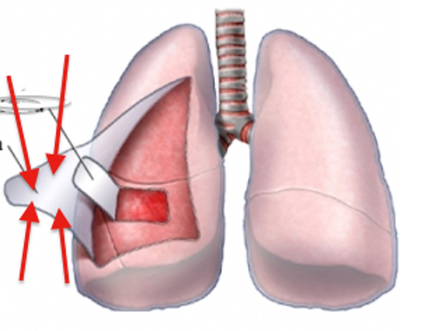
parietal pleura
outer layer of pleura lying closer to the ribs and chest wall
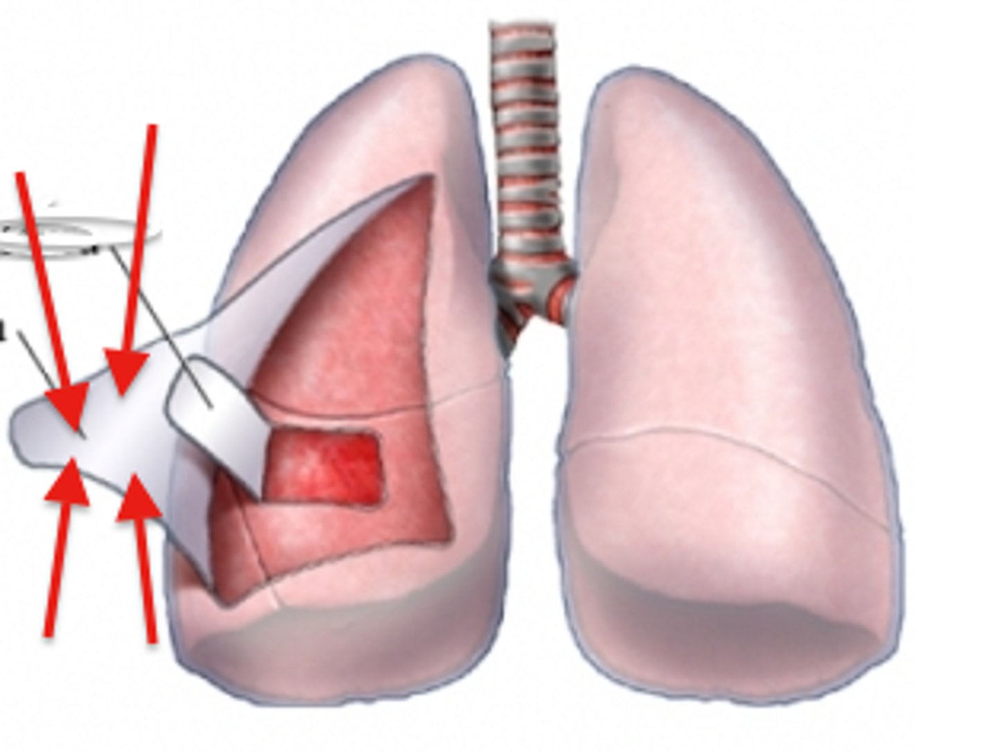
visceral (pulmonary) pleura
covers the external lung surface
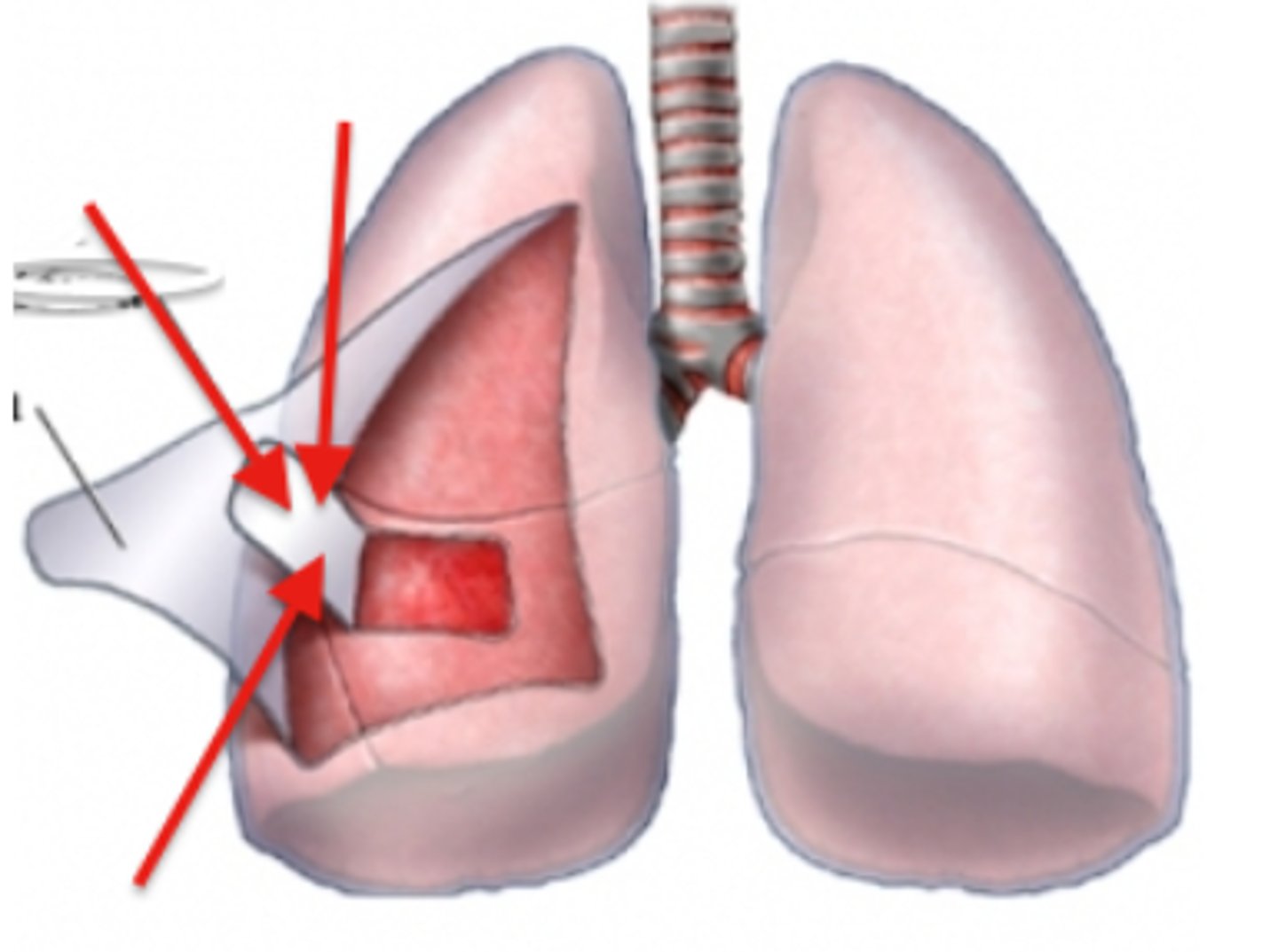
pleural cavity
space between the pleural layers
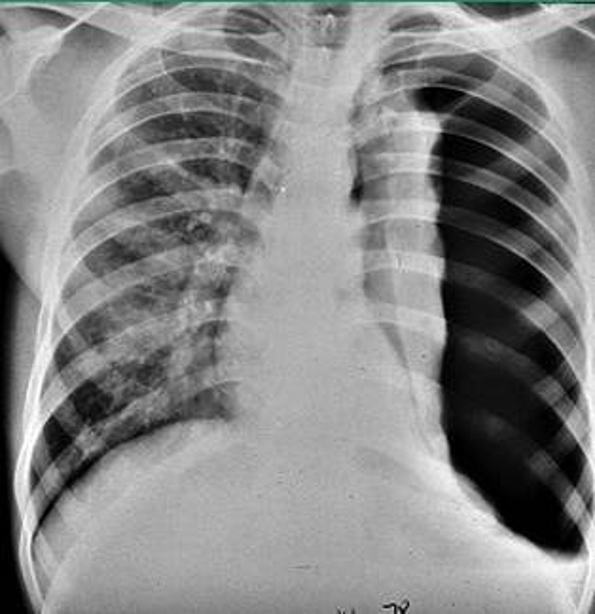
pneumothorax
-condition of air/gas in the pleural cavity
-may cause lung to collapse
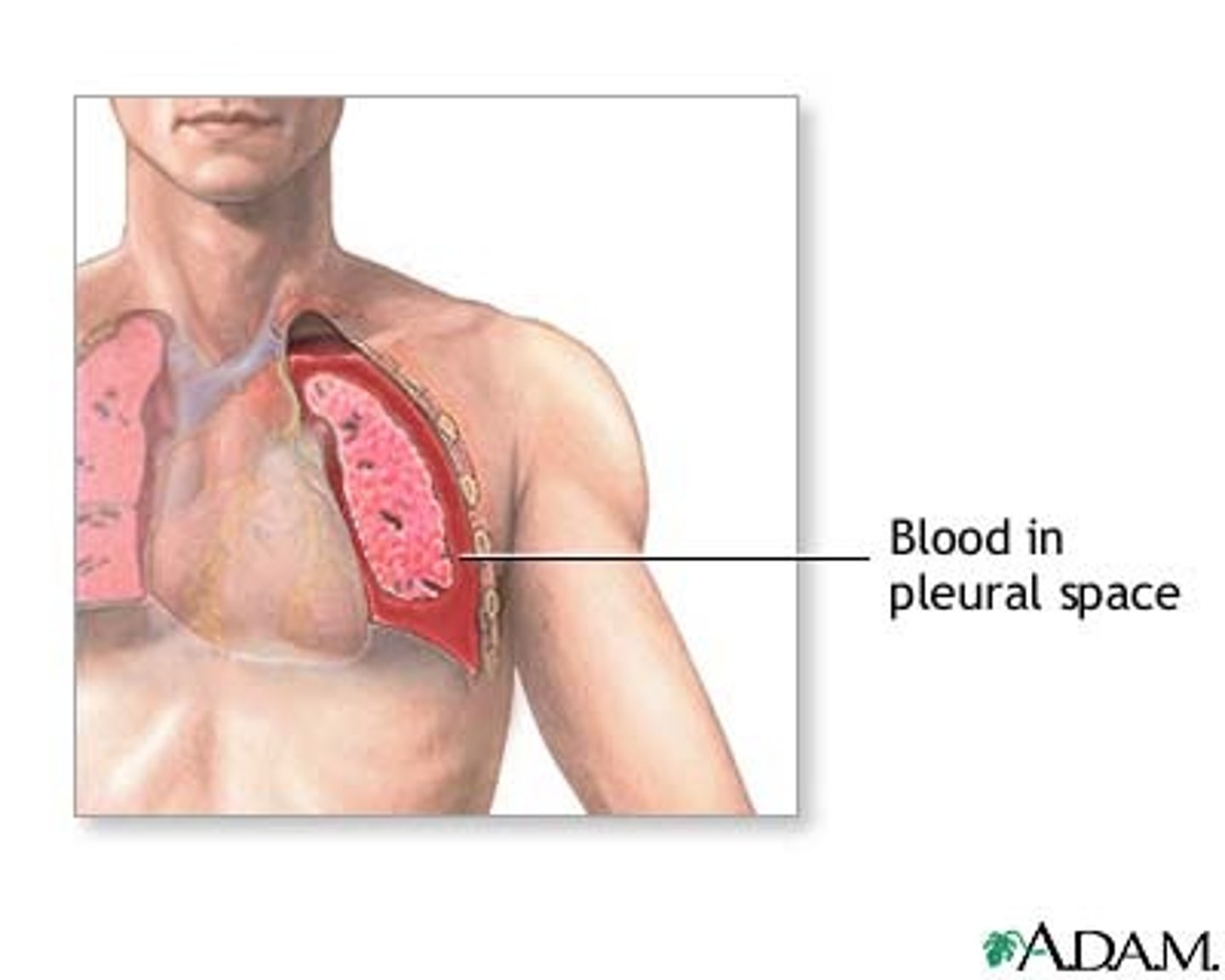
hemothorax
condition of blood accumulating in the pleural cavity
pleural effusion
refers specifically to the fluid that acculmulates in the pleural cavity
what pulmonary anatomy should be seen on a PA chest x-ray?
-Apices
-Carina
-Base of lungs [rests on diaphragm]
-Costophrenic angles
-Hilum
hilum
depression in an organ where blood vessels and nerves enter and leave
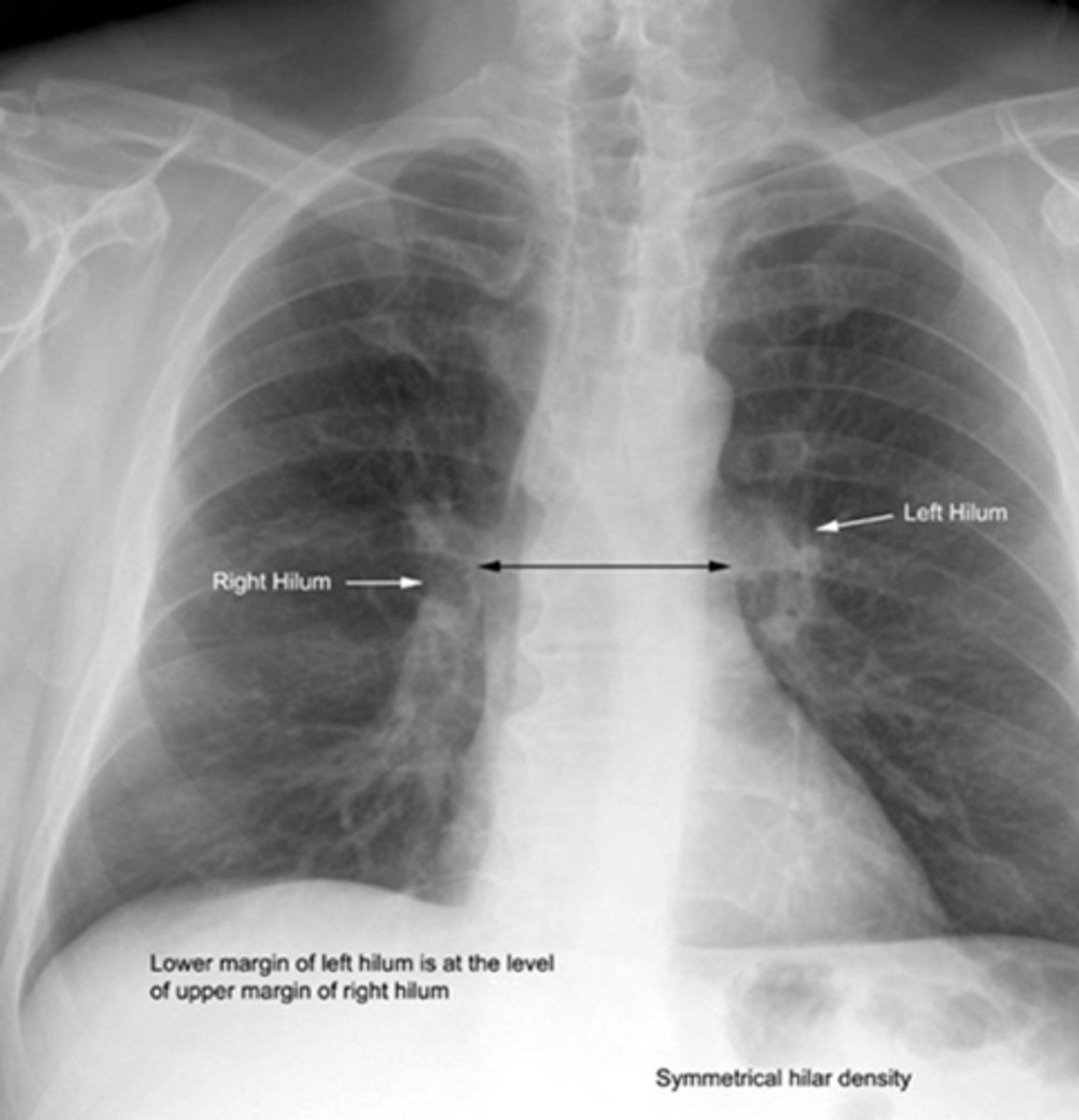
what is uniquely visible during lateral chest x-ray?
the oblique fissure
Mediastinum
-space between the lungs.
-It contains the heart, esophagus, trachea, thymus glands, and great blood vessels.
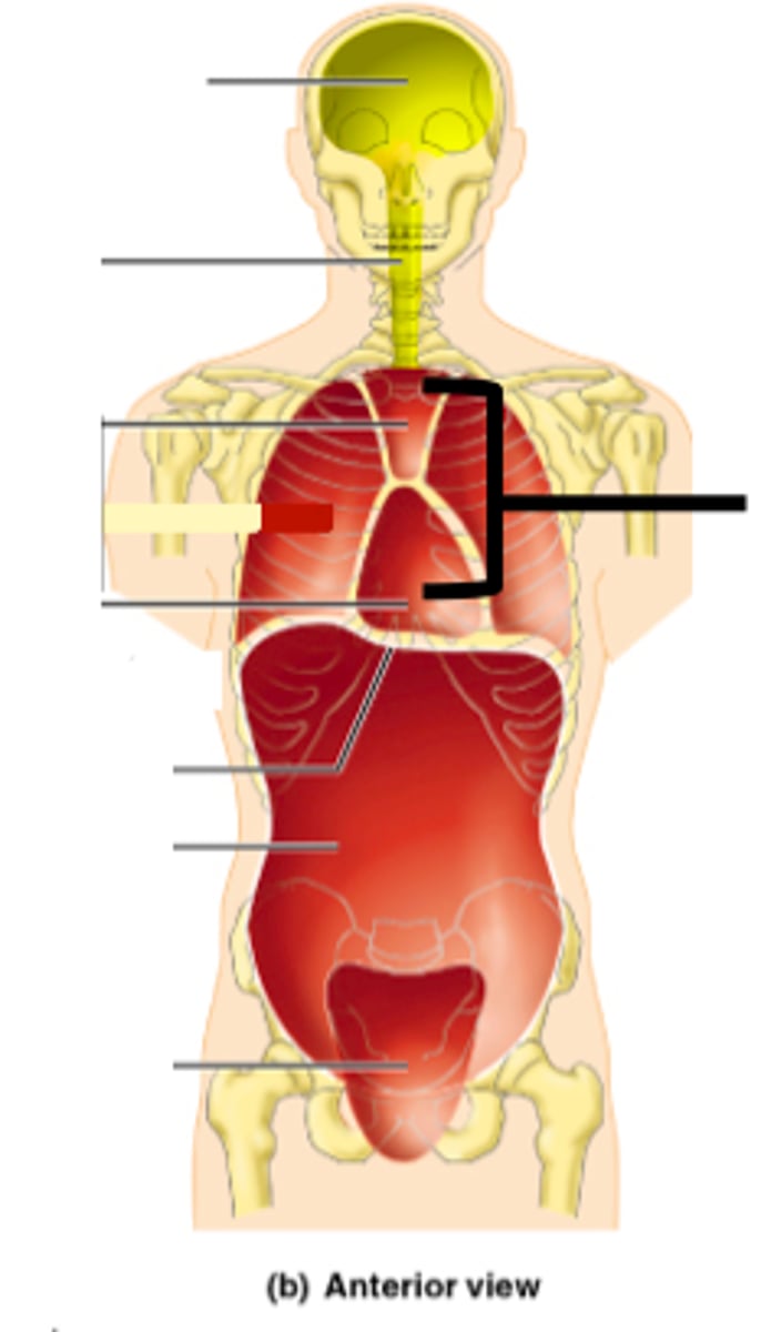
pericardial sac
surrounds the heart and helps prevent overfilling.
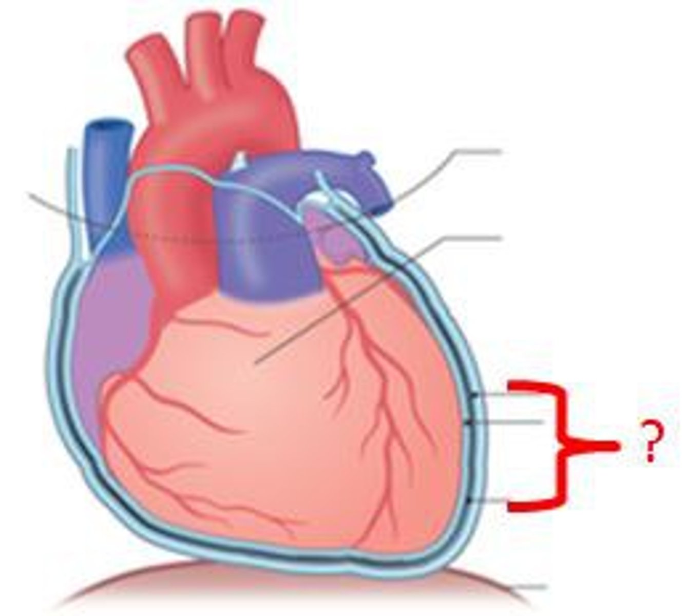
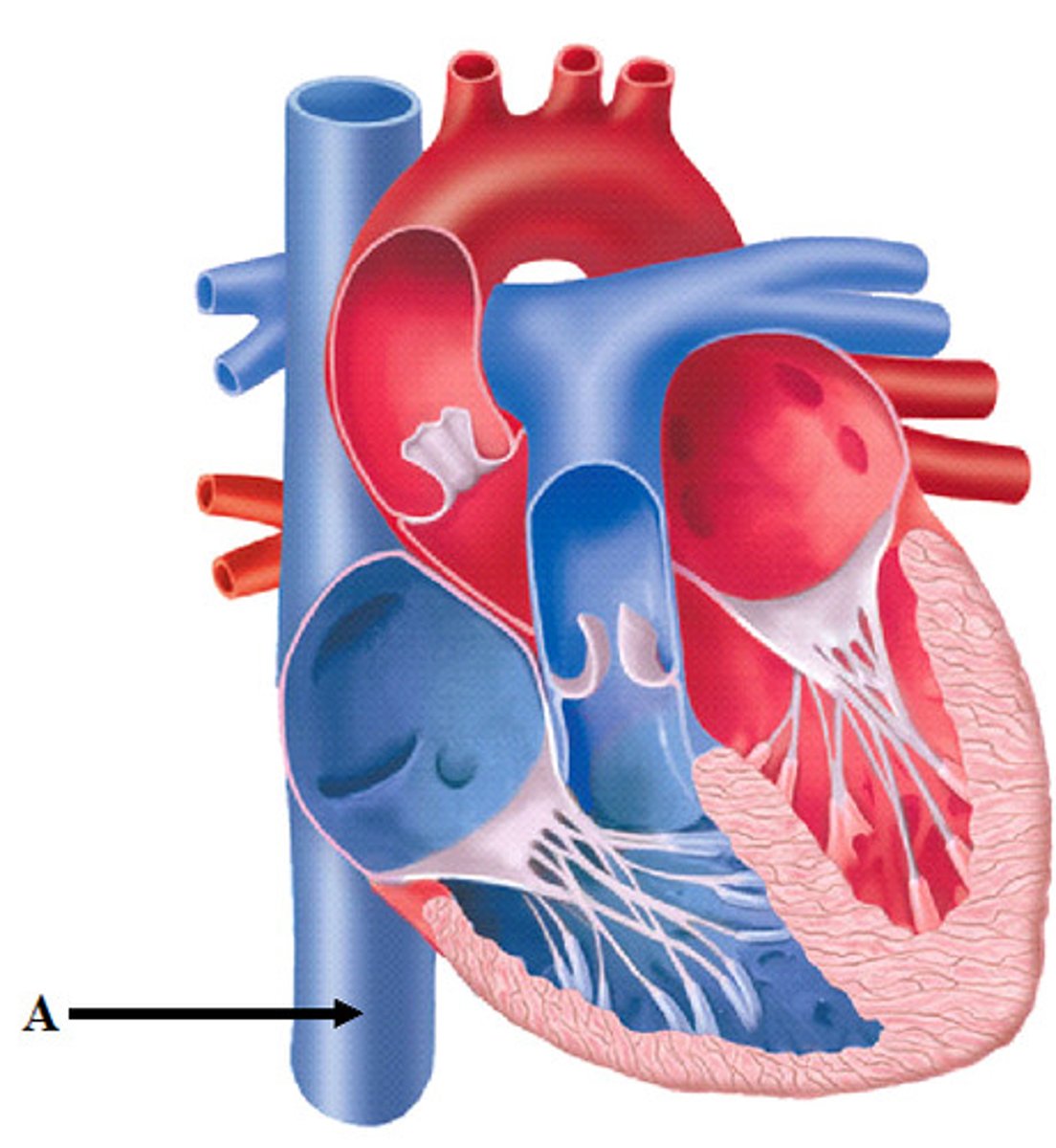
superior vena cava
large vein that returns deoxygenated blood to the heart from the upper body
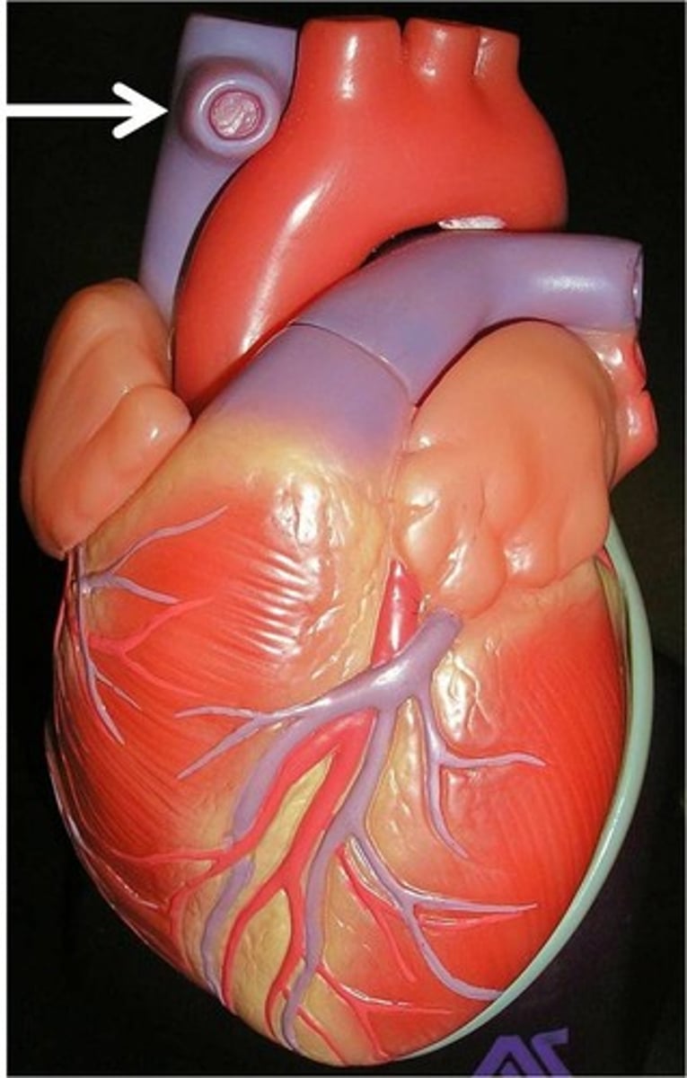
inferior vena cava
large vein that returns deoxygenated blood to the heart from the lower body
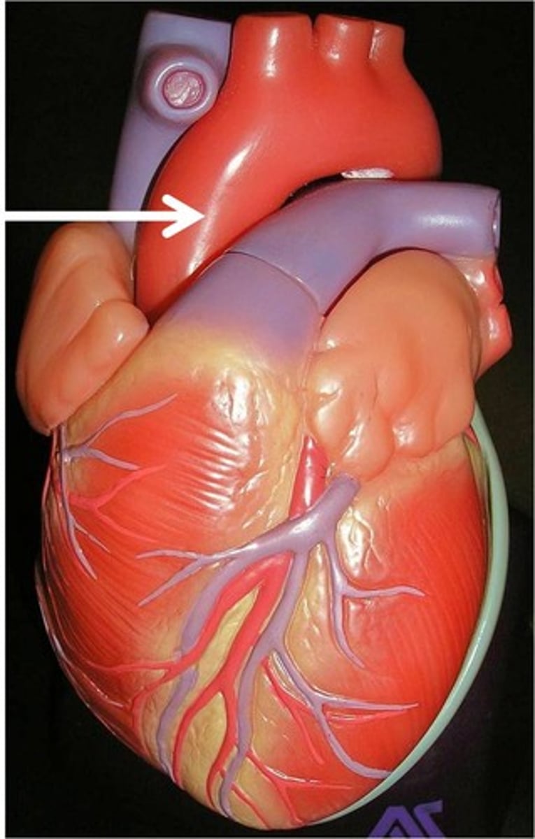
aorta
-The largest artery in the body.
-Carries oxygenated blood from the left ventricle to the rest of the body.
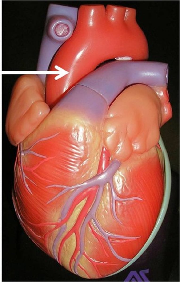
ascending aorta
Branches off the left ventricle; carries oxygen rich blood to parts of the body above the heart
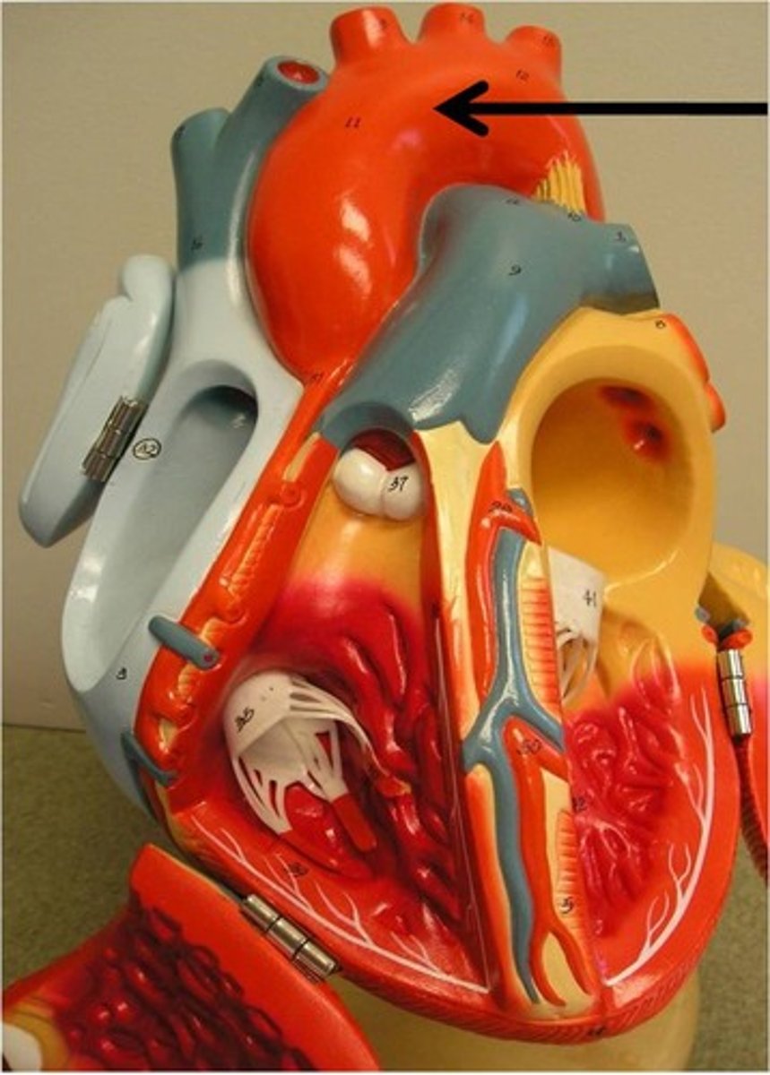
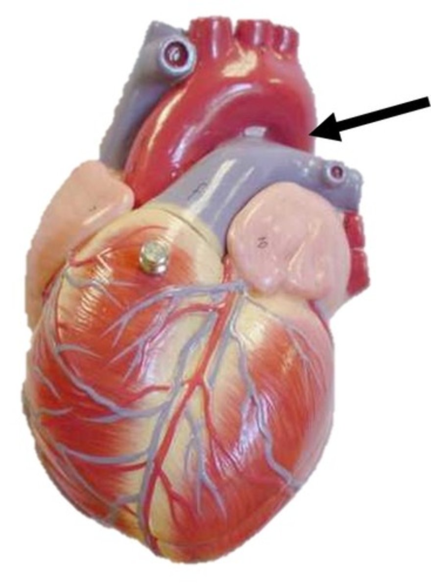
arch of aorta
The most superior portion of the aorta, lying between the ascending and descending segments of the aorta.
descending aorta
the descending part of the aorta that branches into the thoracic and abdominal aortae
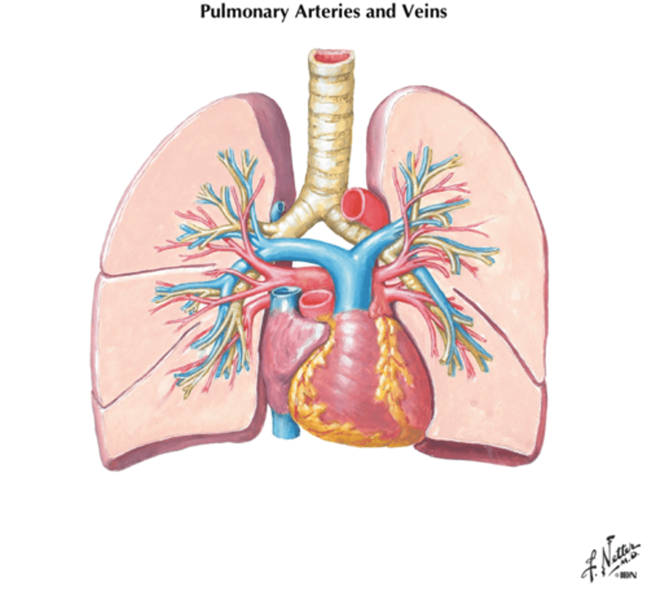
pulmonary arteries & veins
supply blood and return blood to and from all segments of the lungs
body habitus
general appearance/state of a person's body
asthenic
very slender (10%) build
-narrow thorax/chest
-long vertically
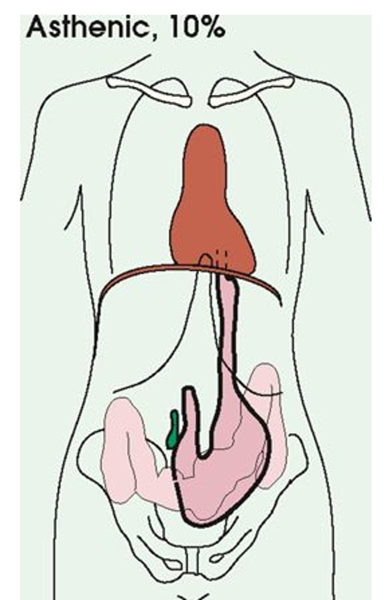
hyposthenic
slender (35%)
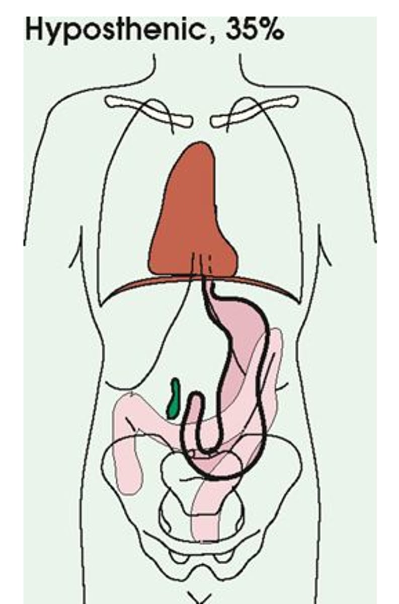
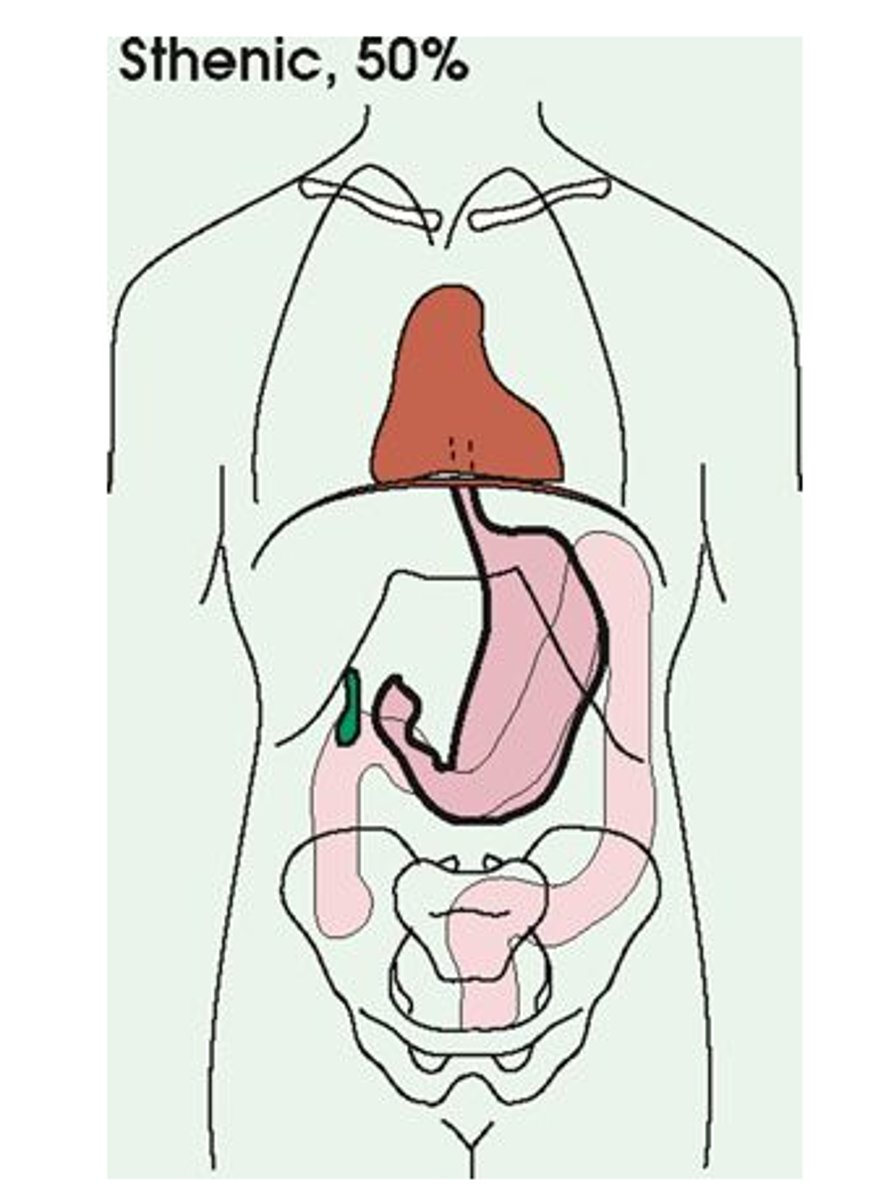
sthenic
Average body habitus (50%)
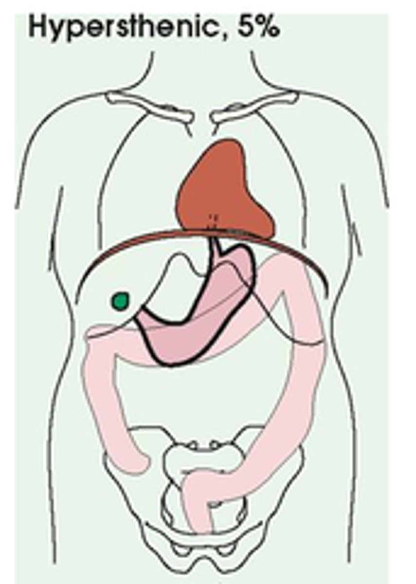
hypersthenic
broad frame (5%)
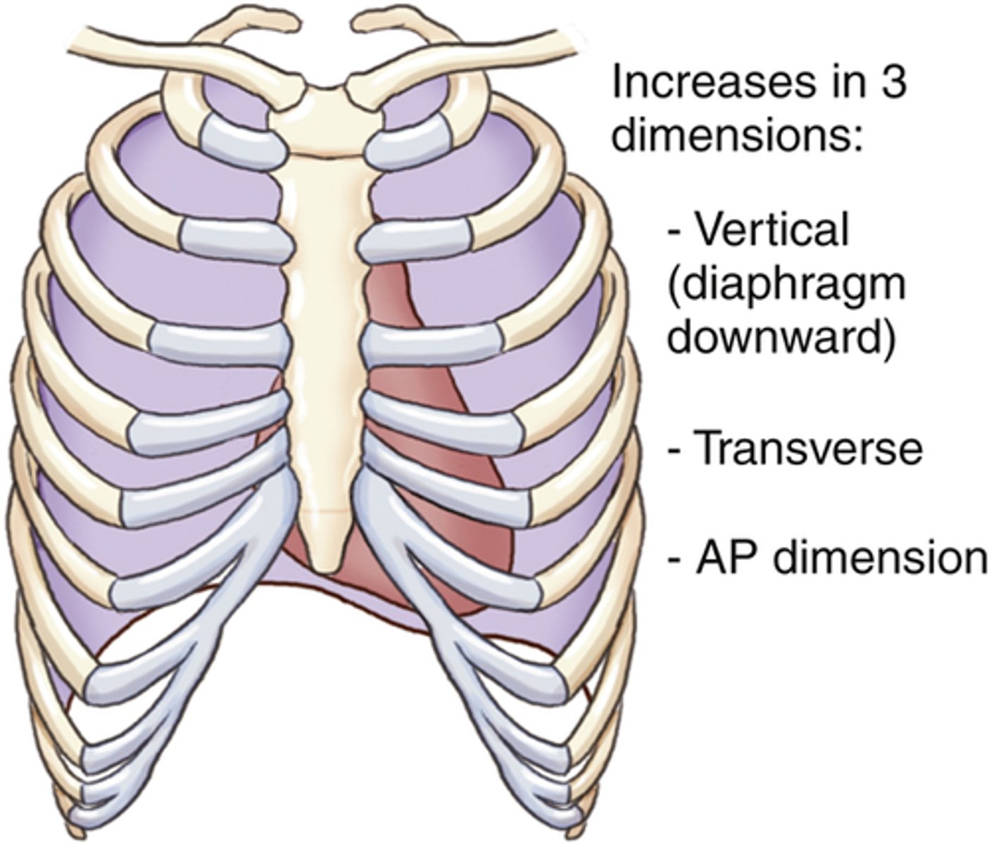
what three dimensions does the thoracic cavity increase with inspiration?
1. vertical diameter
2. transverse diameter
3. anteroposterior diameter
-> returns to normal size with expiration
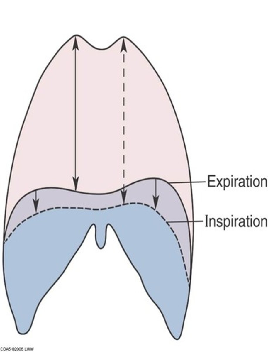
vertical diameter
increases volume by contraction and downward movement of the diaphragm
transverse diameter
ribs swing outward and upward
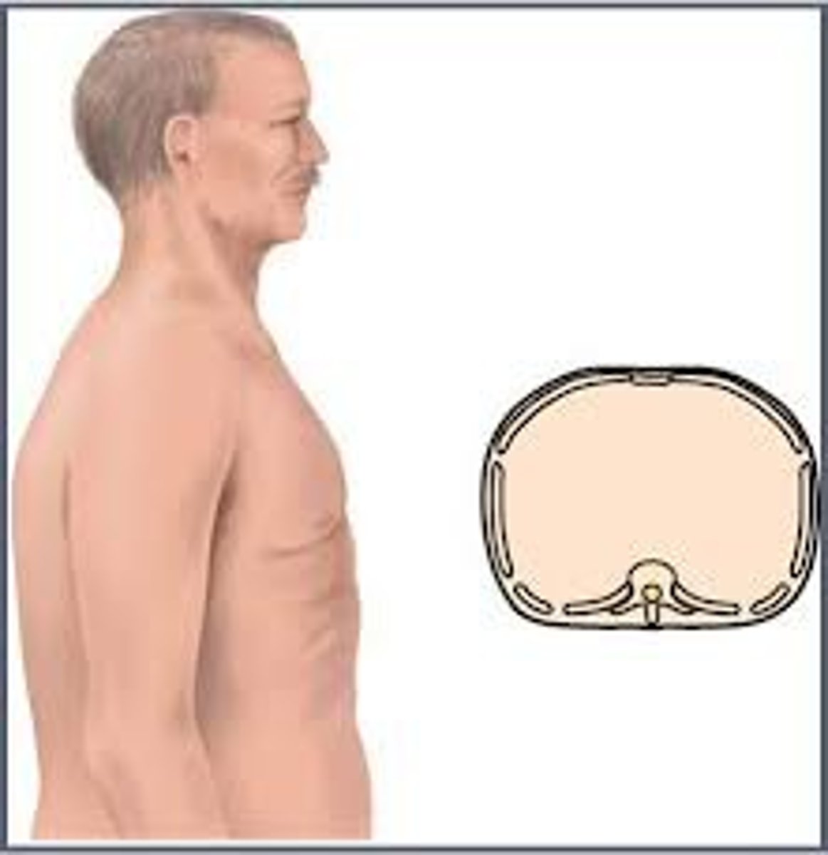
anteroposterior diameter
raise ribs, specifical 2nd-6th ribs
a minimum of ____ in posterior x-ray
10-11 ribs visible
chest x-ray uses ____ contrast
low
kVp of chest x-ray
-adult: 110-125
-pediatric: 70-80
situs inversus/visceral inversion
major organs of the body are on the opposite side
how do you position a newborn for a chest x-ray?
-AP: supine OR PA with Pigg-O-Stat
-Lateral supine or Pigg-O-Stat
Geriatric CR location
some geriatric patients have a smaller lung field; may need to adjust CR to T6-T7
why are chest x-rays done standing?
1. diaphragm moves down farther
2. air and fluid levels can be properly visualized
3. prevents engorgement and hyperemia
engorgement
swollen with fluid
hyperemia
excess of blood in an area of the body
evaluation criteria for PA chest x-ray
-no rotation
-extended chin
evaluation criteria for lateral chest x-ray
-left [unless ordered otherwise]
-no rotation
-no tilt
-raise arms/no superimposition
chest x-ray collimation
1.5-2 inches above vertebra prominens
SID for chest radiography
72 inches
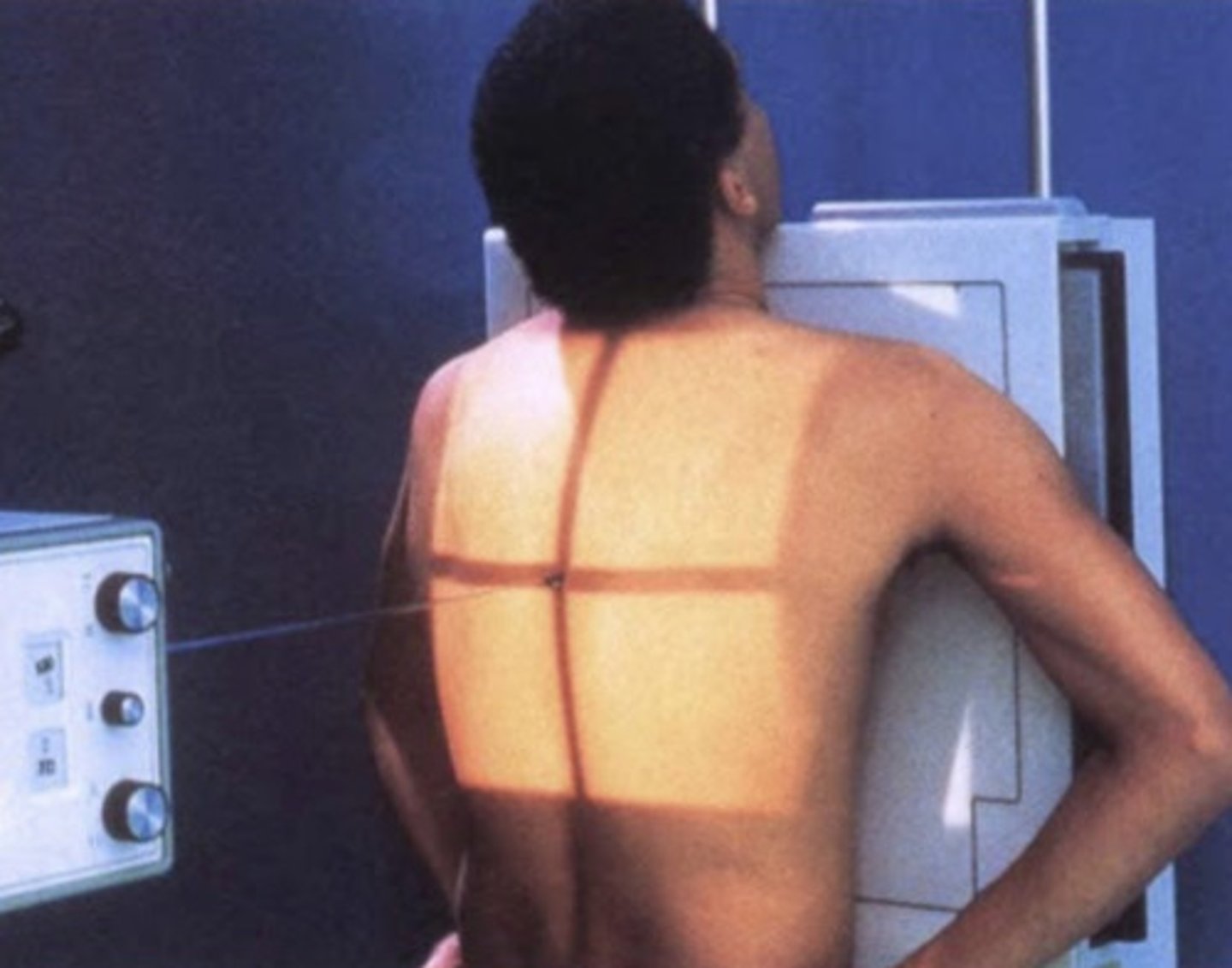
patient positioning for PA chest X-ray
-even footing
-chest towards wall IR
-shoulders rolled forward
-chin up
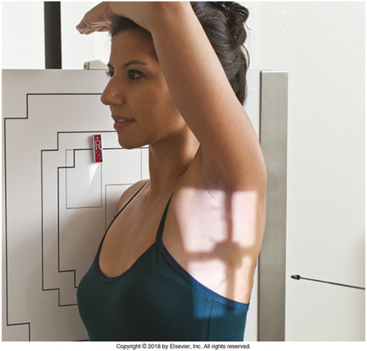
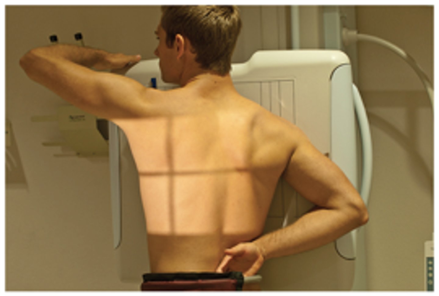
Patient Positioning for Lateral Chest X-ray
-left side towards wall iR
-straight back
-arms raised away from sides
Positioning AP semierect
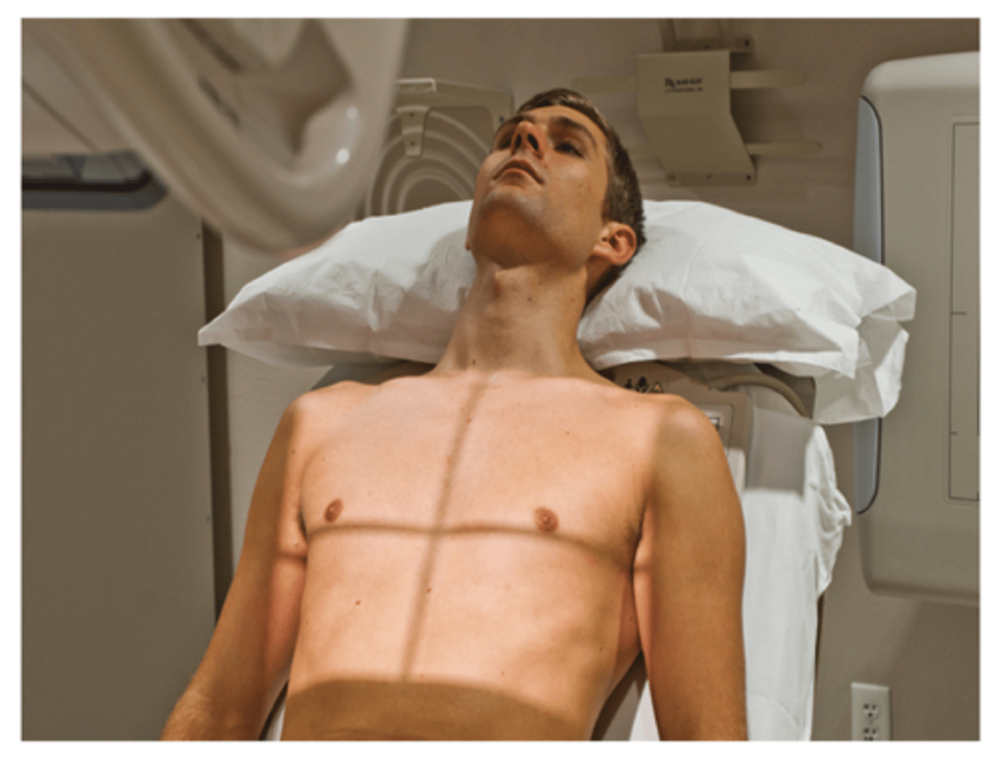
Positioning AP supine
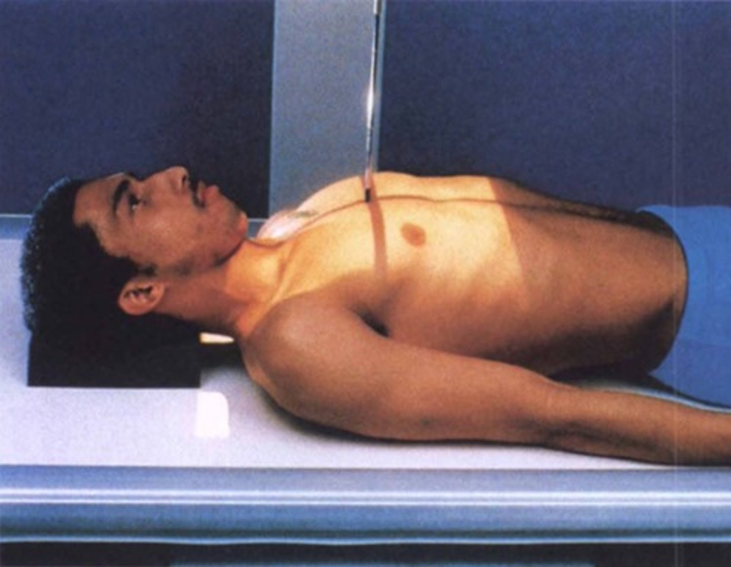
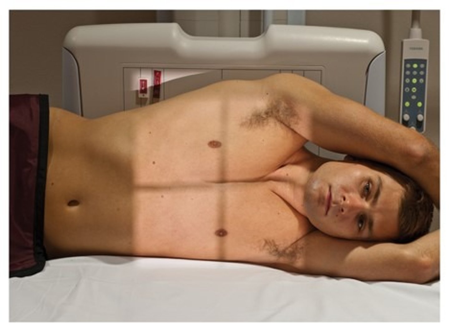
AP lateral decubitus
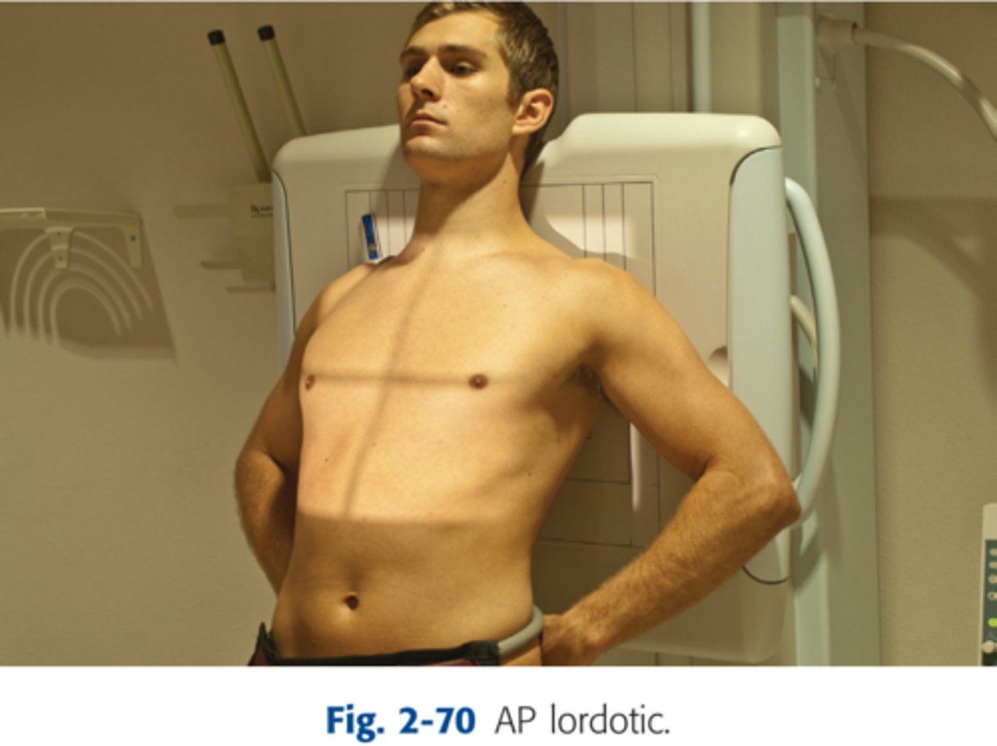
AP Lordotic Chest
-CR- perpendicular to mid sternum, with pt standing at a foot away from the IR leaning back
-Back and head against IR
-If pt cannot stand direct CR 15-20 degrees cephalic
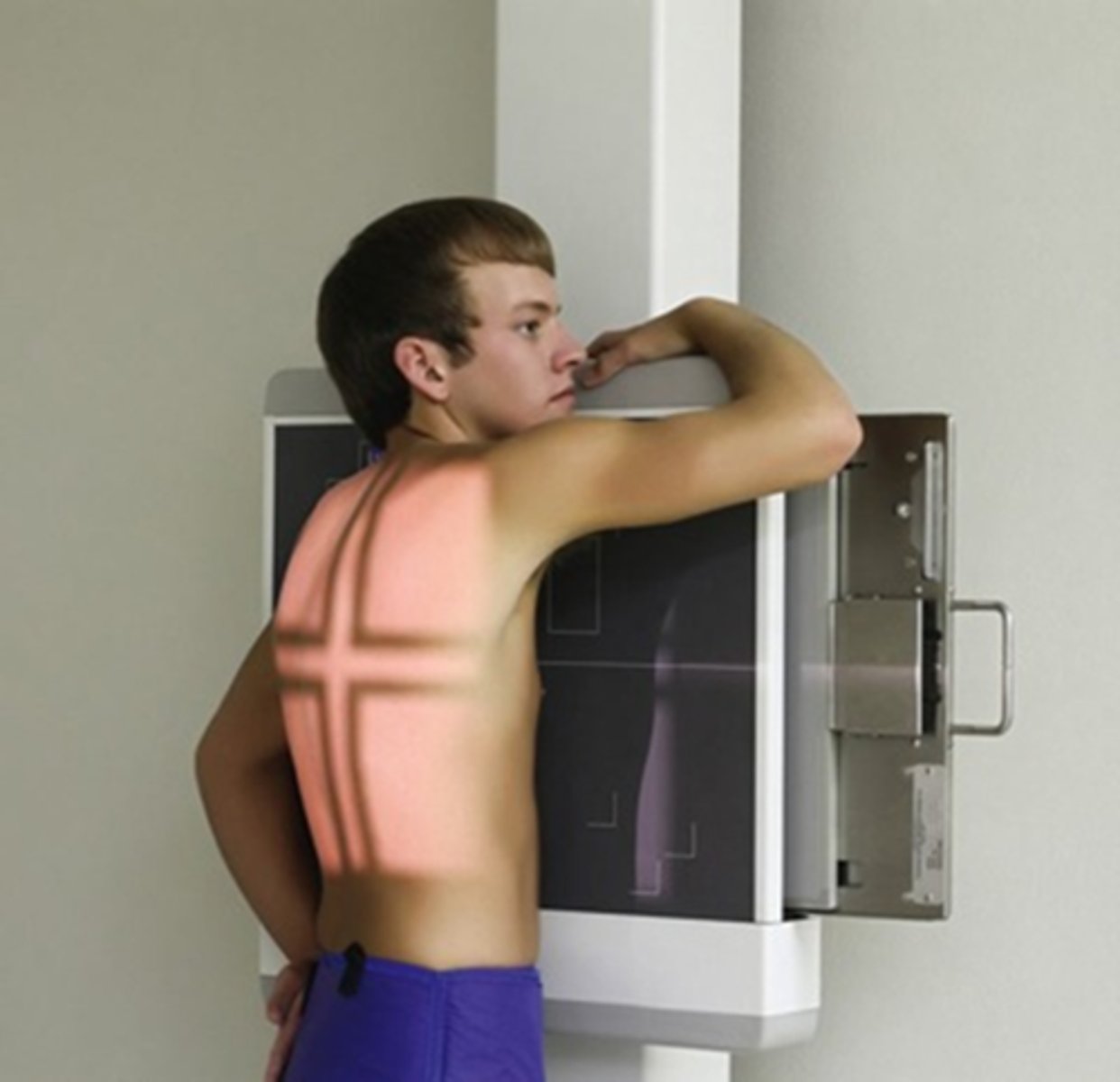
left anterior oblique (LAO)
angle that places left anterior portion of the body closest to the image receptor; usually 45 degree angle
right anterior oblique
angle that places right anterior portion of the body closest to the image receptor; usually 45 degree angle
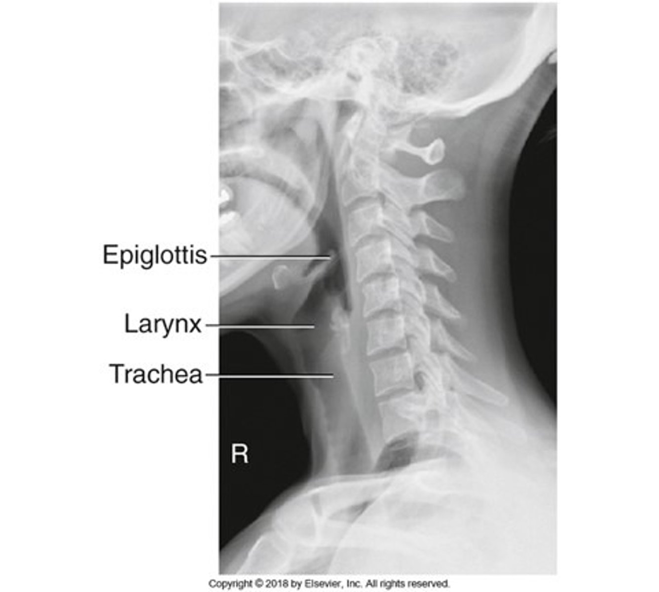
Lateral Upper Airway X-Ray Stats
-CR: C6-C7
-SID: 72"
-10 x 12 inch cassette
-Grid
-kVp: 75-85
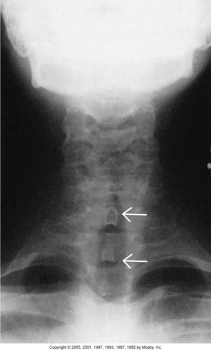
AP Upper Airway X-Ray Stats
-CR: T1-T2
-SID: 40"
-10 x 12 inch cassette
-Grid
-kVp: 75-85
-raise chin
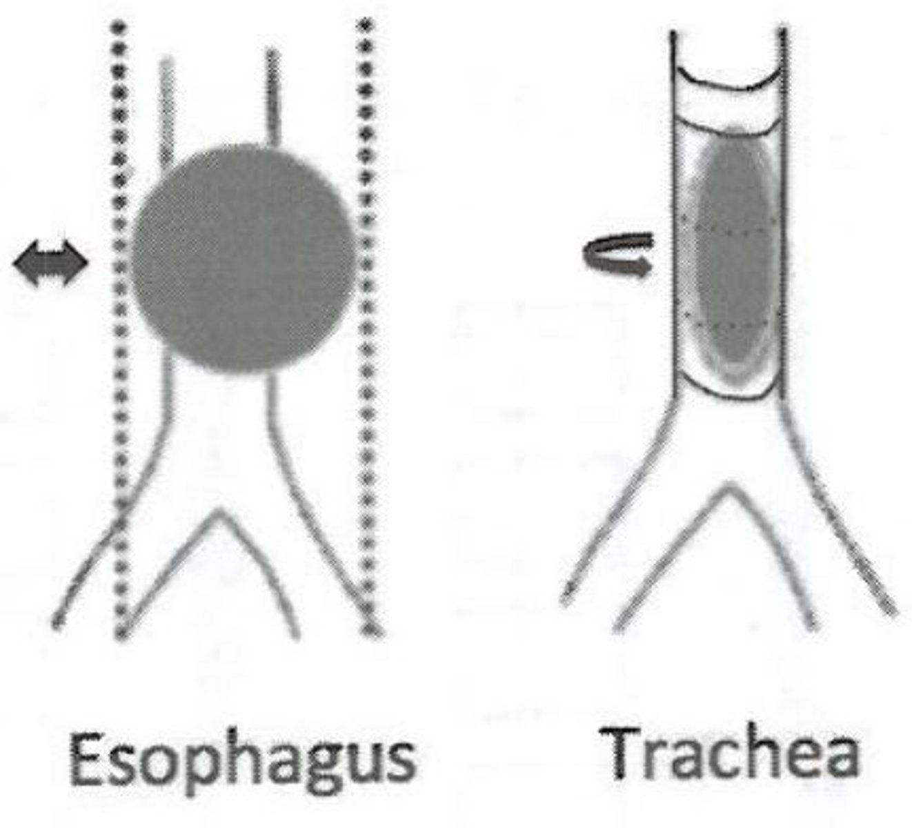
aspiration
-mechanical obstruction, usually when foreign objects are swallowed into bronchial tree
-AP + Lateral Chest, AP + Lateral upper airway X-Rays
Atelectasis
-condition involving a collapsed lung
-collapsed portion appears lighter (more "radiodense") sense there is less air in the lung
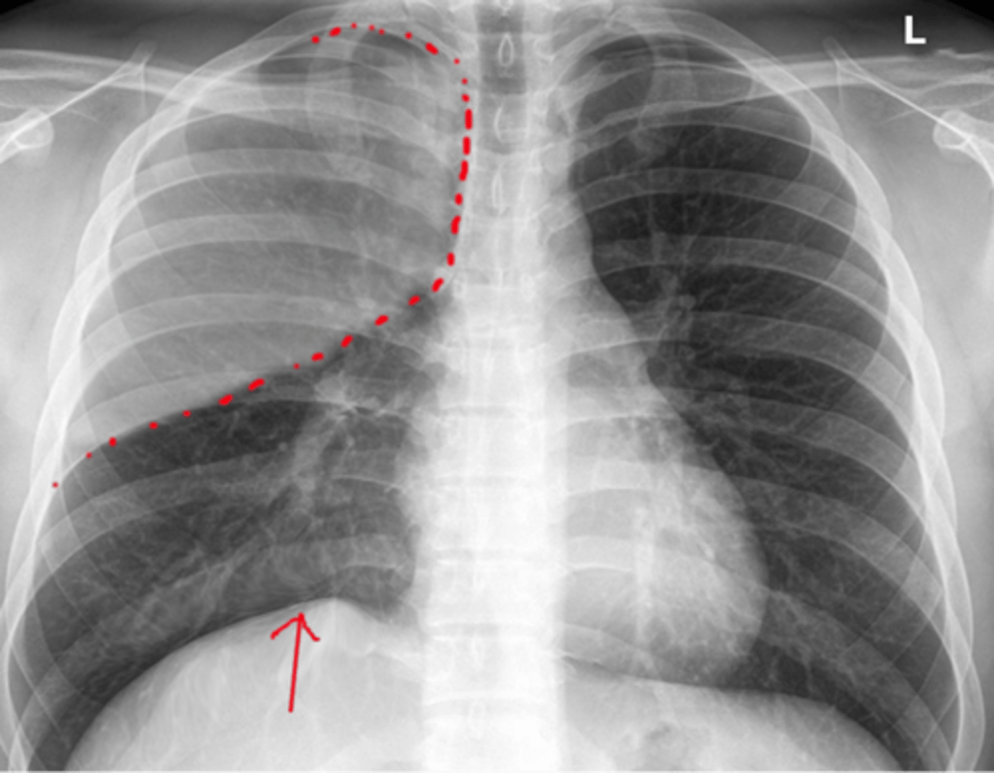
mediastinal shift
shift of central thoracic structures toward one side
Bronchiectasis
abnormal dilation of the bronchi with accumulation of mucus
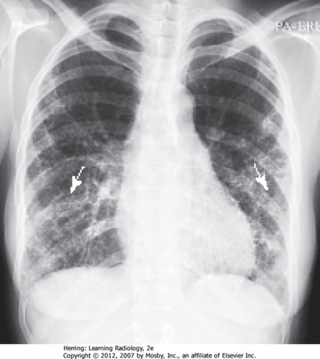
chronic obstructive pulmonary disease (COPD)
-a disease that results in a gradual loss of lung function; increasingly difficult to empty air from the lungs
-only present on x-ray if severe
cystic fibrosis (CF)
-hereditary; causes excessive mucus to clog the bronchioles
-x-ray may show hyperinflated (large) lungs and lots of radiodensity
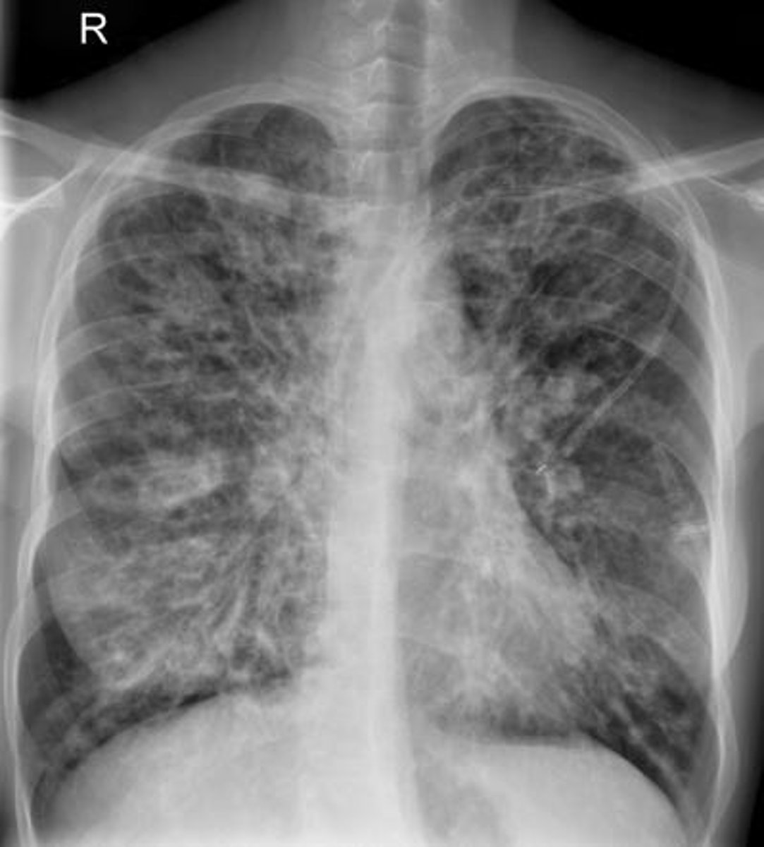
Emphysema (COPD)
-progressive destruction of the alveoli in the lungs; trouble expelling air
-x-rays show increased lung dimension, very radiolucent (dark), depressed diaphragm, and elongated heart shadow
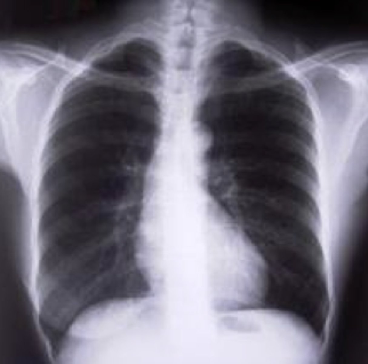
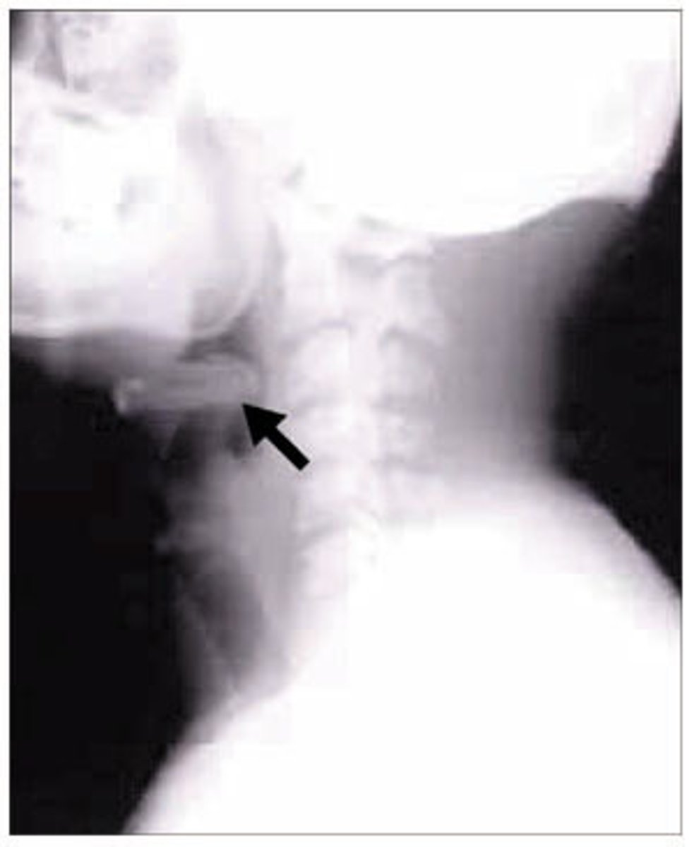
Epiglottitis
-inflammation of the epiglottis
-can be fatal in children 2-5
lung neoplasm
new growth or tumor;
may be benign or malignant
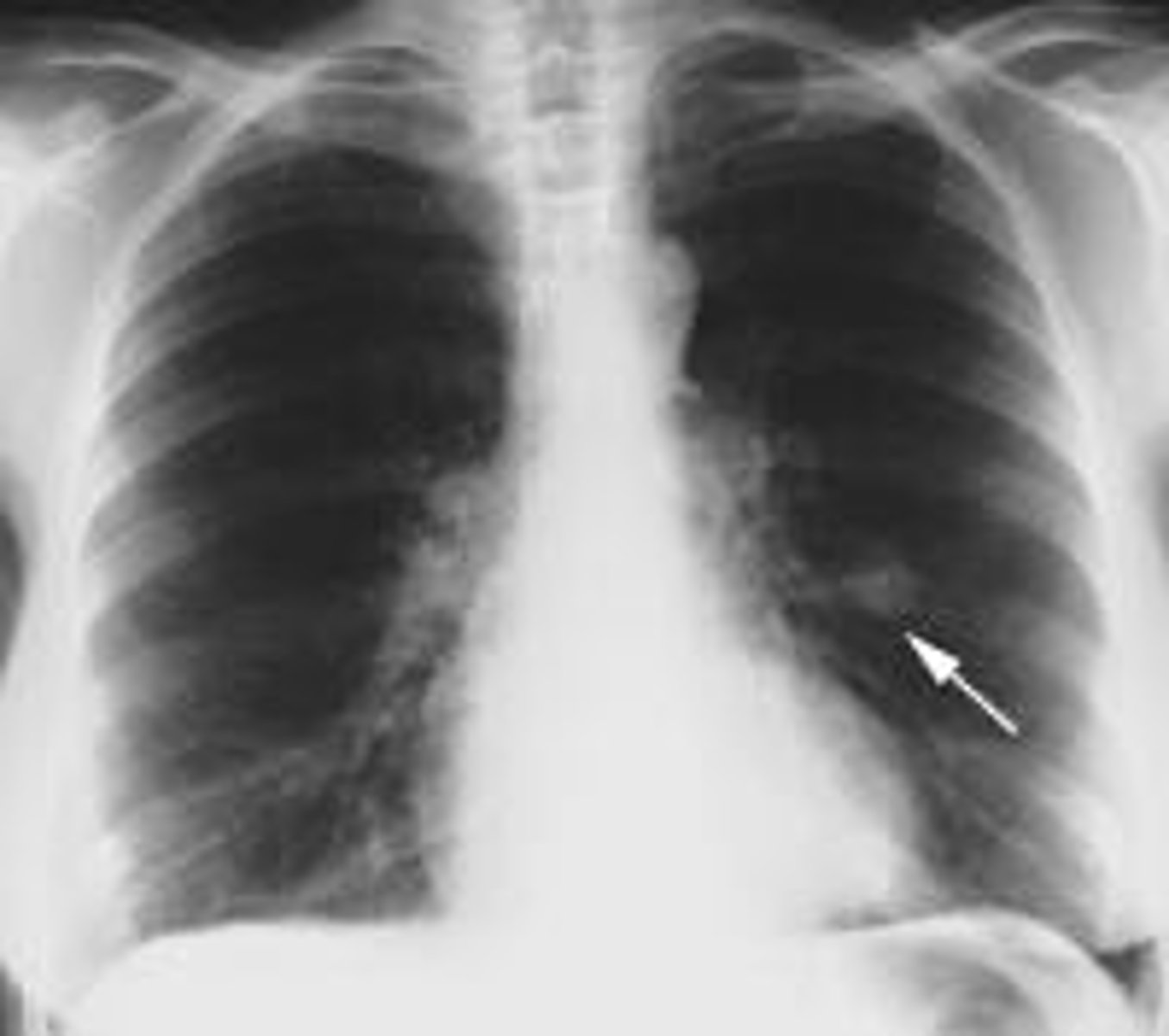
Hamartoma
benign peripheral pulmonary mass
empyema
pus in the pleural cavity