Pulm Path III: Restrictive Lung Diseases
 Call Kai
 Learn
Learn Practice Test
 Spaced Repetition
 Match
Match Flashcards
 Knowt Play
1/26

There's no tags or description
Looks like no tags are added yet.
Name | Mastery | Learn | Test | Matching | Spaced | Call with Kai | Chat |
|---|
No analytics yet
Send a link to your students to track their progress
27 Terms
Describe the core pathogenesis of idiopathic pulmonary fibrosis (IPF).
Repeated alveolar epithelial injury + defective repair → fibroblast proliferation → collagen deposition → progressive interstitial fibrosis.
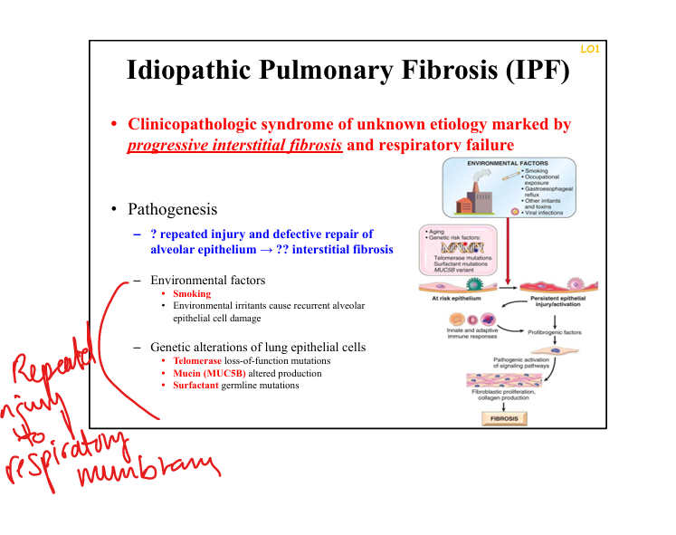
What environmental factors contribute to IPF?
Smoking, occupational irritants,
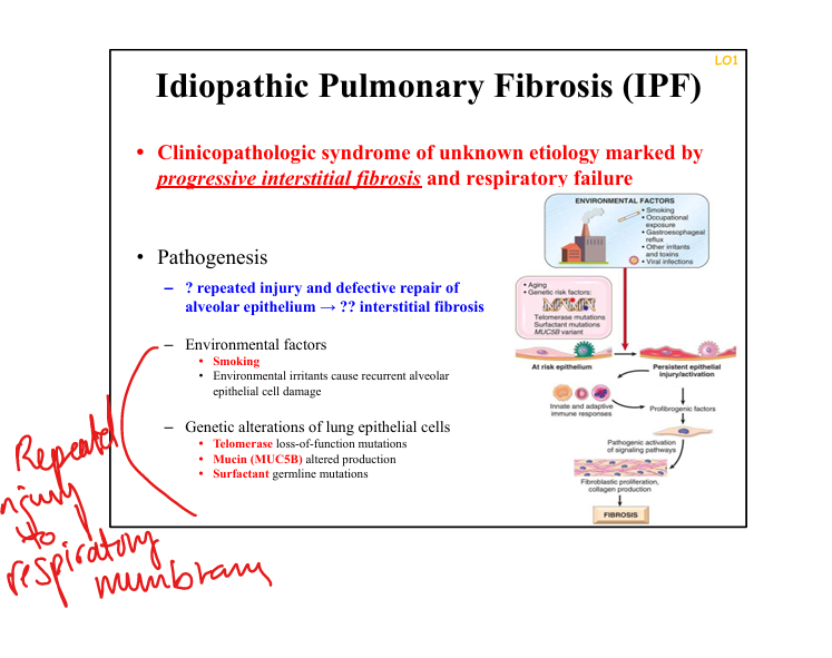
What genetic factors increase risk for IPF?
Telomerase loss‑of‑function mutations, surfactant gene mutations, MUC5B promoter variant.
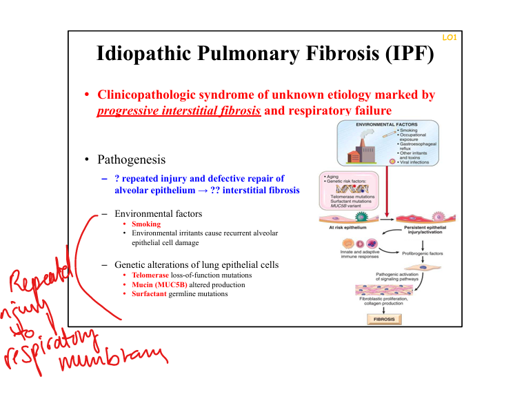
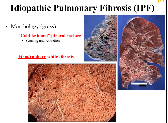
What is the gross morphology of IPF?
Cobblestoned pleural surface with firm, rubbery white fibrosis.
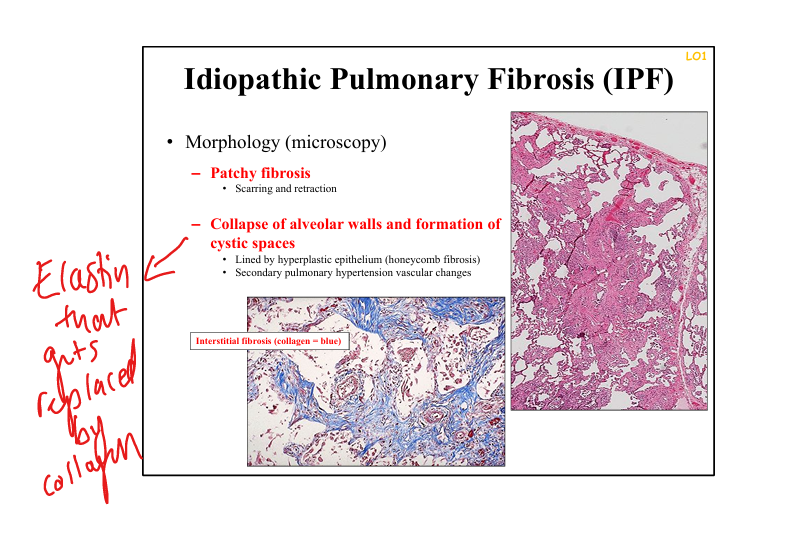
What is the microscopic hallmark of IPF?
Patchy interstitial fibrosis with temporal heterogeneity and honeycomb cysts lined by hyperplastic epithelium.
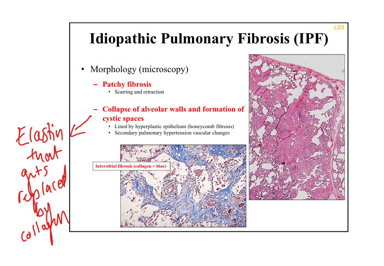
What causes honeycomb lung in IPF?
Collapse of alveolar walls + cystic spaces from end‑stage fibrosis.
Differentiate IPF from emphysema pathologically.
IPF: fibrosis → stiff lungs, honeycombing. Emphysema: loss of elastic fibers → air trapping, hyperexpansion.

What defines granulomatous lung disease?
Presence of granulomas—organized collections of epithelioid macrophages ± giant cells.
What is the immunologic driver of sarcoidosis?
Sustained CD4+ Th1 response → macrophage activation → non‑caseating granulomas.
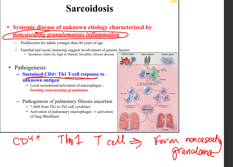
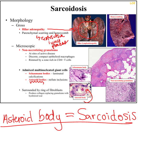
What granuloma features are characteristic of sarcoidosis?
Non‑necrotizing granulomas asteroid bodies.
What organs are commonly involved in sarcoidosis?
Lungs, hilar lymph nodes, skin, eyes, bone marrow, liver, spleen.

What lab abnormalities occur in sarcoidosis?
↑ACE, hypercalcemia due to macrophage 1‑α hydroxylase.
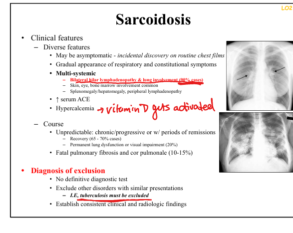
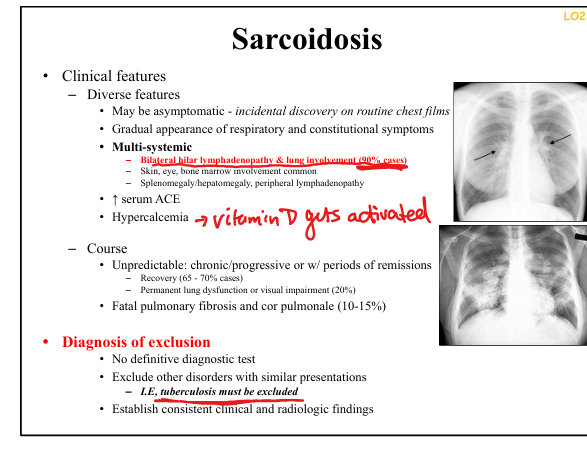
What radiologic finding is classic for sarcoidosis?
Bilateral hilar lymphadenopathy.
What causes hypersensitivity pneumonitis (HP)?
Intense, repeated inhalation of organic antigens (thermophilic bacteria, fungi, animal proteins).
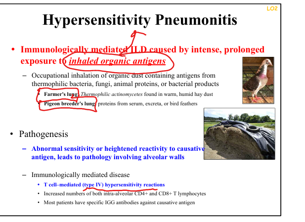
What immune mechanism drives Hypersensitivity Pneumonitis?
Type IV hypersensitivity with CD4+ and CD8+ T‑cell infiltration
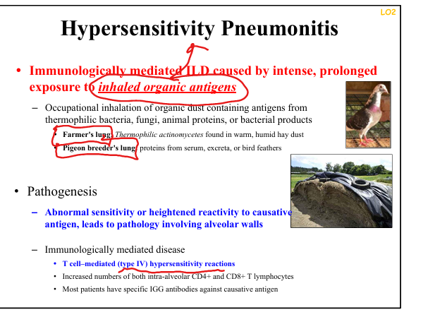
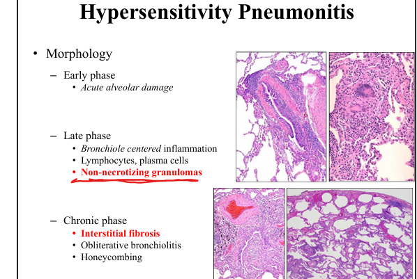
What granulomas occur in HP?
Poorly formed, non‑necrotizing granulomas.

What occurs in chronic HP?
Interstitial fibrosis, obliterative bronchiolitis, honeycombing.
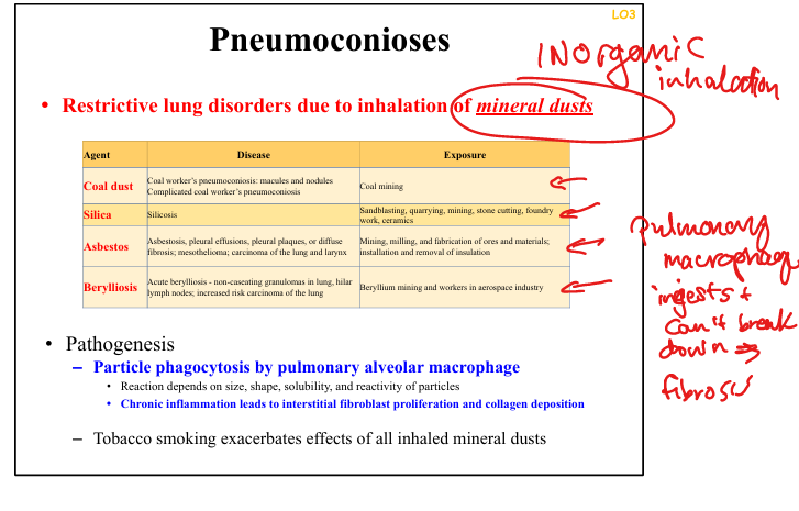
What causes pneumoconioses?
Inhalation of inorganic mineral dusts → macrophage phagocytosis → chronic inflammation → interstitial fibrosis.
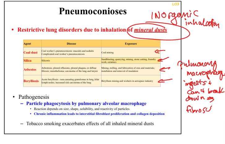
What factors determine pneumoconiosis severity?
Particle size, shape, solubility, reactivity
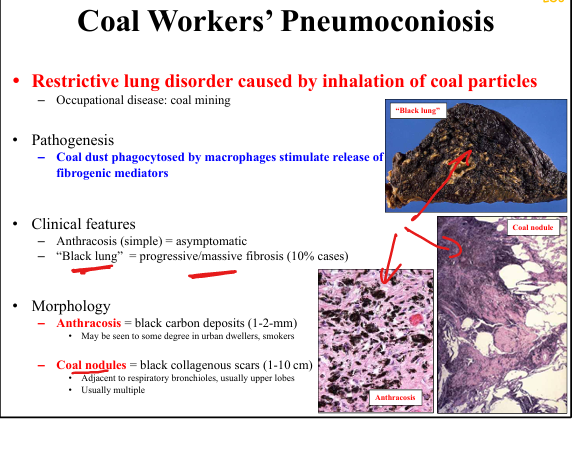
What is anthracosis?
Black carbon pigment deposition in macrophages
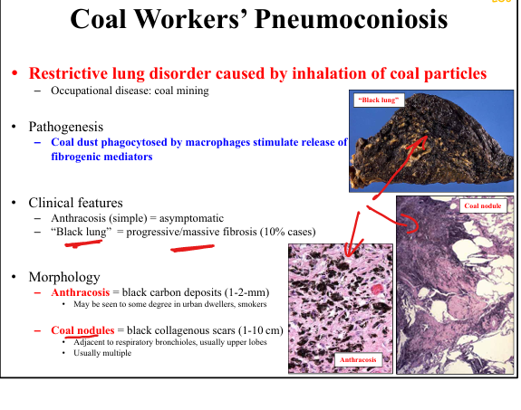
What characterizes coal worker’s pneumoconiosis (CWP)?
Coal macules/nodules
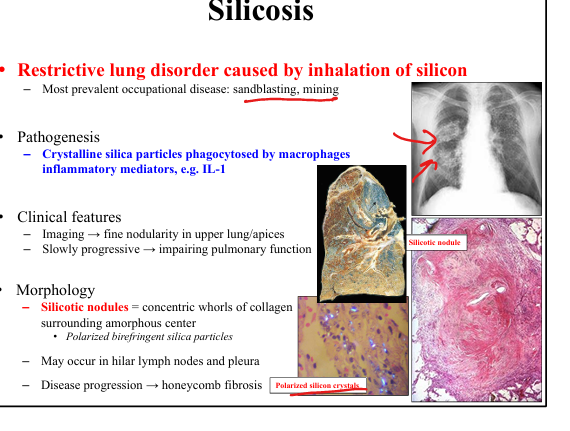
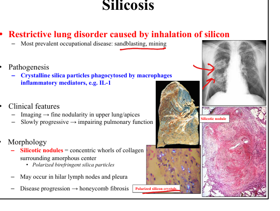
What is the hallmark of silicosis?
Silicotic nodules with concentric collagen whorls and birefringent silica crystals.

Where does silicosis predominantly affect the lung?
Upper lobes/apices.
What complication is associated with silicosis?
Increased susceptibility to TB.
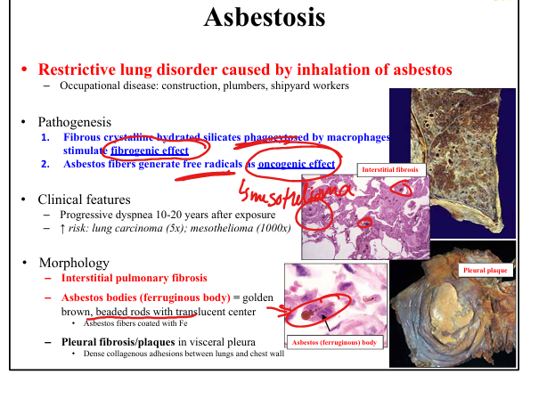
What is the hallmark of asbestosis?
Ferruginous (asbestos) bodies—golden brown rods with translucent centers coated in iron. + pulmonary fibrosis

What cancers are associated with asbestos exposure?
Lung carcinoma (5× risk) and mesothelioma (1000× risk).
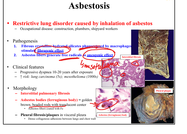
What pleural finding is classic for asbestos exposure?
Pleural plaques—dense collagenous adhesions on parietal pleura.