Special Tests for Hip

0.0(0)
Studied by 1 person
call kaiCall Kai
Locked
learnLearn
examPractice Test
spaced repetitionSpaced Repetition
heart puzzleMatch
flashcardsFlashcards
GameKnowt Play
Card Sorting

1/19

encourage image

There's no tags or description

Looks like no tags are added yet.

Last updated 11:14 PM on 7/21/26
Name
Mastery
Learn
Test
Matching
Spaced
Call with Kai
Chat

No analytics yet

Send a link to your students to track their progress

20 Terms

1
New cards

Hamstring Syndrome

Hamstring Syndrome Provocation Test

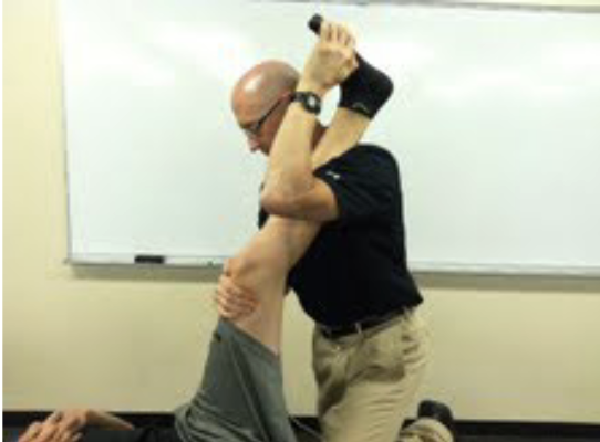
  • How: Pt in supine and examiner stands to the side being tested. Flex the hip to 80–90°, extend the knee to ~15°, dorsiflex the ankle, then ask the patient to resist knee flexion.

  • Positive: Reproduction of the patient's familiar buttock or posterior thigh pain, suggesting sciatic nerve compression/entrapment.

What other test would be positive with hamstring syndrome?

  • Straight Leg Raise (SLR)must also be positive to support a diagnosis of true hamstring syndrome. If the SLR is negative, think hamstring contractile tissue pain rather than sciatic nerve entrapment.
<p><strong>Hamstring Syndrome Provocation Test</strong></p><ul><li><p><strong>How:</strong> Pt in supine and examiner stands to the side being tested. Flex the <strong>hip to 80–90°</strong>, extend the <strong>knee to ~15°</strong>, dorsiflex the ankle, then ask the patient to <strong>resist knee flexion</strong>.</p></li><li><p><strong>Positive:</strong> <strong>Reproduction of the patient's familiar buttock or posterior thigh pain</strong>, suggesting <strong>sciatic nerve compression/entrapment</strong>.</p></li></ul><p><strong>What other test would be positive with hamstring syndrome?</strong></p><ul><li><p><strong>Straight Leg Raise (SLR)</strong> – <strong>must also be positive</strong> to support a diagnosis of <strong>true hamstring syndrome</strong>. If the SLR is negative, think <strong>hamstring contractile tissue pain</strong> rather than sciatic nerve entrapment.</p></li></ul><p></p>
2
New cards

Piriformis Syndrome

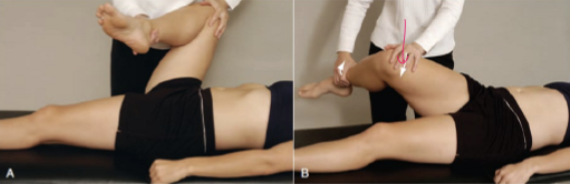
  • How: Pt in supine. Stand on same side. Bring the hip into full flexion, adduction, and external rotation, then apply resisted internal rotation.

  • Positive: Reproduction of the patient's sciatic symptoms, suggesting compression of the sciatic nerve by the piriformis. A positive Straight Leg Raise (SLR) is also required to diagnose true piriformis syndrome.

3
New cards

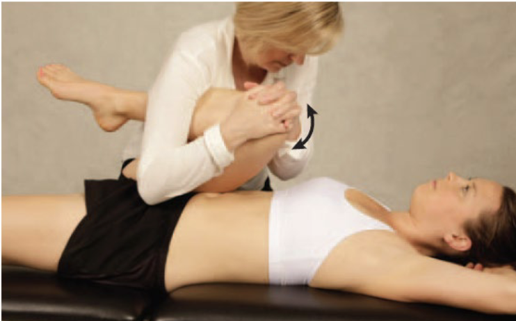
Hip Scour

Hip Scour (Grind) Test (Flexion-Adduction Test)

  • Purpose: anterior medial acatabulum becomes compressed

  • How: Patient supine. Flex and adduct the hip, apply axial compression, then move the hip into abduction while maintaining flexion and compression.

What is (+)?

Could be: Bursa, bone spurs, labrum

Could be tendons: 

  • add longus (felt w/ medial compression)

  • Pectineus

  • Iliopsoas

  • Sartorius 

  • TFL

<p><strong>Hip Scour (Grind) Test (Flexion-Adduction Test)</strong></p><ul><li><p><strong>Purpose</strong><span style="background-color: transparent;">: anterior medial acatabulum becomes compressed</span></p></li><li><p><strong>How:</strong> Patient supine. Flex and adduct the hip, apply <strong>axial compression</strong>, then move the hip into <strong>abduction while maintaining flexion and compression</strong>.</p></li></ul><p><strong>What is (+)?</strong></p><p><span style="background-color: transparent;">Could be: </span>Bursa<span style="background-color: transparent;">, bone spurs, labrum</span></p><p><span style="background-color: transparent;">Could be tendons:&nbsp;</span></p><ul><li><p><span style="background-color: transparent;">add longus (felt w/ medial compression)</span></p></li><li><p><span style="background-color: transparent;">Pectineus</span></p></li><li><p><span style="background-color: transparent;">Iliopsoas</span></p></li><li><p><span style="background-color: transparent;">Sartorius&nbsp;</span></p></li><li><p><span style="background-color: transparent;">TFL</span></p></li></ul><p></p>
4
New cards

Log Roll Test

Log Roll (Passive Supine Rotation) Test

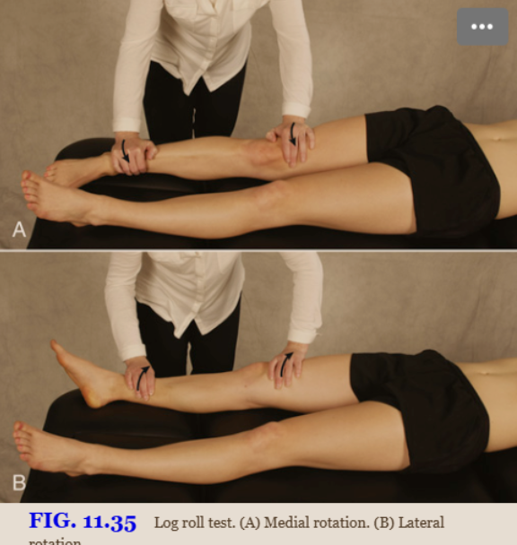
  • How: Patient supine with both legs extended. Passively rotate the femur medially and laterally to end range and compare both hips.

  • If rotational mobility is restricted or painful, → intra-articular hip joint pathology.

  • - A “click” during test → a labral tear (intra-articular) increased lateral rotation → a lax iliofemoral ligament
<p><strong>Log Roll (Passive Supine Rotation) Test</strong></p><ul><li><p><strong>How:</strong> Patient supine with both legs extended. Passively <strong>rotate the femur medially and laterally</strong> to end range and compare both hips.</p></li><li><p><span style="background-color: transparent;">If rotational mobility is restricted or painful, → intra-articular hip joint pathology.</span></p></li><li><p><span style="background-color: transparent;">- A “click” during test → a labral tear (intra-articular) increased lateral rotation → a lax iliofemoral ligament</span></p></li></ul><p></p>
5
New cards

FABER (Patrick) test

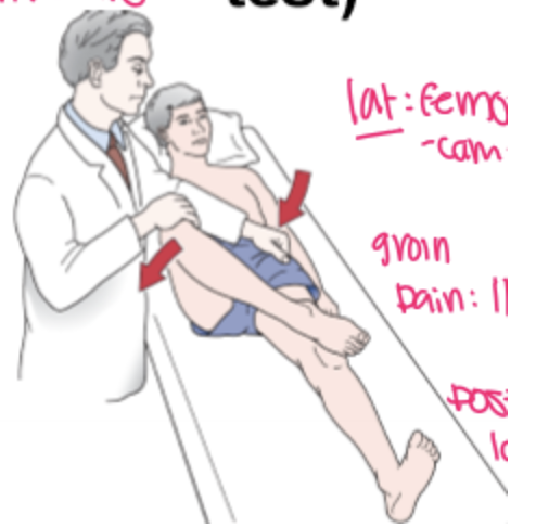
  • How: Patient supine. Place the test leg in the  Flexion, abduction, and external rotation (FABER) position and lower the knee toward the table. This position displaces the anterosuperior part of the femoral head-neck junction to the 12 o’clock position of the acetabular rim.

  • Positive: Pain provocation and/or the test knee remains above the opposite leg (does not reach the table). Pain location helps identify the source:

    • Lateral painSuperolateral/lateral FAI

    • Groin painIliopsoas pathology, psoas impingement, or anterior capsule involvement

    • Posterolateral painIschiofemoral (ischiotrochanteric) impingement

    • Posterior painSacroiliac joint involvement

  • (-) test if  the test leg’s knee falling to the table or being parallel with the opposite leg. 
<ul><li><p><strong>How:</strong> Patient supine. Place the test leg in the <span style="background-color: transparent;"><em>&nbsp;</em><strong><em>Flexion, abduction, and external rotation (</em></strong></span><strong>FABER) position</strong> and lower the knee toward the table. <span style="background-color: transparent;">This position displaces the anterosuperior part of the femoral head-neck junction to the 12 o’clock position of the acetabular rim.</span></p></li><li><p><strong>Positive:</strong> <strong>Pain provocation</strong> and/or the <strong>test knee remains above the opposite leg (does not reach the table)</strong>. Pain location helps identify the source:</p><ul><li><p><strong>Lateral pain</strong> → <strong>Superolateral/lateral FAI</strong></p></li><li><p><strong>Groin pain</strong> → <strong>Iliopsoas pathology, psoas impingement, or anterior capsule involvement</strong></p></li><li><p><strong>Posterolateral pain</strong> → <strong>Ischiofemoral (ischiotrochanteric) impingement</strong></p></li><li><p><strong>Posterior pain</strong> → <strong>Sacroiliac joint involvement</strong></p></li></ul></li><li><p><span style="background-color: transparent;">(<strong>-) test </strong>if&nbsp; the test leg’s knee falling to the table or being parallel with the opposite leg.&nbsp;</span></p></li></ul><p></p>
6
New cards

Anterior Labral Tear (FADIR)

  • How: Patient supine. Start with the hip in full flexion, abduction, and external rotation, then move into extension with internal rotation and adduction.

  • Positive: Pain, reproduction of the patient's symptoms, a click, or apprehension, indicating an

    • Anterosuperior impingement (FAI),

    • Anterior labral tears

    • Iliopsoas tendinitis.

<p></p><ul><li><p><strong>How:</strong> Patient supine. Start with the hip in <strong>full flexion, abduction, and external rotation</strong>, then move into <strong>extension with internal rotation and adduction</strong>.</p></li><li><p><strong>Positive:</strong> <strong>Pain, reproduction of the patient's symptoms, a click, or apprehension</strong>, indicating an </p><ul><li><p><strong>Anterosuperior impingement (FAI),</strong></p></li><li><p><strong>Anterior labral tears</strong></p></li><li><p><strong>Iliopsoas tendinitis</strong>.</p></li></ul></li></ul><p></p>
7
New cards

Posterior Labral Tear

Posterior Labral Tear Test

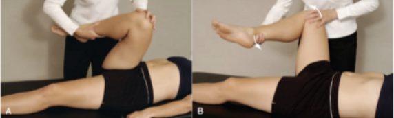
  • How: Patient supine. Start with the hip in full flexion, adduction, and internal rotation, then move into extension with abduction and external rotation.

  • Positive: Groin pain, reproduction of the patient's symptoms, with or without a click, indicating a posterior labral tear, anterior hip instability, or posterior inferior impingement.
<p><strong>Posterior Labral Tear Test</strong></p><ul><li><p><strong>How:</strong> Patient supine. Start with the hip in <strong>full flexion, adduction, and internal rotation</strong>, then move into <strong>extension with abduction and external rotation</strong>.</p></li><li><p><strong>Positive:</strong> <strong>Groin pain, reproduction of the patient's symptoms, with or without a click</strong>, indicating a <strong>posterior labral tear, anterior hip instability, or posterior inferior impingement</strong>.</p></li></ul><p></p>
8
New cards

Gluteal Bursa

Gluteal Bursa Test

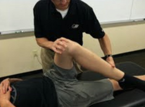
  • How: Patient supine. Bring the hip into flexion + adduction + external rotation (gluteus medius) or flexion + adduction + internal rotation (gluteus maximus).

  • Positive: Pain with flexion, adduction, and external rotation indicates gluteus medius bursitis; pain with flexion, adduction, and internal rotation indicates gluteus maximus bursitis.
<p><strong>Gluteal Bursa Test</strong></p><ul><li><p><strong>How:</strong> Patient supine. Bring the hip into <strong>flexion + adduction + external rotation</strong> (gluteus medius) or <strong>flexion + adduction + internal rotation</strong> (gluteus maximus).</p></li><li><p><strong>Positive:</strong> <strong>Pain with flexion, adduction, and external rotation</strong> indicates <strong>gluteus medius bursitis</strong>; <strong>pain with flexion, adduction, and internal rotation</strong> indicates <strong>gluteus maximus bursitis</strong>.</p></li></ul><p></p>
9
New cards

Iliopectineal Bursa

Iliopectineal Bursa Test

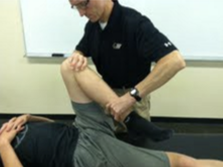
  • How: Patient supine. Bring the hip into hyperflexion and external rotation, stressing the iliopectineal bursa (located between the ASIS and pubic symphysis).

  • Positive: Pain provoked indicates iliopectineal bursitis.

Biomechanically, hyperflexion with external rotation compresses the iliopectineal bursa between the iliopsoas tendon/anterior hip structures and the anterior hip joint (capsule/pelvis). If the bursa is inflamed, this compression reproduces anterior groin pain, making the test positive
<p><strong>Iliopectineal Bursa Test</strong></p><ul><li><p><strong>How:</strong> Patient supine. Bring the hip into <strong>hyperflexion and external rotation</strong>, stressing the <strong>iliopectineal bursa</strong> (located between the <strong>ASIS and pubic symphysis</strong>).</p></li><li><p><strong>Positive:</strong> <strong>Pain provoked</strong> indicates <strong>iliopectineal bursitis</strong>.</p></li></ul><p>Biomechanically, <strong>hyperflexion with external rotation compresses the iliopectineal bursa between the iliopsoas tendon/anterior hip structures and the anterior hip joint (capsule/pelvis)</strong>. If the bursa is inflamed, this <strong>compression reproduces anterior groin pain</strong>, making the test positive</p><p></p>
10
New cards

Stinchfield’s Test

Stinchfield Test (Resisted Straight Leg Raise)

  • How: Patient supine. Raise the leg 20–30° with the knee straight while the examiner applies downward resistance above the knee.

  • Positive: Groin or anterior hip pain.

  • Indicates: Hip joint pathology or iliopsoas pathology.

11
New cards

Ely’s Test

Ely's Test

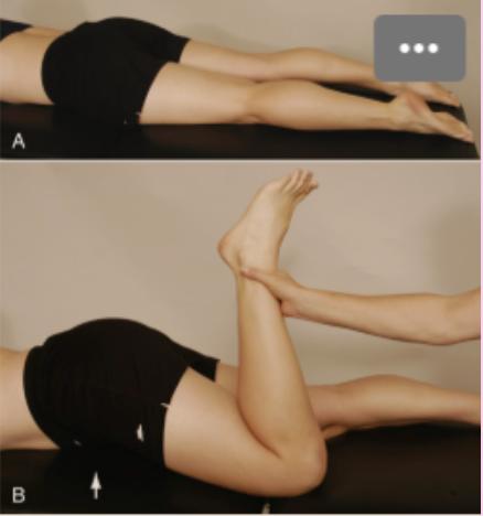
  • How: Patient prone. Passively flex the knee while observing the hip.

  • Positive: Passive knee flexion causes spontaneous ipsilateral hip flexion, indicating a tight rectus femoris.

Another test / Clinical presentation:

  • Resisted Knee Extension (RROM)pain with resisted knee extension suggests rectus femoris involvement.
<p><strong>Ely's Test</strong></p><ul><li><p><strong>How:</strong> Patient prone. Passively <strong>flex the knee</strong> while observing the hip.</p></li><li><p><strong>Positive:</strong> <strong>Passive knee flexion causes spontaneous ipsilateral hip flexion</strong>, indicating a <strong>tight rectus femoris</strong>.</p></li></ul><p><strong>Another test / Clinical presentation:</strong></p><ul><li><p><strong>Resisted Knee Extension (RROM)</strong> – <strong>pain with resisted knee extension</strong> suggests <strong>rectus femoris involvement</strong>.</p></li></ul><p></p>
12
New cards

Ober’s Test

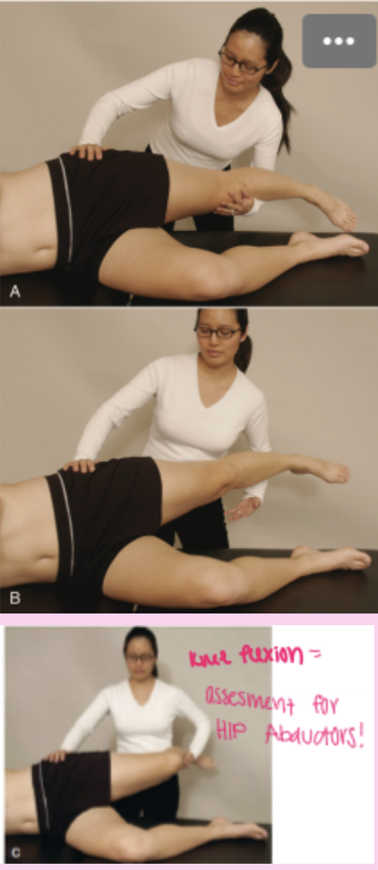
  • How: Patient side-lying. Passively abduct and extend the hip with the knee straight, stabilize the pelvis, then lower the leg toward the table.

  • Positive: The leg remains abducted and does not fall to the table, indicating TFL/ITB contracture.

Note:

 Also tests: the gluteus medius and minimus muscles as well as the hip joint capsule. 

Thus if the test is found to be positive, these should be differentially diagnosed
<p></p><ul><li><p><strong>How:</strong> Patient side-lying. Passively <strong>abduct and extend the hip with the knee straight</strong>, stabilize the pelvis, then <strong>lower the leg toward the table</strong>.</p></li><li><p><strong>Positive:</strong> <strong>The leg remains abducted and does not fall to the table</strong>, indicating <strong>TFL/ITB contracture</strong>.</p></li></ul><p>Note:</p><p><span style="background-color: transparent;"><em>&nbsp;Also tests: the gluteus medius and minimus muscles as well as the hip joint capsule.&nbsp;</em></span></p><p><span style="background-color: transparent;"><em>Thus if the test is found to be positive, these should be differentially diagnosed</em></span></p><p></p>
13
New cards

90-90 SLR

90–90 Straight Leg Raise Test (Hamstring Contracture Test)

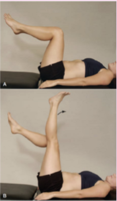
  • How: Patient supine with both hips flexed to 90°. The patient actively extends one knee while maintaining 90° hip flexion.

  • Positive: A popliteal angle <125° (or inability to come within 20° of full knee extension) indicates tight hamstrings.
<p><strong>90–90 Straight Leg Raise Test (Hamstring Contracture Test)</strong></p><ul><li><p><strong>How:</strong> Patient supine with <strong>both hips flexed to 90°</strong>. The patient <strong>actively extends one knee</strong> while maintaining 90° hip flexion.</p></li><li><p><strong>Positive:</strong> <strong>A popliteal angle &lt;125°</strong> (or inability to come within <strong>20° of full knee extension</strong>) indicates <strong>tight hamstrings</strong>.</p></li></ul><p></p>
14
New cards

Piriformis

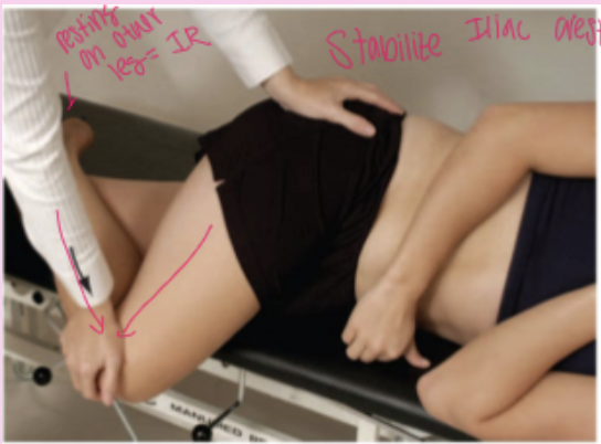
  • How: Patient side-lying with the test hip flexed to 60°, adducted, and internally rotated (FAIR position). Stabilize the pelvis and apply downward pressure to the knee.

  • Positive: Pain in the piriformis muscle, indicating piriformis tightness. If the piriformis compresses the sciatic nerve, the patient may also have buttock pain and sciatica.
<p></p><ul><li><p><strong>How:</strong> Patient side-lying with the test hip <strong>flexed to 60°, adducted, and internally rotated (FAIR position)</strong>. Stabilize the pelvis and apply <strong>downward pressure to the knee</strong>.</p></li><li><p><strong>Positive:</strong> <strong>Pain in the piriformis muscle</strong>, indicating <strong>piriformis tightness</strong>. If the piriformis compresses the sciatic nerve, the patient may also have <strong>buttock pain and sciatica</strong>.</p></li></ul><p></p>
15
New cards

Thomas

  • How: Patient supine. Flex one hip to the chest to flatten the lumbar spine and stabilize the pelvis, while observing the opposite leg.

  • Positive: The opposite (straight) leg rises off the table, indicating a hip flexion contracture. If the leg abducts instead of lifting, it suggests a tight iliotibial band (TFL/ITB).
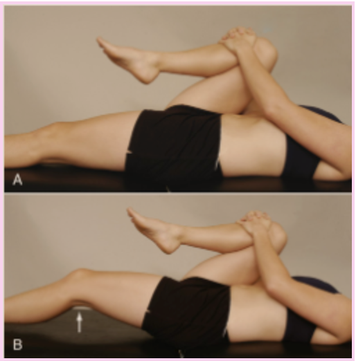
<ul><li><p><strong>How:</strong> Patient supine. Flex one hip to the chest to <strong>flatten the lumbar spine and stabilize the pelvis</strong>, while observing the opposite leg.</p></li><li><p><strong>Positive:</strong> <strong>The opposite (straight) leg rises off the table</strong>, indicating a <strong>hip flexion contracture</strong>. If the leg <strong>abducts instead of lifting</strong>, it suggests a <strong>tight iliotibial band (TFL/ITB)</strong>.</p></li></ul><p></p>
16
New cards

Craig Test for femoral version

  • Purpose: Assesses femoral anteversion or retroversion.

  • How: Patient prone with the knee flexed to 90°. Palpate the greater trochanter while internally and externally rotating the hip until the trochanter is most prominent laterally, then estimate the angle.

  • Positive:

    • Anteversion >15° (commonly 30–35°) → excessive femoral anteversion

    • ≈0° or external rotationfemoral retroversion.

Clinical presentation:

  • Excessive anteversion: Toeing-in, squinting patellae, >60° of hip internal rotation, and decreased external rotation.

  • Retroversion: Decreased internal rotation and increased external rotation.
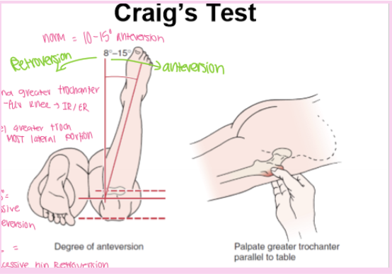
<ul><li><p><strong>Purpose:</strong> Assesses <strong>femoral anteversion or retroversion</strong>.</p></li><li><p><strong>How:</strong> Patient prone with the <strong>knee flexed to 90°</strong>. Palpate the <strong>greater trochanter</strong> while internally and externally rotating the hip until the trochanter is <strong>most prominent laterally</strong>, then estimate the angle.</p></li><li><p><strong>Positive:</strong></p><ul><li><p><strong>Anteversion &gt;15°</strong> (commonly <strong>30–35°</strong>) → <strong>excessive femoral anteversion</strong></p></li><li><p><strong>≈0° or external rotation</strong> → <strong>femoral retroversion</strong>.</p></li></ul></li></ul><p><strong>Clinical presentation:</strong></p><ul><li><p><strong>Excessive anteversion:</strong> <strong>Toeing-in</strong>, <strong>squinting patellae</strong>, <strong>&gt;60° of hip internal rotation</strong>, and <strong>decreased external rotation</strong>.</p></li><li><p><strong>Retroversion:</strong> <strong>Decreased internal rotation</strong> and <strong>increased external rotation</strong>.</p></li></ul><p></p>
17
New cards

Fulcrum Test

  • How: Patient sits with the knees flexed over the edge of the table. Place your forearm under the thigh as a fulcrum and apply gentle downward pressure on the knee while moving the fulcrum along the femur.

  • Positive: Sharp localized pain and apprehension when the fulcrum is positioned under the fracture site, indicating a femoral stress fracture.
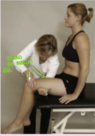
<p></p><ul><li><p><strong>How:</strong> Patient sits with the <strong>knees flexed over the edge of the table</strong>. Place your forearm under the thigh as a <strong>fulcrum</strong> and apply <strong>gentle downward pressure</strong> on the knee while moving the fulcrum along the femur.</p></li><li><p><strong>Positive:</strong> <strong>Sharp localized pain and apprehension</strong> when the fulcrum is positioned under the <strong>fracture site</strong>, indicating a <strong>femoral stress fracture</strong>.</p></li></ul><p></p>
18
New cards

Patellar-Pubic Percussion Test

  • Purpose: Assesses for periacetabular, iliopubic, ischiopubic ramus, and femoral fractures.

  • How: Patient supine. Place the bell of the stethoscope over the pubic symphysis, then percuss each patella and compare the pitch and loudness between sides.

  • Positive: A duller sound on the affected side, indicating bone pathology (e.g., hip or femoral fracture).
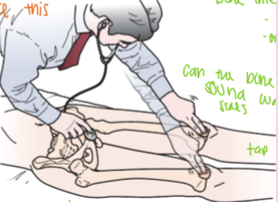
<ul><li><p><strong>Purpose:</strong> Assesses for <strong>periacetabular, iliopubic, ischiopubic ramus, and femoral fractures</strong>.</p></li><li><p><strong>How:</strong> Patient supine. Place the <strong>bell of the stethoscope over the pubic symphysis</strong>, then <strong>percuss each patella</strong> and compare the <strong>pitch and loudness</strong> between sides.</p></li><li><p><strong>Positive:</strong> <strong>A duller sound on the affected side</strong>, indicating <strong>bone pathology (e.g., hip or femoral fracture)</strong>.</p></li></ul><p></p>
19
New cards

True Leg Length

  • Purpose: Assesses true leg length discrepancy.

  • How: Measure from the ASIS to the medial (or lateral) malleolus, ensuring the pelvis and lower limbs are in comparable positions.

  • Positive: A difference >1–1.5 cm suggests a true leg length discrepancy (smaller differences may be normal but can still be symptomatic).

Clinical presentation / Problems caused by leg length discrepancy:

  • Pelvic obliquity (pelvic tilt)

  • Functional scoliosis

  • Abnormal gait

  • Hip, knee, or low back pain due to altered biomechanics.
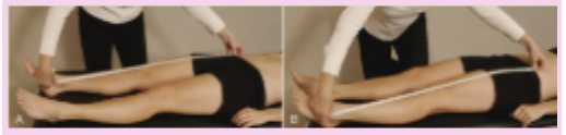
<ul><li><p><strong>Purpose:</strong> Assesses <strong>true leg length discrepancy</strong>.</p></li><li><p><strong>How:</strong> Measure from the <strong>ASIS to the medial (or lateral) malleolus</strong>, ensuring the pelvis and lower limbs are in comparable positions.</p></li><li><p><strong>Positive:</strong> <strong>A difference &gt;1–1.5 cm</strong> suggests a <strong>true leg length discrepancy</strong> (smaller differences may be normal but can still be symptomatic).</p></li></ul><p><strong>Clinical presentation / Problems caused by leg length discrepancy:</strong></p><ul><li><p><strong>Pelvic obliquity (pelvic tilt)</strong></p></li><li><p><strong>Functional scoliosis</strong></p></li><li><p><strong>Abnormal gait</strong></p></li><li><p><strong>Hip, knee, or low back pain</strong> due to altered biomechanics.</p></li></ul><p></p>
20
New cards

Sign of the Buttock

Sign of the Buttock Test

  • Purpose: Assesses for serious buttock pathology (non-capsular hip lesion).

  • How: Perform a Straight Leg Raise (SLR). If pain or restriction occurs, flex the knee while maintaining the hip position, then attempt to flex the hip further.

  • Positive: Hip flexion does not increase when the knee is flexed, indicating pathology in the buttock (e.g., bursitis, tumor, or abscess) and a non-capsular pattern of the hip.
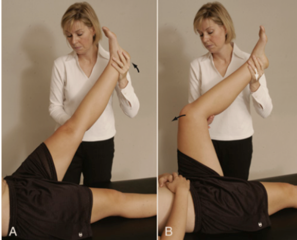
<p><strong>Sign of the Buttock Test</strong> </p><ul><li><p><strong>Purpose:</strong> Assesses for <strong>serious buttock pathology</strong> (non-capsular hip lesion).</p></li><li><p><strong>How:</strong> Perform a <strong>Straight Leg Raise (SLR)</strong>. If pain or restriction occurs, <strong>flex the knee</strong> while maintaining the hip position, then attempt to <strong>flex the hip further</strong>.</p></li><li><p><strong>Positive:</strong> <strong>Hip flexion does not increase when the knee is flexed</strong>, indicating <strong>pathology in the buttock</strong> (e.g., <strong>bursitis, tumor, or abscess</strong>) and a <strong>non-capsular pattern of the hip</strong>.</p></li></ul><p></p>