General and Oral Histology and Embryology Image Collection
 Call Kai
 Learn
Learn Practice Test
 Spaced Repetition
 Match
Match Flashcards
 Knowt Play
1/43

Earn XP
Description and Tags
A comprehensive set of vocabulary flashcards covering the histology of various tissues and organ systems based on the Belgrade Faculty of Dental Medicine image collection.
Name | Mastery | Learn | Test | Matching | Spaced | Call with Kai | Chat |
|---|
No analytics yet
Send a link to your students to track their progress
44 Terms
Simple squamous epithelium
A single layer of thin, flat cells with scarce cytoplasm and prominent nuclei; examples include the endothelium when stained with silver nitrate at 400× magnification.
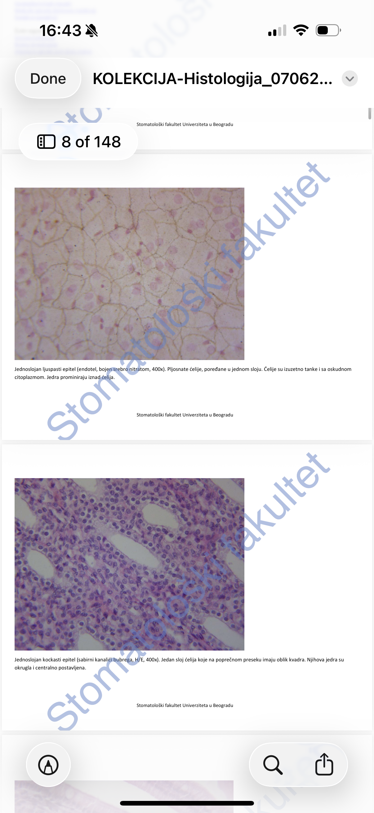
Simple cuboidal epithelium
A single layer of cells that appear square-shaped in cross-section with round, centrally located nuclei, found in the collecting ducts of the kidney.
Simple columnar epithelium
Cylindrical cells with oval nuclei located in the basal third of the cell, often featuring microvilli and goblet cells on the surface, typical in intestinal villi.
Pseudostratified bipolar epithelium
Consists of two cell types: basal cells (stem cells with oval euchromatic nuclei) and principal cylindrical cells (with non-motile stereocilia), both attached to the basal lamina.
Respiratory epithelium (Pseudostratified tri-layered)
Contains seven cell types attached to the basal lamina, including ciliated cells with kinocilia, brush cells with microvilli, goblet cells, basal stem cells, endocrine cells, wedge-shaped cells, and Langerhans cells.
Transitional epithelium (Urothelium)
A pseudostratified epithelium in the urinary bladder composed of three layers: stratum basale (cuboidal stem cells), stratum intermedium (club-shaped cells), and stratum superficiale (umbrella or Dogiel cells).
Stratified squamous non-keratinized epithelium
Keratinocytes arranged in three layers: stratum basale, stratum intermedium, and stratum superficiale, found in the esophagus.
Stratified squamous keratinized epithelium
Multiple rows of keratinocytes divided into stratum basale, spinosum, granulosum, lucidum (only on palms/soles), and corneum (split into stratum conjunctum and disjunctum).
Glandular epithelium (Exocrine)
Example of a simple tubular gland shaped like a straight tube without an excretory duct, alongside unicellular goblet cells that synthesize mucins to form mucus.
Mesenchymal tissue
Embryonal connective tissue consisting of star-shaped cells with numerous cytoplasmic processes, high differentiation potential, and a matrix with sparse reticular fibers.
Mucous connective tissue
Also known as Wharton's jelly, found in the umbilical cord (funiculusumbilicalis); it consists of fibroblasts, thin collagen fibers, and a large amount of ground substance.
Loose connective tissue
The most widespread connective tissue in the organism, found in the lamina propria, submucosa, and tunica adventitia, containing cells and an extracellular matrix.
Tendon (tendo)
Parallel collagen fibers organized into primary and secondary bundles with central endotenonium (loose connective tissue) for nutrition.
White (unilocular) adipose tissue
Specialized connective tissue made of adipocytes where the nucleus is pushed to the periphery by a large fat vacuole.
Reticular connective tissue
Specialized tissue forming the stroma of hematopoietic and lymphopoietic organs, consisting of reticular cells and extracellular matrix.
Neutrophil granulocyte
A type of white blood cell seen in blood smears with a nucleus segmented into 3 to 5 parts.
Eosinophil granulocyte
A blood cell characterized by a bilobed nucleus and eosinophilic cytoplasm.
Red bone marrow
Hematopoietic tissue containing megakaryocytes, which are large bright cells with lobated nuclei that produce platelets.
Hyaline cartilage
Cartilage surrounded by perichondrium (dense irregular connective tissue); contains chondroblasts and mature chondrocytes located inside lacunae (hondroplasti).
Elastic cartilage
Cartilage rich in type II collagen and red-stained elastic fibers, with chondrocytes arranged individually or in isogenous groups.
Fibrous cartilage
A mix of hyaline cartilage and dense connective tissue properties, lacking a perichondrium; it contains type I collagen fibers.
Osteon
The basic unit of compact bone consisting of bone lamellae concentrically arranged around Haversian canals.
Direct (Endesmal) ossification
Bone formation where trabeculae of new bone are surrounded by mesenchyme, with osteoblasts on the periphery and osteocytes in lacunae.
Epiphyseal plate zones (Indirect ossification)
The five zones seen in endochondral ossification, ordered: resting cartilage, growth, hypertrophy, calcification, and ossification.
Multipolar neuron
A cell containing a soma, many branched dendrites, and one long axon; the cell body features Nissl substance (rough ER) and an axon hillock (Deiterscone).
Myelin sheath
Consists of Schwann cell membrane layers, featuring Schmidt-Lanterman incisures (cytoplasm pockets) and Ranvier nodes (interruptions).
Skeletal muscle tissue
Long cylindrical parallel cells with multiple peripheral nuclei under the sarcolemma and transverse striations.
Cardiac muscle tissue
Short, branched cells with one or two central nuclei and intercalated discs (discusintercalatus) connecting the cells.
Smooth muscle tissue
Spindle-shaped myocytes with an ellipsoidal central nucleus and homogenous sarcoplasm without striations.
Endometrium
Separated into stratum basale (permanent) and stratum functionale (changes thickness); consists of simple columnar epithelium and a stroma of fibroblasts and glands.
Aorta (Elastic artery)
Features a tunica intima, an internal elastic membrane, a tunica media with many elastic laminae, an external elastic membrane, and an adventitia.
Capillaries
Vessels whose walls consist of an endothelial layer (tunica intima), basal membrane, and pericytes (acting as the media).
Thymus
A primary lymphatic organ with a lobulated structure consisting of a cortex dominated by thymocytes and a medulla containing Hassall's corpuscles.
Spleen (Lien)
The largest lymphatic organ, containing white pulp (PALS and follicles) and red pulp (Bilroth's cords and sinusoids).
Thyroid gland (Glandulathyroidea)
Endocrine organ with follicles lined by thyrocytes and parafollicular (C) cells, containing colloid in the follicle cavity.
Adrenal gland cortex zones
The three concentric layers from surface to deep: zona glomerulosa, zona fasciculata, and zona reticularis.
Dentin
Mineralized tissue featuring S-shaped primary curves of dentinal tubules and lateral branches (ramilaterales).
Enamel prisms
The basic morphological unit of enamel, appearing as discrete wavy lines; separated by interprestmatic substance.
Periodontium
Specialized connective tissue between the cementum and alveolar bone, containing Sharpey's fibers.
Liver lobule
The classical unit of hepatic structure where hepatocytes are arranged in Remak's lamellae radiating from a central vein.
Islets of Langerhans (Insulae)
The endocrine clusters of cells (insulociti) found within the pancreas.
Cornea layers
The five layers of the cornea: epithelium, Bowman's membrane, stroma (keratocytes and collagen), Descemet's membrane, and endothelium.
Retina layers
Layered structure including the pigment epithelium, rods and cones (bacillorumetconorum), and various granular, reticular, and ganglion layers.
Organ of Corti (Organumspiralecorti)
The organ of hearing located in the membranous cochlear canal, containing scala media (endolymph), scala vestibuli, and scala tympani (perilymph).