Physiology of Bacterial Growth
1/32
Earn XP
Description and Tags
Lecture 1
Name | Mastery | Learn | Test | Matching | Spaced |
|---|
No study sessions yet.
33 Terms
Prokaryotes in general
mirco-organsims
no nucleus
single-celled (although some exceptions)
Age
Oldest cellular life
3.8 billion years ago
Where found
All ecosystems
deep-sea thermal vents
dust particles
On organisms:
muutualistically
pathogen
Archaea
Inhabit only extreme environemnts
but some are found in many environmnets
Bacterial cell stucrure
Size: 2-5 micrometers
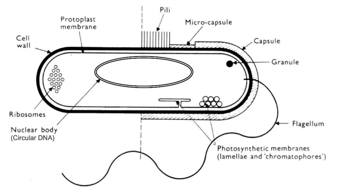
Essential bacterial structural elements
Nucleaur body- single circular chromosome
2000 genes
Cytoplasms
Metabolic acitivity
ribosomes etc
Protoplast/ cell membrane
major barrier between cell and environment
Cell wall
petidoglycan
confer shape
osmotic protection
Non essential extra stutures
Granules
storage polymers
made of polyphosphate or glycogen
Photosynthetic membrane
some do photosynthesis
with bacteriochlorophyll (like plants)
or
Arachea use bacteriorhodopsin proton pump
Capsule
polymer sugar or amino acids
surround cell
Pili
protein filaments 0.1 micrometre long
attachment
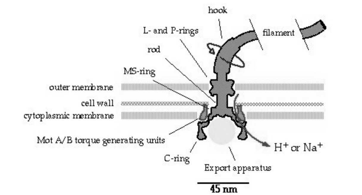
Flagellum
protein filament
3-20 micromemtres
long for mobility
Flagellum motion how
Driven by H+ or Na+ driven rotary motor
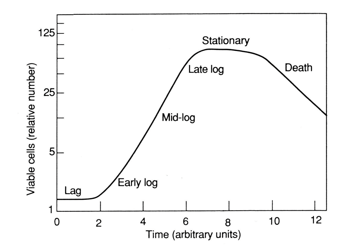
Kinetics of Bacterial growth
Binary fission
divides symmetrically
two daughter cells
Optimal conditions:
exponential increase
doubling time= 10-20 mins
10²2 cells in 24 hours
Bacterial growth graph
Lag: adapting to environement
Log phase: exponential growth
Stationary: stops due to lack of nutrients or toxic waste
Death: exponential loss of viable cells
Nutrient acquisition in bacteria
Active transport
low molecular weight solutes across membrane
E.g lactose H+ symport or phosphate H+ symport
Release enzymes
degrade polymeric substances
so small enough to be transported in
Release toxins
plants and animal pathogens
Increase nutrient availability
Adaptive reponses
chemotaxis- moving towards nutrient sources
Induction of high affinity transport systems
Novel thing of getting iron in: Siderophores
Nutrient requirements for bacteria
Carbon: energy source
Hydrogen: organic nutrients
Phosphorus: Pi
Nitrogen: NH4+ or amino acids
Sulphur: SO42-
Ions: K+,MG2+ and Cl- and trace Fe2+, (MoO4)2-
Acquisition of Fe2+
Free iron in environments is low: 10^-17
release siderophores
e.g enterobactin
Bind iron with high affinity
then acitively transported back into cell
iron released for use
How can permeability of bacterial cell envelope differ?
Can have Gram positive and Gram-negative
permeability is different for each
found with differential staining
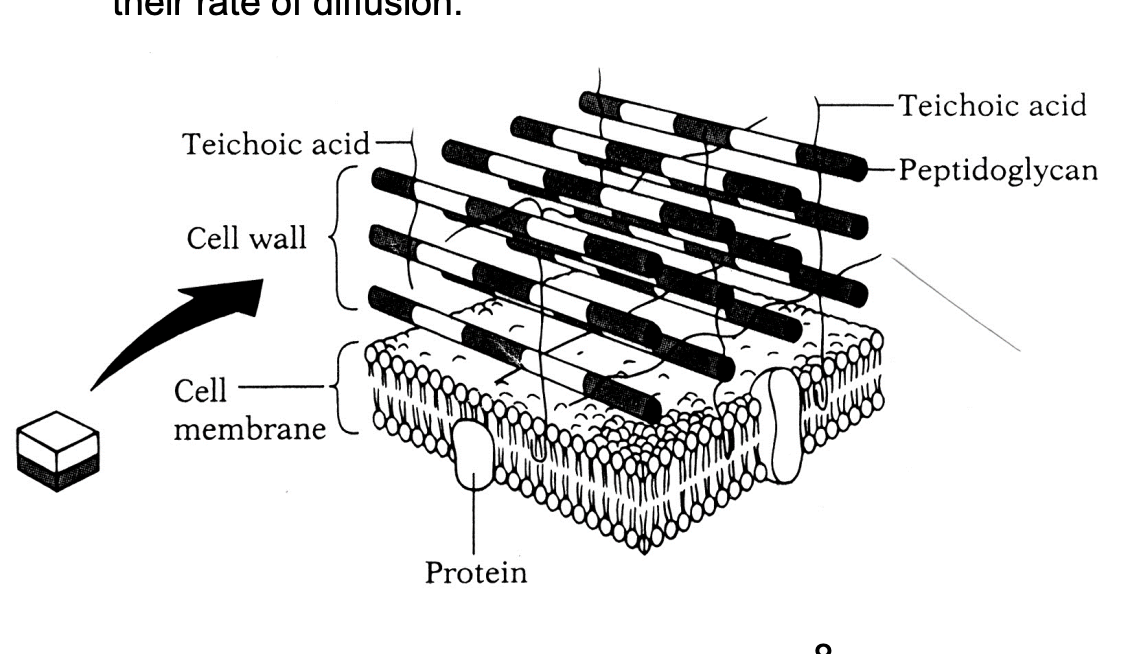
Gram-positive bacteria
50 layers of peptidoglycan
interspersed with teichoic acids
Permeability:
freely permeable to molecules of < 1000 Da
Most stuff gets through
Only limited by rate of diffusion
Gram-negative bacteria strucutre
Outer and inner membrane with cell wall inbetween
Creates na additional compartments: periplasm
space between outer and cell wall
Cell wall thinner:
3-5 sheets of peptidoglycan
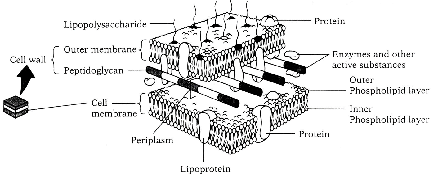
Gram negative advantages
Advantage over gram positive:
limit access of delertious mocleues
antibiotics and detergenes
extra compartment: periplasms
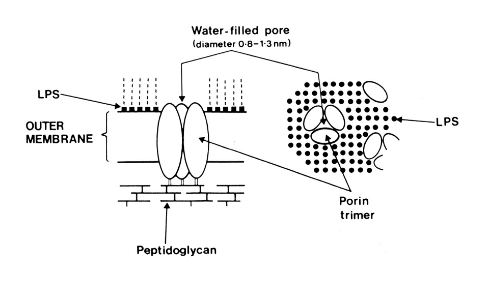
Gram negative permeability how?
Outermembane specific proteins
e.g Porins
Trimeric with water filled pore of 0.8-1.3 nm diameter
allows hydrophobic moelcules of <700 Da
No ion-gradeint linked active transport
Example strucutre of porin
OmpF and OmpC
Role of peptidoglycan
Structural component
Maintain shape
osmotic protectant
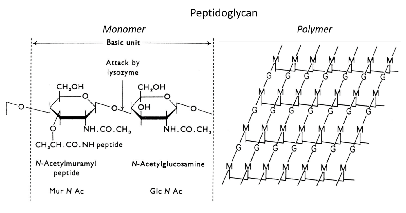
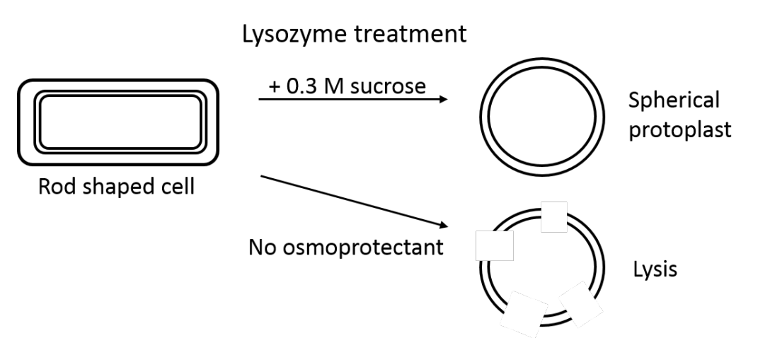
Evidence for peptidoglycan role
Purified peptidogclycan reatins shape of cells
Treatment of bacteria with lysozyme
cleaves peptidoglycan off
when put in low hypotonic solution:
lysis
Challenge to bacteria growth
Must expand the cell walls
But
Grow in dilute aqueous environments
hypotonic environment: under high osmotic pressure
How get over this problem: how grow?
Autolysins
always breaking down the outside layers
Biosynthetic enzymes
crosslink new peptiglycan as inner layers
incorporated in realxed state
Eventually gets to the outside layers
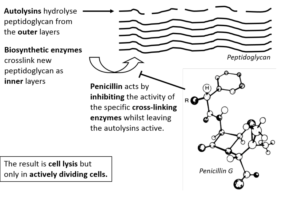
How penecillin works
Inhibits acitivity of cross-link enzymes:
no more cell wall made
bacteria eventually autolyse all cell wall off
Grow Peptidoglycan in Gram-positive rod- shaped bacterium STEP 1
Enxtention of linear petidoglycan between two rigid poles
incorporated in relaxed sate in inner surface
outer layers cleaved
Internal osmotic pressure pushes poles apart
Result: Linear growth of the wall

Grow Peptidoglycan in Gram-positive rod- shaped bacterium STEP 2
synthesis of double thickness cross wall
constricts protoplast membrane
divides cytoplasm in two
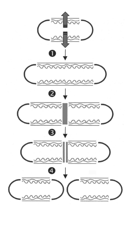
Grow Peptidoglycan in Gram-positive rod- shaped bacterium STEP 3
-
Cleavage of the wall before it is fully cross-linked
internal osmotic pressure of the cell pushes out the cross wall
forms a hemispherical pole

Grow Peptidoglycan in Gram-positive rod- shaped bacterium STEP 4
Final cross-linking of the poles
to form rigid structures
NB: this is only part of the cell division process
DNA rep
DNA seg
cytosolic and envelope components

Ways bacteria are unicellular BUT can work together
Quorum sensing
Rapid Electrical signalling
Quorum sensing
Release chemical signals
homoserine, lactone
Monitor own Population density
Take co-ordinated action together only when a critical cell density is reached
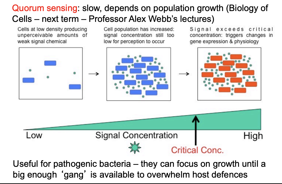
Why have Quorum sensing?
Controls toxin and effector production by pathogentic bacteria
tell when big enough to be able to sufficiently overwhelm e.g plant defences
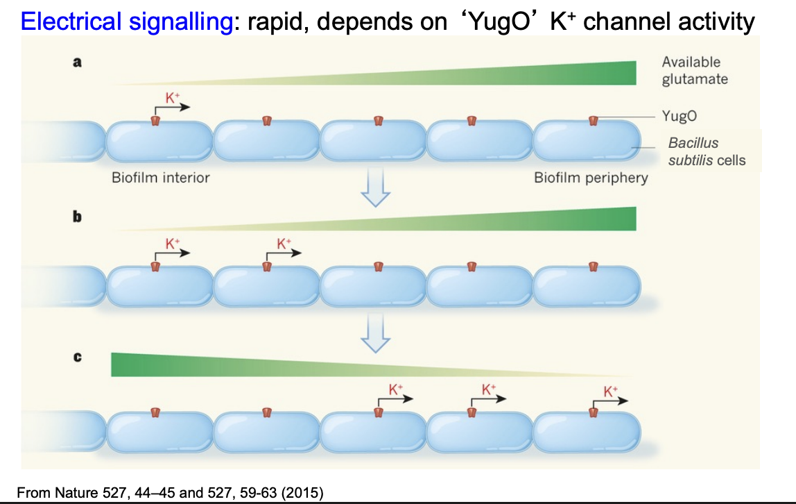
Rapid electrical signalling
form communities of biofilms
e.g tooth decay
What they do?
ensure enven distribution of food supply
electrical signalling mediated by K+ channels in plasma membrane
Rapid electrical signalling how work- glutamic acid
Cells at edge find glutamate
cells at centre lack glutamate
These activate K+ ion extrusion
Change in trans-membrane voltage around neighbouring cells
… which activates YugO channels
expanding wave of increased K+ created
inhibits glutamate uptake by edge cells
More to the centre
This can then reverse as the edge cells get hungry- oscillations
OVERALL: even distribution of resource