🌊 Fluid Mosaic Model
1/30
There's no tags or description
Looks like no tags are added yet.
Name | Mastery | Learn | Test | Matching | Spaced | Call with Kai |
|---|
No analytics yet
Send a link to your students to track their progress
31 Terms
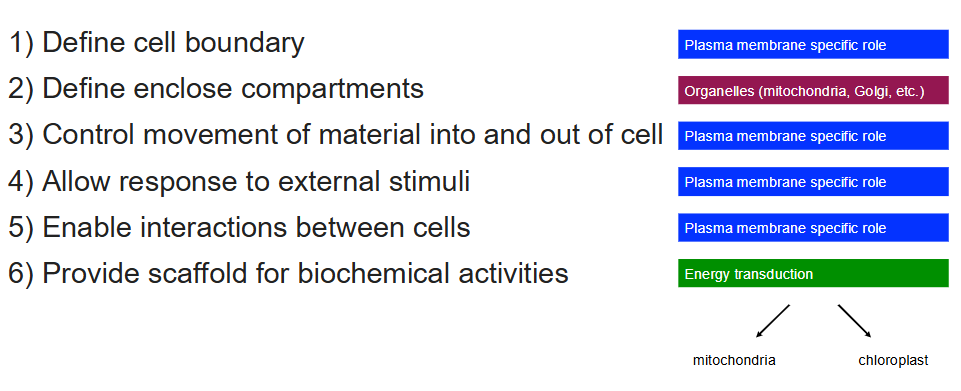
Function of Biological Membranes
Define cell boundary - PM
Define enclosed compartments - Organelles
Control movement of material into and out of cell - PM
Allow response to external stimuli - PM
Enable interactions between cells - PM
Provide scaffold for biochemical activities - Mitochondria/chloroplasts
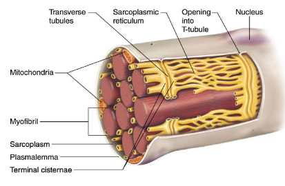
The Plasma Membrane
Most studied cell membrane
PM = Plasma membrane
SR = Sarcoplasmic reticulum - Found in muscle cells, similar to ER, plays crucial role in cation movement (essential for muscle contraction), contains t-tubulus to regular ion movement
Red Blood Cells
Particularly useful as model for study of membrane structures since they don’t contain nuclei or internal membranes
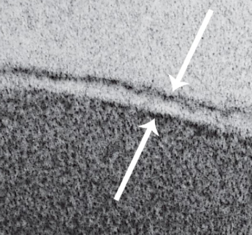
Trilaminar Structure
Made up of phospholipid bilayer
Dark outer layers = phosphate heads (hydrophilic)
Light middle layer = fatty acid tails (hydrophobic)
Found in plasma membrane and membranes of organelles
Provides selective permeability
Maintains fluidity and flexibility of the membrane
Supports membrane proteins for transport, signaling, and structure
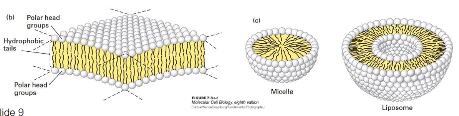
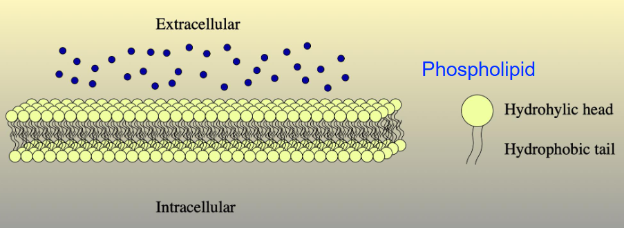
Phospholipid Bilayer
Phospholipid bilayer
6 nm thick
Polar heads
Non-polar cells
Examples
Micelle - Solid
Liposome - Fluid-filled center
Phospholipids
Molecules naturally adopt most stable conformation
Phospholipids are amphipathic
Micelles are phospholipids with one hydrophobic tail
Phospholipids have two hydrophobic tails and a hydrophilic head
In water, phospholipids spontaneously arrange into a bilayer
Tails face inward (away from water)
Heads face outward (toward water, since water is polar)
This forms the basic structure of the plasma membrane
Provides stability, selective permeability, and compartmentalization
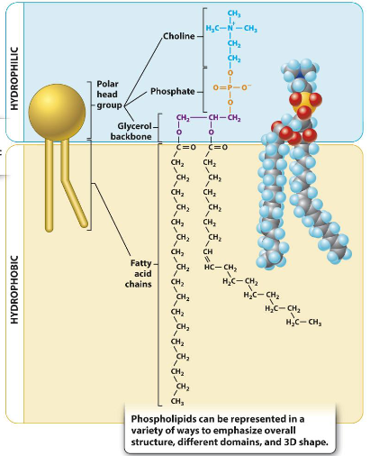
Phospholipid Structure
Left: Schematic drawing
Middle: Chemical formula
Right: Space-filling model
Made of glycerol backbone
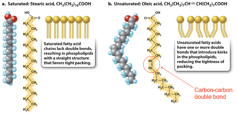
sn-1 (C1) & sn-2 (C2) positions: two fatty acid chains (hydrophobic tails, can be saturated and straight or unsaturated and bent)
sn-3 (C3) position: phosphate group linked to head group (hydrophilic)
Forms amphipathic molecule (both hydrophobic & hydrophilic parts)
Key building block of biological membranes
Phosphate Head Groups
Phosphatidylethanolamine (PE) – Ethanolamine head group
Phosphatidylcholine (PC) – Choline head group
Phosphatidylserine (PS) – Serine head group
Phosphatidylinositol (PI) – Inositol sugar head group
Sphingomyelin (SM) – Choline head group with Sphingosine backbone
Phospholipid Synthesis
Occurs at interface of cytosol and outer endoplasmic reticulum (ER) membrane
ER has enzymes for phospholipid synthesis and distribution
Synthesis is multistep, involves many specialized proteins
Transported to membrane via vesicles or lipid transfer proteins
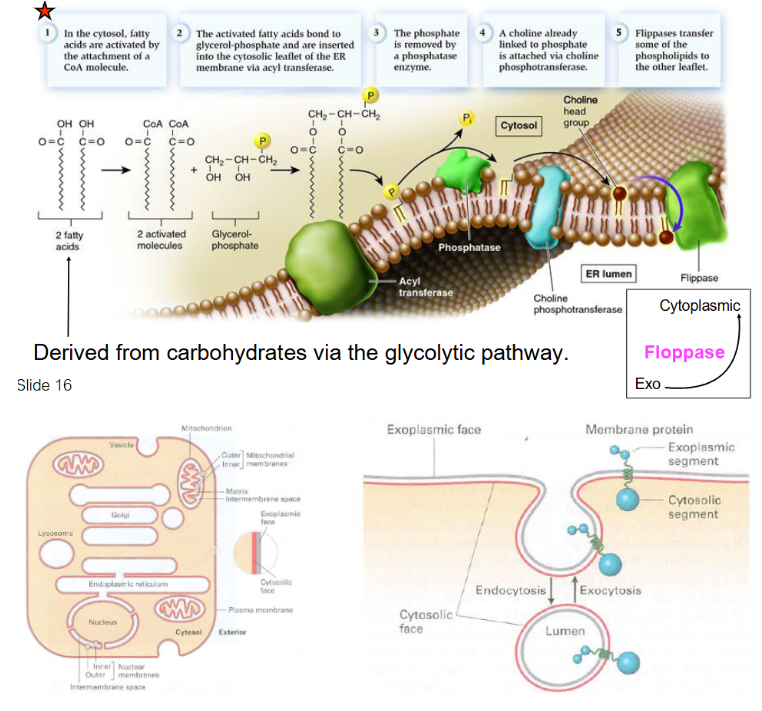
Phospholipid Synthesis Steps
In cytosol, fatty acids from carbs via glycolytic pathway are activated by attachment of CoA molecule
Activated fatty acids bond to glycerol-phosphate and are inserted into inner leaflet of ER membrane via acyl transferase
Phosphate is removed by phosphatase
Choline for head group is already linked to a phosphate and is attached via choline phosphotransferase
Flippases/floppases transfer some phospholipids to other leaflet
Eventually, a vesicle buds off from ER, containing phospholipids destined for the cytoplasmic cellular membrane on its exterior leaflet and phospholipids destined for the exoplasmic cellular membrane on its inner leaflet
Vesicle fuses with plasma membrane, delivering phospholipids
Phospholipids from outer leaflet are incorporated into plasma membrane’s outer layer, while those in inner leaflet remain on cytoplasmic side
ER makes phospholipids and inserts them into its own membrane
Vesicles then transport the phospholipids to the plasma membrane, where they fuse and deliver the lipids
Once at the plasma membrane, flippases help position the phospholipids in the correct leaflet orientation.
Inner leaflet faces cytoplasm
Outer leaflet faces ECF
Flippase and Floppase
Flippases: Enzymes that move phospholipids from the outer leaflet to the inner leaflet of a membrane. (Think of i in flippase meaning final is inner leaflet)
Floppases: Enzymes that move phospholipids from the inner leaflet to the outer leaflet of a membrane. (Think of o in floppase meaning final is outer leaflet)
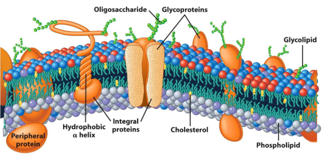
Fluid Mosaic Model of Biological Membranes
Fluid – Individual lipid molecules move, allowing membrane flexibility
Mosaic – Diverse components like proteins, carbohydrates, and cholesterol embedded in lipid layer, creating patchwork structure
Proposed by Seymour Jonathan Singer and Garth Nicolson in 1972
Considered most accurate model of plasma membrane
Plasma membrane viewed as two-dimensional liquid that restricts diffusion of membrane components
Different proteins are embedded in phospholipid bilayers
Components are mobile
Components can interact
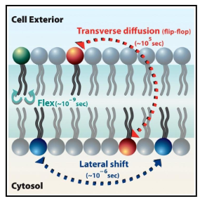
Dynamics of the Plasma Membrane
Lipids move easily, laterally, within leaflet
Lipids' movement to other leaflet is difficult and slow
Membrane proteins diffuse within the bilayer:
Movement of proteins is restricted
Rapid movement is spatially limited
Long-range diffusion is slow
Biochemical modification can alter protein mobility, which is important for signal transduction
Transverse diffusion (flip-flop): Takes 105 seconds; movement from one leaflet to the other
Flexion: Takes 10⁻⁹ seconds; bending or flexing of hydrophobic tail within leaflet
Lateral shift: Takes in 10⁻⁶ seconds; rapid movement of within same leaflet
Frye-Edidin Experiment: Evidence for Fluid Mosaic Model
Goal: To prove membrane proteins are mobile, supporting the Fluid Mosaic Model
Procedure:
Mouse cell had blue proteins
Human cell had green proteins
Cells were forced to fuse together
Findings:
Immediately after fusion: Proteins stayed on their original sides.
After a short time: Proteins mingled and diffused across the unified membrane.
Conclusion: Proved that membrane proteins are mobile, supporting the Fluid Mosaic Model.
Structure of Biological Membranes
Common properties:
6 nm thick (with water)
Stable
Flexible
Capable of self-assembly (due to amphipathic nature)
Differences:
Different membranes have different lipids and proteins
This gives each membrane a specific function
Differences occur between cells and within a single cell
Lipid Rafts
Compartmentalize cell processes, form signal hubs that help proteins interact more efficiently
Important for membrane trafficking, signal transduction, and protein sorting
Debates on exact function
Some believe that they serve as platforms for signaling/trafficking
Others argue that they are experimental artifacts
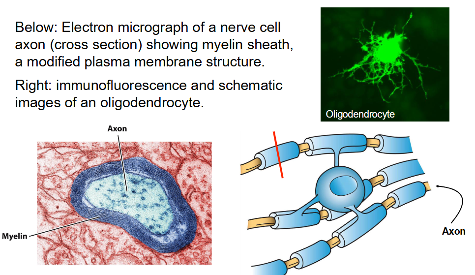
Myelin Sheath and Oligodendrocyte Membrane Structure
Electron micrograph: Nerve cell axon cross-section showing myelin sheath, a modified plasma membrane structure
Right: Immunofluorescence and schematic images of an oligodendrocyte
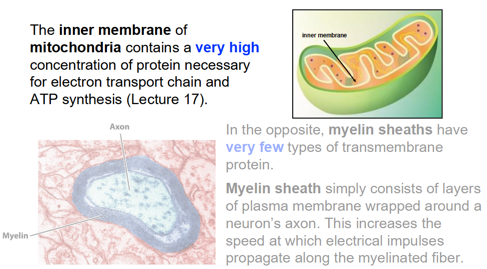
Differences Between Mitochondrial Inner Membrane and Myelin Sheath
Mitochondrial inner membrane: High concentration of proteins for ETC and ATP synthesis
Myelin sheath: Few types of transmembrane proteins, consists of layers of plasma membrane wrapped around axon to increase speed of electrical impulse propagation
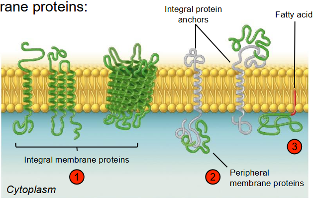
Membrane Proteins
Integral membrane proteins
Span through entire lipid bilayer
Embedded within membrane
Peripheral membrane proteins
On membrane surface
Do not penetrate bilayer
Lipid-anchored proteins
Covalently attached to lipid
Lipid inserts into bilayer
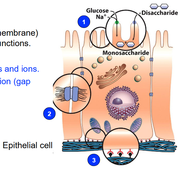
Integral Protein Functions
Transport nutrients and ions across membrane
Example: Channels, Carriers, Pumps
Maintain homeostasis and ion gradients
Enable cell-cell communication
Example: Gap junction proteins
Allow ions, small molecules, and electrical signals to pass between cells
Anchor cells to other cells or structures
Example: Integrins
Help form tissues and maintain shape
Symmetry of Biological Membranes
Biological membranes are asymmetrical
The two leaflets have distinct lipid compositions
Outer leaflet contains glycolipids and glycoproteins (lipids and proteins with carbohydrates attached to them)
Carbohydrates are always on the extracellular side
Fluidity of Biological Membranes
Membrane fluidity is crucial for cell function
Determined by nature of lipids in the membrane:
Unsaturated lipids increase fluidity
Saturated lipids reduce fluidity
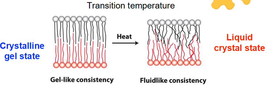
Temperature affects membrane fluidity
Warming increases fluidity → liquid crystal state
Cooling decreases fluidity → crystalline gel state
Transition temperature:
The temperature at which the membrane shifts from a crystalline gel to a liquid crystal state
Consistency of the membrane changes with temperature:
Heat → fluid-like consistency
Cooling → gel-like consistency
Changing Membrane Fluidity
In response to temperature changes, lipid composition of membranes can be changed by:
Desaturation of lipids
Exchange of lipid chains
Balance Between Ordered and Disordered Structure in Membranes
Allows for:
Mechanical support and flexibility
Membrane assembly and modification
Dynamic interactions between membrane components (e.g. proteins can come together reversibly)
Too rigid:
Membrane may become less flexible, affecting ability of cells to change shape or move
Transport of materials across membrane can become inefficient
Protein functions may be impaired due to restricted movement or interaction
Too fluid:
Membrane may lose integrity, making it more prone to leakage
Cell signaling could be disrupted
Membrane proteins may not be properly anchored or may move too freely, impairing their function
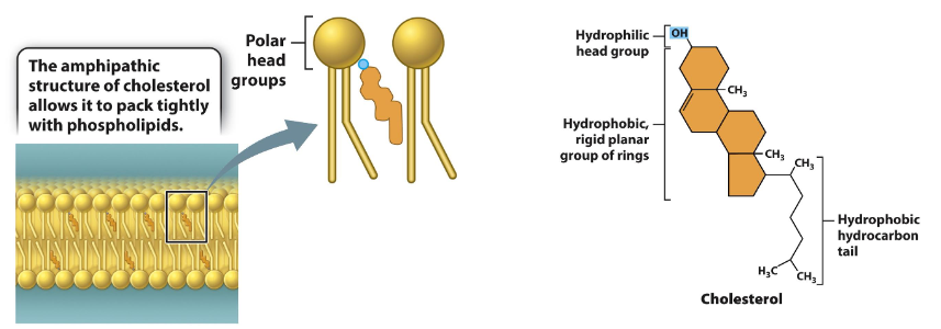
Cholesterol
Cholesterol at high temperatures:
Stabilizes membrane
Raises melting point, preventing membrane from becoming too fluid
Cholesterol at low temperatures:
Prevents phospholipids from clustering together
Maintains membrane fluidity by preventing stiffening
Alters packing and flexibility of lipids:
Cholesterol fits between lipid molecules in the membrane, disrupting their regular packing and changing the membrane's properties.
In a liquid crystal membrane (high fluidity):
Adding cholesterol will decrease fluidity because it stabilizes the membrane by restricting movement of lipid molecules.
In a crystalline gel membrane (low fluidity):
Adding cholesterol will increase fluidity because it prevents lipids from packing too closely, thus allowing for more movement.
Integral Proteins
Membranes are made of both lipids and proteins.
Proteins can make up to 50% of the mass of the membrane (e.g., in red blood cells).
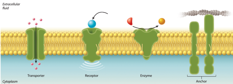
Functions of membrane proteins:
Transporters: Move ions or other molecules across the membrane.
Receptors: Receive signals from the environment.
Enzymes: Catalyze chemical reactions.
Anchors: Attach to other proteins to help maintain cell structure and shape.
Fluorescent Recovery After Photobleaching (FRAP)
Purpose: Measure the mobility of proteins in the membrane
Background:
Proteins in the membrane are tagged with a fluorescent dye
A laser beam is used to bleach a small region of the membrane, making it nonfluorescent
Hypothesis:
If proteins move, the bleached spot will regain fluorescence as unbleached proteins move into the area
If proteins don’t move, the bleached spot will remain nonfluorescent
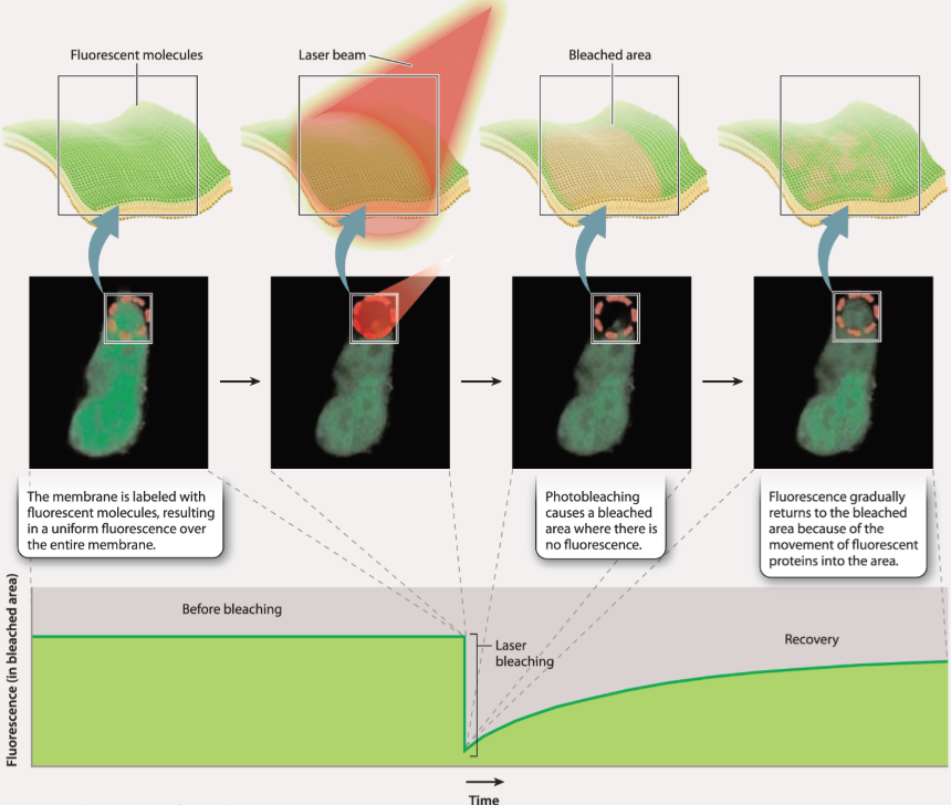
Experiment and Results:
Step 1: Membrane is labeled with fluorescent molecules, and the entire membrane is initially fluorescent
Step 2: A laser is used to bleach a section of the membrane, leaving a nonfluorescent spot
Step 3: The bleached area is clearly visible with no fluorescence
Step 4: Over time, fluorescence gradually returns to the bleached area as proteins move into it
Conclusion:
The gradual recovery of fluorescence indicates that proteins move in the plane of the membrane
Passive Transport and Diffusion
Diffusion: Random movement of molecules
Molecules move in their environment (e.g., in water at room temperature, molecules move at 500 m/sec)
Molecules collide frequently, affecting chemical reactions
Concentration gradient: Molecules move from higher to lower concentration
Movement continues until concentrations are equal
Once concentrations are equal, molecules still move, but no net movement
Some molecules diffuse freely across the plasma membrane due to concentration differences
Oxygen and carbon dioxide move into and out of the cell this way
Hydrophobic molecules like triacylglycerols diffuse through the membrane due to lipid bilayer being hydrophobic
Some molecules that can’t move across the lipid bilayer directly move passively through protein transporters
This is called facilitated diffusion
Diffusion and facilitated diffusion result from random motion of molecules and concentration differences
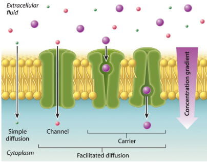
Facilitated diffusion: Molecule moves through a membrane transporter
Simple diffusion: Molecule moves directly through the lipid bilayer
Membrane transporters:
Channels: Provide opening for molecules to pass depending on shape and charge
Some are gated and open in response to chemical or electrical signals
Carriers: Bind to and transport specific molecules
Two conformations: One open to one side of the cell, the other open to the other side
Binding of molecule induces conformational change, allowing transport across the lipid bilayer
Active Transport and Sodium-Potassium Pump
Passive transport only works if concentration gradient is right:
Nutrients: Higher outside, lower inside (needs to be taken in)
Wastes: Higher inside, lower outside (needs to be exported)
Many molecules required by the cell are not highly concentrated in the environment
Some molecules can be synthesized by the cell
Others must be taken up from the environment
Cells need to move substances from areas of lower concentration to higher concentration, which is active transport
Active transport requires energy
Most of the cell’s energy goes into maintaining concentration differences inside and outside the cell
Proteins in the plasma membrane carry out this function
During active transport, substances move through transport proteins embedded in the membrane
Some proteins act as pumps, using energy directly to move substances in or out of the cell
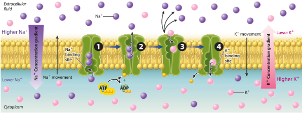
Example: Sodium-potassium pump
Sodium is kept at lower concentrations inside the cell, potassium at higher concentrations inside
Sodium moves out, potassium moves in against their concentration gradients
Energy for this movement comes from ATP
Primary active transport: Uses energy directly (ATP) to move substances
Antiporters: Move ions in opposite directions (e.g., sodium-potassium pump)
Symporters/cotransporters: Move two molecules in the same direction
Secondary Active Transport and Electrochemical Gradients
Small ions can’t cross the lipid bilayer directly
Transport proteins build up ion concentration on one side of the membrane
Creates a concentration gradient that stores potential energy
This energy can drive movement of other substances across the membrane against their concentration gradient
Example: Protons are pumped across the membrane using ATP
Results in higher proton concentration on one side, lower on the other
Creates a chemical gradient (concentration gradient)
This stored potential energy is similar to a dam or battery
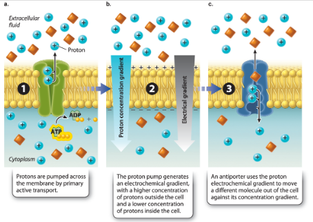
Secondary Active Transport (Fig. 5.13)
Primary active transport: Protons are pumped across the membrane using ATP, creating an electrochemical gradient
Electrochemical gradient: Combination of chemical gradient (concentration difference) and electrical gradient (charge difference)
Protons move from areas of high to low concentration and charge, driven by the electrochemical gradient
Antiporter: Uses the proton electrochemical gradient to move other molecules against their concentration gradient
Electrochemical gradient:
Chemical gradient: Concentration difference of ions (e.g., protons)
Electrical gradient: Charge difference across the membrane
Both gradients favor movement of protons back across the membrane
Secondary active transport:
Protons move down their electrochemical gradient, driving the movement of another molecule against its concentration gradient
The movement of the coupled molecule is powered by the proton gradient, not ATP
Primary vs Secondary Active Transport:
Primary active transport: Uses ATP directly to move molecules
Secondary active transport: Uses energy stored in the electrochemical gradient to move molecules
Common strategy in cells:
Sodium electrochemical gradient: Used to transport glucose and amino acids into cells
Proton electrochemical gradient: Moves molecules and synthesizes ATP
Active Transport and Cell Size Maintenance
Cell size maintenance: Cells use active transport to maintain size and composition
Red blood cells in different solutions:
Hypertonic solution: Higher solute concentration outside the cell, water moves out, cell shrinks
Hypotonic solution: Lower solute concentration outside the cell, water moves in, cell bursts (lysis)
Isotonic solution: Equal solute concentration inside and outside, water moves in and out equally, cell shape remains normal
Sodium-potassium pump:
Helps maintain isotonicity by moving ions across the membrane
Active transport of ions keeps intracellular fluid at equal concentration with extracellular fluid
Paramecium in freshwater:
Extracellular environment is hypotonic compared to the cell’s interior, causing risk of bursting due to water entering by osmosis
Contractile vacuoles: Take up excess water and expel it to the external environment
Some use aquaporins to take in water
Others use proton pumps to take in protons first, with water following by osmosis