Hearing Anatomy and Physiology
1/66
Earn XP
Description and Tags
Flashcards about the anatomy and physiology of hearing, covering the structure of the ear, how hair cells transduce information, and the auditory pathway.
Name | Mastery | Learn | Test | Matching | Spaced |
|---|
No study sessions yet.
67 Terms
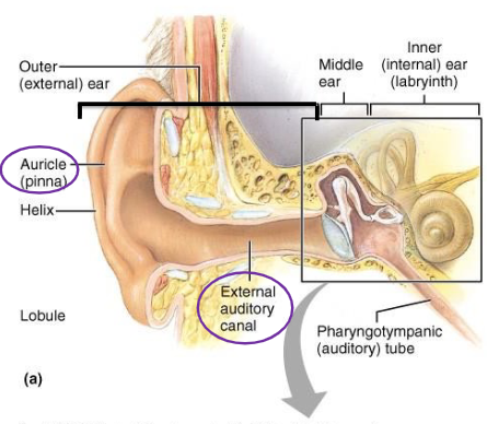
Pinna
Directs air waves into the external auditory canal.

External auditory canal
Air-filled passage in the outer ear that contains hairs and ceruminous glands.
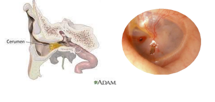
Ceruminous glands
Glands in the external auditory canal that secrete cerumen (ear wax).
Tympanic membrane
Also known as the eardrum; marks the beginning of the middle ear.

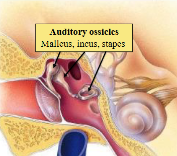
Middle ear
Air-filled cavity containing the malleus, incus, and stapes.

Malleus, Incus, Stapes
Auditory ossicles located in the middle ear.

Auditory ossicles
Connects the tympanic membrane to the oval window of the cochlea.
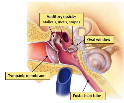
Eustachian tube
Opening of the middle ear that connects to the throat, allowing for pressure equalization.

Cochlea and Vestibular apparatus
Fluid-filled structures of the inner ear responsible for hearing and balance.
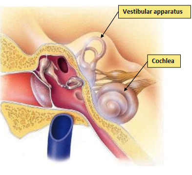
Cochlea
Spiral-shaped organ in the inner ear containing receptors for hearing.

Cochlea
Contains the organ of Corti (receptors for hearing).

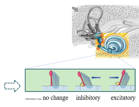
Hair cells
Receptors for hearing found in the cochlea; distortion of cilia results in changes of membrane potential.
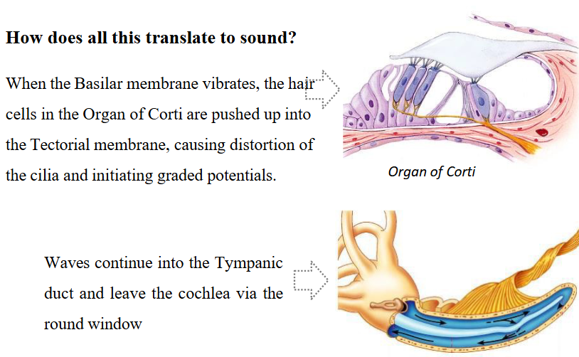
Organ of Corti
Contains hair cells important for hearing
Unwound Cochlea
Tube with a central membranous structure containing three ducts: Scala vestibuli, Scala media, and Scala tympani.
Vestibular membrane
Membrane that separates the scala vestibuli and scala media
Basilar membrane
Membrane in the cochlea where the Organ of Corti is located.
Tectorial membrane
Membrane overlying the hair cells in the Organ of Corti that causes distortion of hair cells.
Sound waves and cochlear fluid
How the cochlea translates air to sound, and the process of air pressure waves to fluid waves
Frequency
Determines pitch of sound; high pitch corresponds to high frequency and low pitch to low frequency.
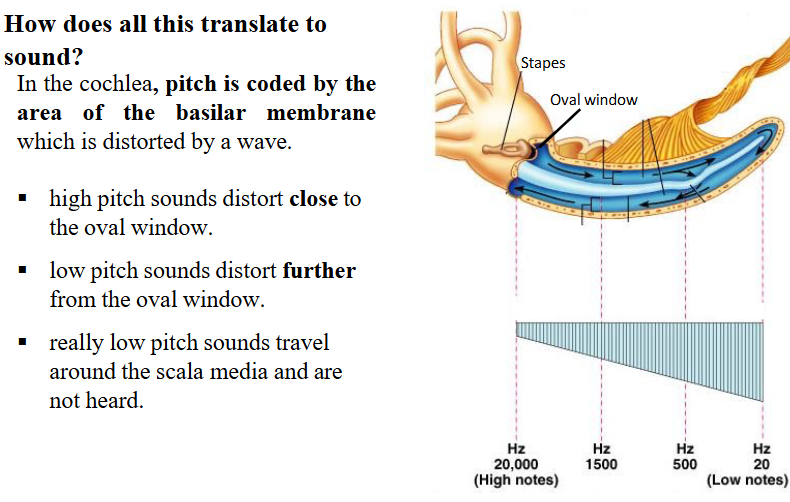
Pitch in the cochlea
Coded by the area of the basilar membrane which is distorted by a wave.
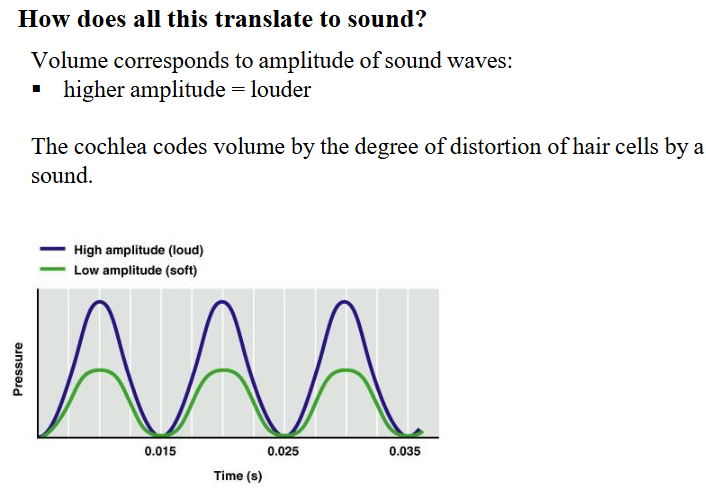
Amplitude
Determines volume of sound; higher amplitude corresponds to louder volume.
Auditory Pathway
The route that auditory information travels.
Spiral ganglia
Nerve fibers pass through to become the cochlear branch of the vestibulocochlear nerve
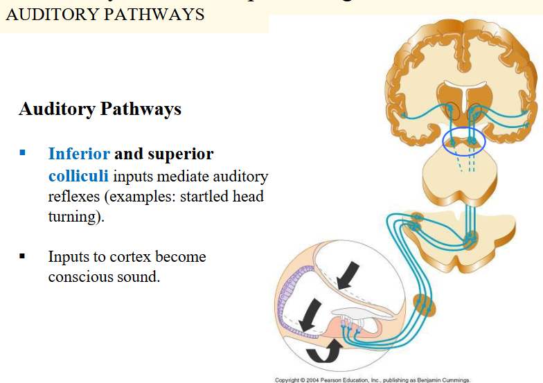
Inferior and superior colliculi
Auditory reflexes are mediated through inputs to these structures
Deafness
Types include conduction and Sensorineural
Conduction deafness
Interference of soundwave conduction caused by ear wax, perforated ear drum, fused ossicles
Sensorineural deafness
Damage to neural structures caused by damage to hair cells, cochlear nerve, or auditory cortex
Explain how the components of the ear contribute to transferring pressure waves from the air to pressure waves in a fluid
The outer ear collects sound waves, funneling them through the ear canal to the eardrum, causing it to vibrate. These vibrations are transmitted via the ossicles (malleus, incus, stapes) to the oval window of the cochlea, converting the mechanical energy into fluid pressure waves in the inner ear.
Describe the three regions of the ear and their function
The ear is divided into three regions: the outer ear (collects sound waves), the middle ear (transmits vibrations), and the inner ear (transduces vibrations to neural signals). Each region plays a crucial role in hearing.
Explain how hair cells transduce information
Hair cells transduce sound vibrations into electrical signals through the movement of their stereocilia, which opens ion channels and generates receptor potentials. This electrochemical process ultimately leads to the release of neurotransmitters at the synapse with the auditory nerve, conveying sound information to the brain.
Explain how the cochlear transduces pressure waves into sound
The cochlea transduces pressure waves by using fluid displacement to move the basilar membrane, stimulating hair cells in the organ of Corti. This mechanical movement creates electrical signals that travel through the auditory nerve to the brain, interpreting the sound.
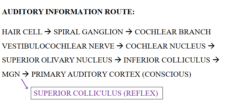
Describe the route of auditory information processing.
Auditory information processing involves sound waves entering the outer ear, traveling through the middle ear, and entering the cochlea in the inner ear where hair cells transduce the vibrations into electrical signals. These signals are then sent via the auditory nerve to the brain for interpretation.
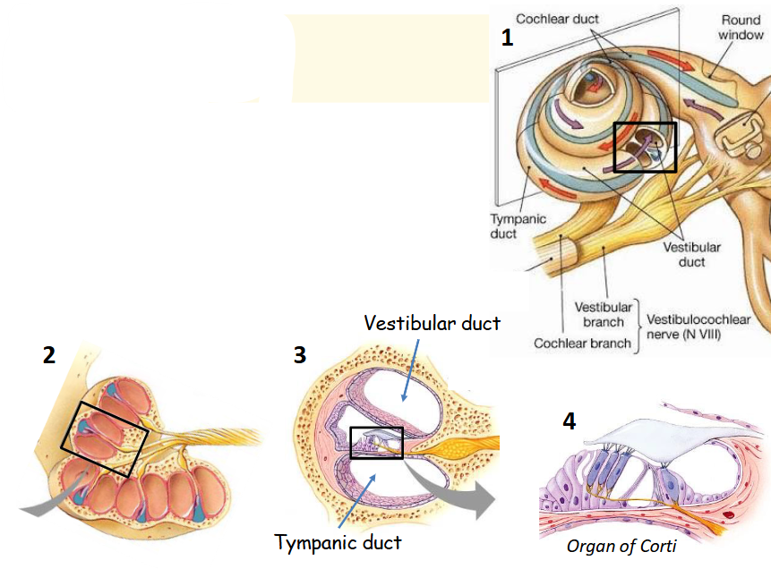
What makes up the labyrinth?
Cochlea and vestibular apparatus together make up the bony and membranous labyrinth
Where does the cochlear, vestibular branches and vestibulocochlear nerve originate?
Cochlear and vestibular branches of vestibulocochlear nerve originate here
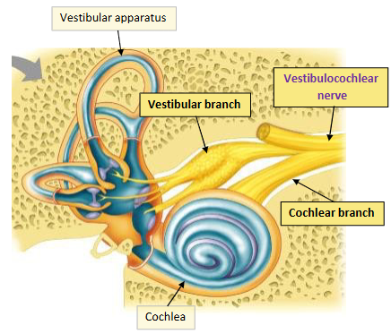
What type of hearing receptors does the cochlear contain?
Organ of Corti
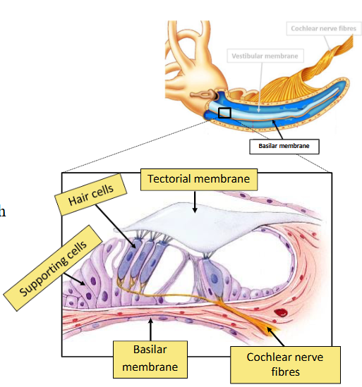
What is the organ of Corti?
Hair cells (Receptor cells)
What is meant by distortion of cilia?
Distortion of cilia results in change of membrane potential. The greater the amount of distortion, the greater the change in membrane potential.
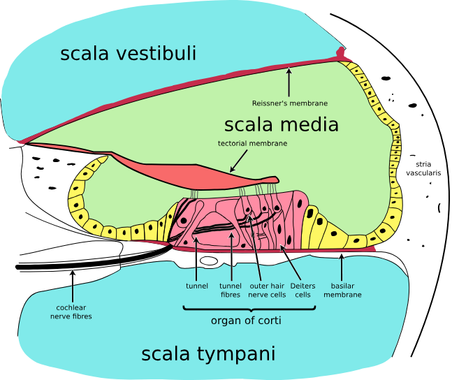
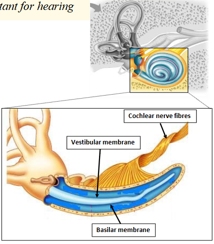
What happens if you unwind the cochlea?
If you unwind the spiral of the cochlea à it is a tube with a central membranous structure making three ducts
What are the 3cochlea ducts called?
§ Scala vestibuli (vestibular duct) begins at the oval window filled with perilymph.
§ Scala media (cochlear duct) filled with endolymph.
§ Scala tympani (Tympanic duct) end at the round window filled with perilymph
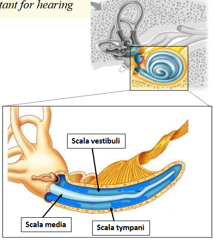
What are the cochlea ducts separated by?
§ Vestibular membrane
§ Basilar membrane
§ Cochlear nerve fibres run length of cochlea
What does the organ of Corti sit on?
Organ of Corti sits on the basilar membrane in scala media
Look at this diagram
What is the organ of Corti composed of?
Is composed of hair cells surrounded by supporting cells
sitting on the basilar membrane.
The hair cells are associated with cochlear nerve fibres.
The tectorial membrane overlies the hair cells which causes distortion of hair cells
What are soundwaves?
= pressure waves in the air
How does sound occur?
Travel down external auditory canal to tympanic membrane and cause it to vibrate.
Vibration causes auditory ossicles to rock.
Stapes sets up vibration in the oval window, which sets up waves in the cochlear fluid.
Transferred waves from air to fluid filled cavity.
Amplified by transfer from tympanic membrane to oval window ~ (x20)
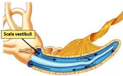
How does all this translate to sound?
1. Waves in the cochlear fluid travel along the Scala vestibule.
2. At a given point waves penetrate vestibular membrane and enter the scala media.
3. Waves pass through the basilar membrane, causing it to vibrate
How does all this translate to sound?
How does all this translate to sound?
What is Sound described as?
Sound is described in terms of pitch and volume
What does pitch correspond too?
Pitch corresponds to frequency of sound waves:
§ high pitch = high frequency
§ low pitch = low frequency
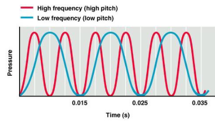
How does frequency determine pitch?
How does amplitude affect volume of sound?
Describe the auditory pathways
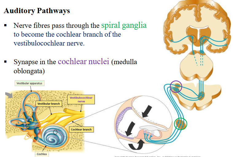
Look at this image of auditory pathways

Look at this image of auditory pathways
What is the auditory information route?
What are types of deafness?
Conduction deafness
Sensorineural deafness
What is Sensorineural deafness
damage to neural structures
What causes sensorineural deafness?
§ hair cells
§ cochlear nerve
§ auditory cortex
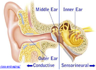
What is Conduction deafness
interference of soundwave conduction
What causes Conduction deafness?
§ ear wax
§ perforated ear drum
§ fused ossicles