bone & cartilage
 Call Kai
 Learn
Learn Practice Test
 Spaced Repetition
 Match
Match Flashcards
 Knowt Play
1/144

There's no tags or description
Looks like no tags are added yet.
Name | Mastery | Learn | Test | Matching | Spaced | Call with Kai | Chat |
|---|
No analytics yet
Send a link to your students to track their progress
145 Terms
What method do long bones use to form?
Endochondral ossification, where cartilage is replaced by bone.
First step to Endochondral ossification
Mesenchymal stem cells- unspecialized stem cells gather in an area
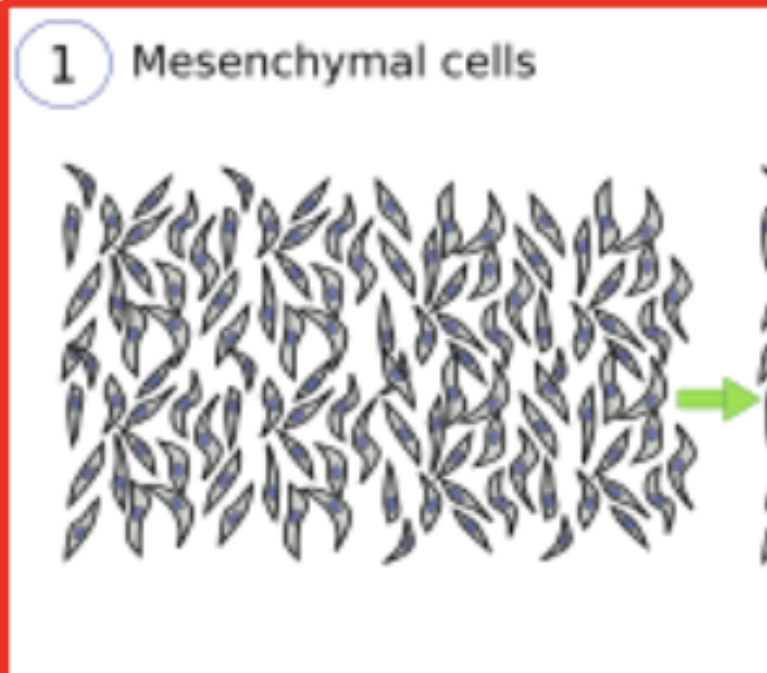
Whats going on here?
step 1) mesenchymal stem cells are clustering together
What happens after mesenchymal cells cluster together?
Unspecialized stem cells clump together tightly to form a blastema

What’s going on here
Step 2) The blastema, (clumps), begins to form through differentiation of mesenchymal stem cells
What happens after the blastema forms?
The clump turn into chondrocytes, making a cartilage model of the bone
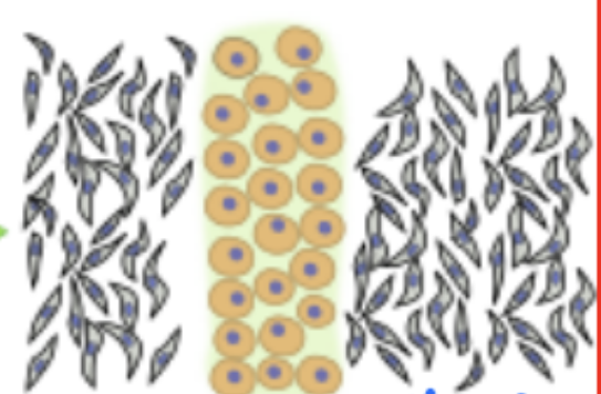
Whats going on here
step 3) The clump turns into chondrocytes, a cartilage model of the bone
What happens after blastema turn into chondrocytes, that made a cartilage model of the bone
Bone collar forms, a ring of bone tissue that forms around the cartilage shaft
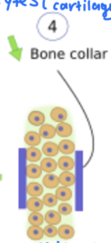
What’s going on here?
Bone collar formation
What happens after the bone collar forms?
Hypertrophic chondrocytes, start differentiation

Whats going on here?
Step 5) Hypertrophic chondrocytes, differentiation
What happens after hypertrophy of chondrocytes?
Invasion of blood vessels, bringing osteoblasts with them

Whats going on here?
Invasion of blood vessels, brings osteoblasts with them
What happens after blood vessels invade and bring osteoblasts with them?
Matrix condenses and calcifies, swollen cartilage cells die off and allow blood vessels to go into cartilage

What’s going on here
Death of chondrocytes, space allows blood vessels to go into cartilage
What happens after chondrocytes die off?
bone spreads outward from primary ossification center along cartilage columns, bone grows, cartilage is pushed to the side

What’s going on here?
Bone spreads outward from primary ossification center, cartilage pushed out
What happens after bone grows from the primary ossification center?
Secondary ossification center starts at the ends (epiphyses)

What’s going on here?
Chondrocytes at epiphyses (not the ones that died off in middle differentiate into osteoblasts - Secondary Ossification Center
What happens after secondary ossification center?
osteoblasts start laying down bone matrix on calcified cartilage (growth plate between primary and secondary ossification center)
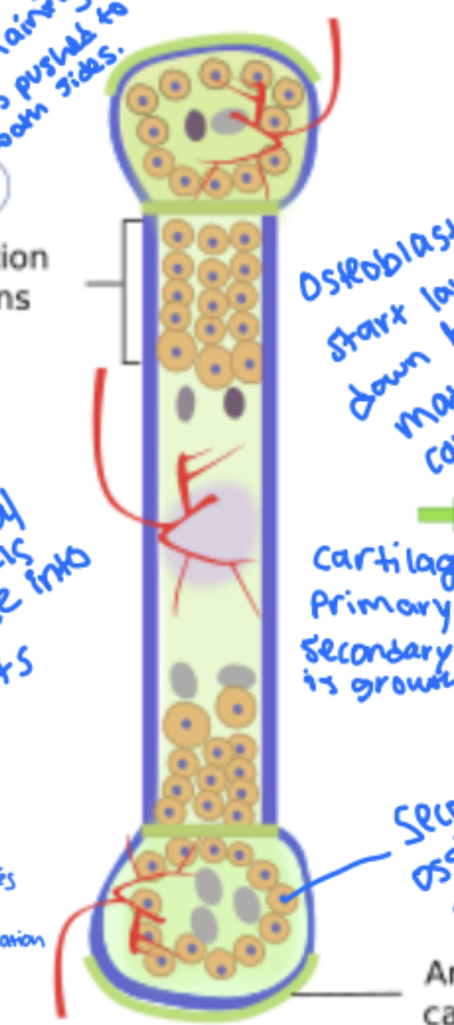
What’s going on here?
Osteoblasts lay down bone matrix in growth plate.
What happens after osteoblasts lay down bone matrix?
Bone will become calcified and stop growing

What’s going on here?
Bone will become calcified and stop growing
What is endochondral ossification:
Forming bone from inside of cartilage
What is intramembranous Bone Formation
Bone forms directly from mesenchymal tissue without cartilage
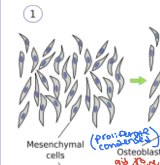
IBF: Step 1 of Intramembranous bone formation
Mesenchymal cells proliferate
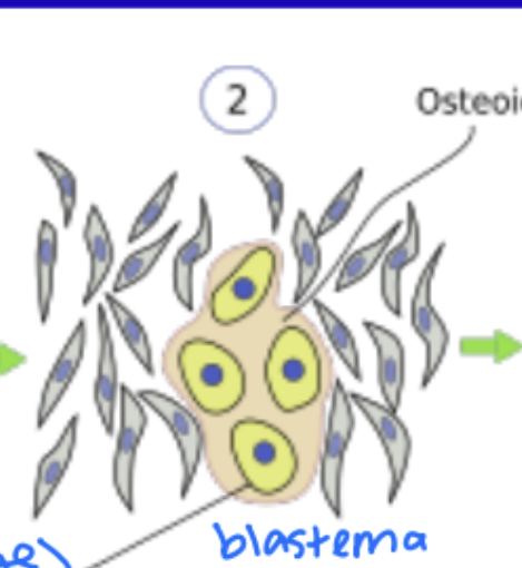
IBF: What happens after mesenchymal cells proliferate?
the mesenchymal cells cluster together (proliferate/condense) into a blastema, and directly differentiate into osteoblasts

IBF: What happens after differentiation into osteoblasts?
The osteoblasts start secreting osteoid (uncalcified bone matrix), some already trapped as osteocytes
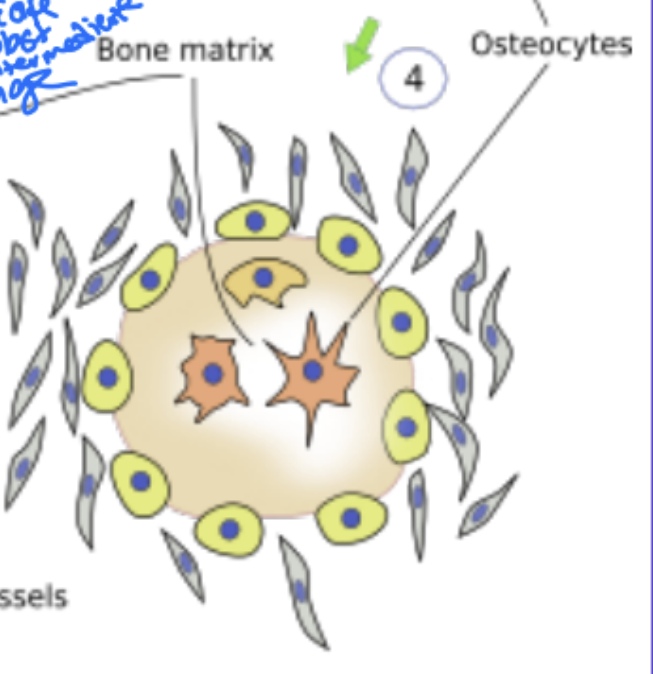
IBF: What happens after osteoblasts secrete osteoid
Osteocytes clearly established

IBF: What happens after osteocytes clearly established?
The bone matrix continues expanding (irregular, "spongy" looking shape), with osteocytes scattered throughout, blood vessels running through it, and osteoclasts arriving to start reshaping/remodeling the new bone.
What are cells of cartilage?
Chondrocytes, big and round, live in lacunae
Characteristic of cartilage
rigid and resilient
Is cartilage innervated?
No
Is cartilage vascular?
No
What is on surface of cartilage
Perichondrium, nerve and blood supply is here
Function of cartilage:
support of soft tissues( lubricate locomotion)
Cartilage plays as a what during endochondral bone formation?
Template
What is the appearance of cartilage?
glossy appearing because of hyaline cartilage
Chondrocytes reside in
lacunae within cartilage matrix.
Types of cartilage
Elastic, Hyaline, Fibro
Characteristic of elastic cartilage
same as hyaline cartilage, with elastic fibers
Fibrocartilage
Type 1 collagen + hyaline cartilage
Hyaline cartilage in brain
Synchondroses
Where is hyaline cartilage found as a template?
Embryonic skeleton
Where in respiratory system is hyaline cartilage found?
Trachea
What is the “end of lip” name hyaline cartilage?
Costal cartilage
Where hyaline cartilage found in joints?
Articular surfaces
Whats the place between primary and secondary ossification center where hyaline cartilage is found?
Epiphyseal growth plate?
What are the two main layers of the perichondrium, and what does each contain?
The outer layer is the fibrous connective tissue layer containing fibroblasts and type I collagen. The inner layer is the cellular/chondrogenic layer containing chondroprogenitor cells (and chondroblasts), which go on to form cartilage
What type of collagen do fibroblasts in the outer perichondrium secrete, and how does this differ from the collagen produced deeper in the cartilage?
Fibroblasts secrete type I collagen, which is structural/fibrous. Deeper in the cartilage, the matrix is made of type II collagen (typical of hyaline cartilage) along with proteoglycans — suited for cushioning and flexibility rather than pure structural support
What are the flattened cells located just beneath the fibrous outer layer, and what do they eventually differentiate into?
These are chondroblasts. They eventually mature into chondrocytes once surrounded by the matrix they secrete.
True or false: The perichondrium's chondroblasts are the major driving force behind cartilage growth
False. Chondroblasts secrete only a small amount of collagen matrix and are not the major driving force — the deeper chondrocytes contribute far more to matrix production and growth.
What term is used for the stem-like cells found in the inner connective tissue layer that are committed to becoming cartilage cells
Chondroprogenitor cells?
What are the two mechanisms of cartilage growth, and which cell population drives each one
Appositional growth — new cells/matrix added at the surface, driven by chondroblasts differentiating from chondroprogenitor cells. Interstitial growth — existing chondrocytes within the matrix divide and secrete additional matrix from the inside

What this
Hyaline Cartilage

What this
Chondroblasts
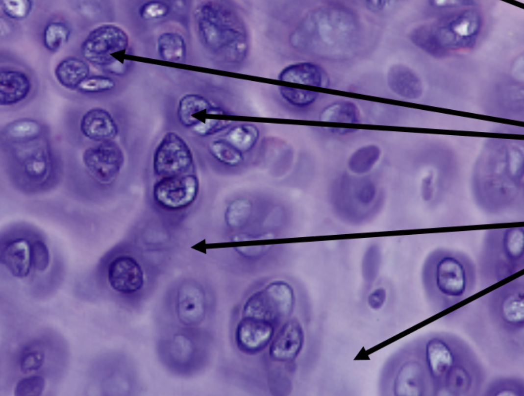
What this
Chondrocytes
Why is there lots of cytoplasm in hyaline cartilage?
For secretion
Composition of hyaline cartilage
80% type 2, 9% proteoglycans (aggrecans)
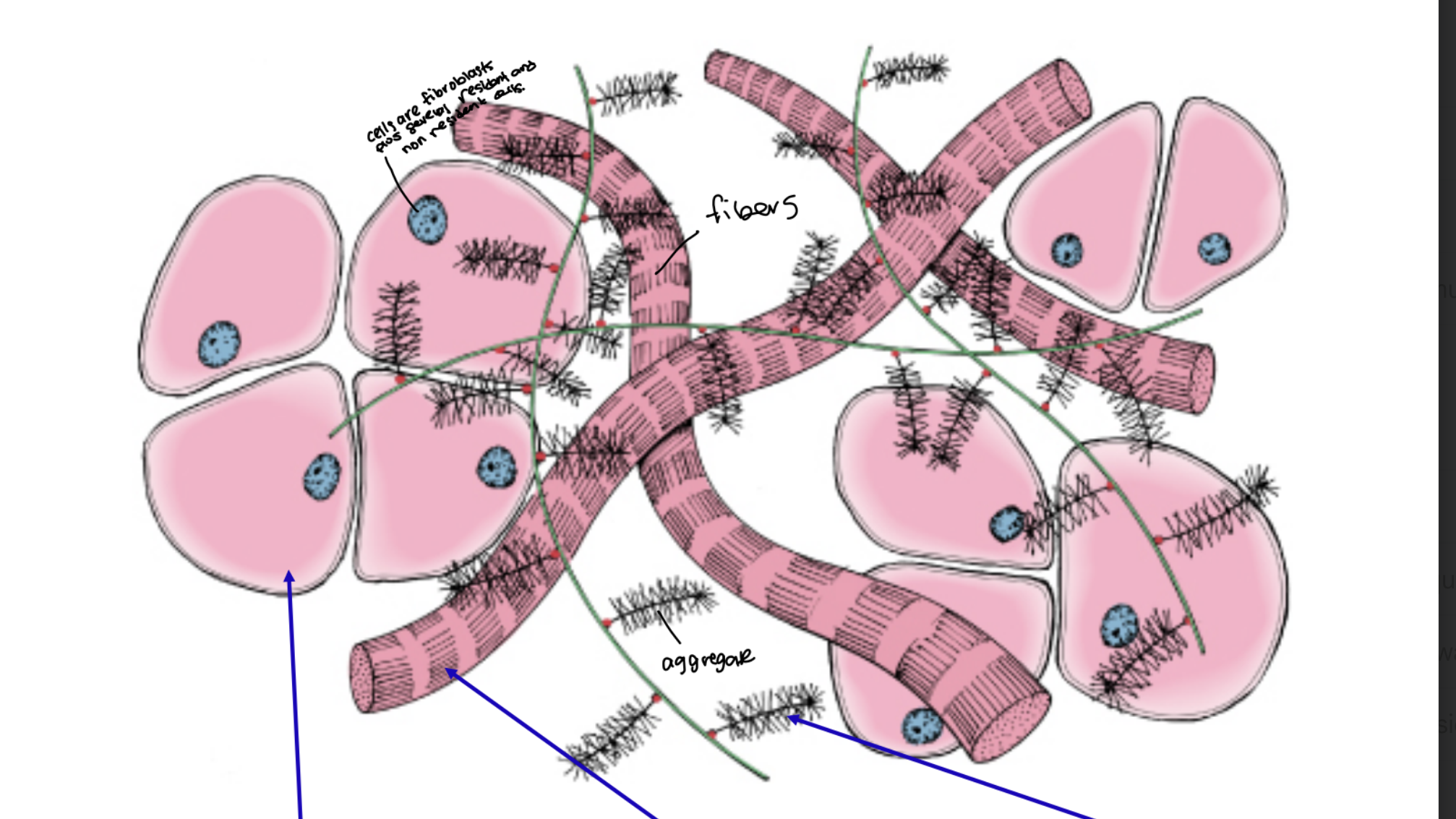
What that
Hyaline Cartilage ECM

What that/ what collagen
Fibers/ type 2

What that
aggrecan

What that
Chondrocytes
What does ground substance do in Hyaline cartilage ECM?
Resists compression
What do collagen fibers do in Hyaline Cartilage ECM?
Resist tension
What is the territorial matrix, and how does it appear on H&E staining?
The territorial matrix is the matrix immediately surrounding each chondrocyte. It appears darker stained on H&E histology.
What is the interterritorial matrix, and how does it appear on H&E staining compared to the territorial matrix?
The interterritorial matrix is the matrix located farther away from the chondrocytes, between the territorial zones. It stains lighter compared to the territorial matrix.
Which matrix type (territorial or interterritorial) would you expect to provide more cushioning/resilience, and which would contribute more to overall tissue rigidity?
the denser territorial matrix (closer to the cell, more condensed) contributes more to localized rigidity/support right around the cell, while the interterritorial matrix (less concentrated, more spread out) contributes to the tissue's overall flexibility/resilience under tension.
What matrix is this
Territorial

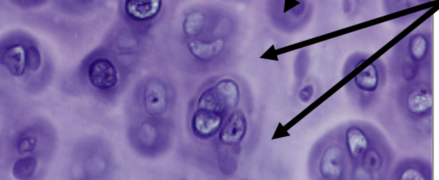
What matrix is this
Interterritorial matrix
What are isogenous groups in hyaline cartilage?
Isogenous groups in hyaline cartilage are clusters of chondrocytes (sisters) that originate from a single progenitor cell and are surrounded by a shared matrix. They play a role in the maintenance and repair of cartilage tissue.

What that
Isogenous groups in hyaline cartilage
Cartilage goes through what kinda growth?
Interstitial growth
Is a perichondrium required for chondroblasts
no
Are chondroblasts needed for cartilage?
no
What kinda cells can perichondrium produce?
Progenitor cells and chondroblasts
Explain how appositional growth (outside to inside) is different from the growth happening "from inside to outside"
Appositional growth (outside to inside) adds new material at the surface via the perichondrium's progenitor cells/chondroblasts, expanding the tissue inward from the edge. The inside-to-outside growth you noted instead refers to interstitial growth — existing chondrocytes already embedded within the cartilage dividing and secreting matrix from within, pushing the tissue outward from inside rather than adding cells at the surface
Perichondrium is characterized by
appositional growth
Whats happening in appositional growth?
Chondroprogenitor cells become
chondroblasts. Chondroblasts mature into chondrocytres
What is interstitial growth, and where does it occur (cartilage or bone)?
Interstitial growth occurs within cartilage, not bone. It's growth that happens from within the existing tissue rather than from an outer layer.
As cells divide during interstitial growth, what happens to the matrix and the cells themselves?
The cells separate from each other as they divide, and they accumulate/secrete matrix between themselves and other cells, which pushes them away from other cells.
What is appositional growth, and from which structure do the growth-driving cells originate?
Appositional growth is when cartilage grows from the outside. The growth-driving cells come from the perichondrium.
What are the immature cells from the perichondrium called, and what is their function?
These cells are called chondroblasts. They are immature cells that make new cartilage.
Where cartilage gets their nutrients?
Since cartilage itself has no blood vessels, the nutrients have to come from somewhere — and that somewhere is the perichondrium, which does contain blood vessels
Hyaline cartilage examples
epiphyseal growth plate, articular cartilage, formation of initial skeleton
What are synchondroses?
Joint composed of hyaline cartilage formed back to back to expand size of cranial base
Properties of elastic cartilage
gives cartilage elastic properties in addition to resilience of hyaline cartilage
What is elastic cartilage surrounded by?
Perichondrium, so appositional and interstitial growth
Elastic cartilage is composed of
Type II collagen
Locations of elastic cartilage
Pinna of ear, eustachian tube, epiglottis, larynx, external auditory meatus

What that
Elastic cartilage
What type of collagen is Fibrocartilage
Type 1
Location of fibrocartilage
attachment of tendons and ligaments to bones, intervertebral discs, symphysis pubis
What type of growth is fibrocartilage?
Only interstitial, no perichondrium because of high stress
What is fibrocartilage a hybrid of?
Dense regular connective tissue + hyaline cartilage
How are the cells aligned in fibroblasts?
in single rows and isogenous groups

What that
Fibrocartilage

What that
Dense regular connective tissue
Why can’t cartilage really repair itself
Because it is avascular, but some limited repair can occur in presence of perichondrium