MOVEMENT
1/76
There's no tags or description
Looks like no tags are added yet.
Name | Mastery | Learn | Test | Matching | Spaced |
|---|
No study sessions yet.
77 Terms
Vertebrae muscles include…
smooth muscles
skeletal or striated muscles
cardiac muscles
Smooth Muscles
control the digestive system and other organs
found in the intestines and other organs, consisting of long thin cells
Skeletal or Striated Muscles
controls movement of the bidy in relation to the environment
consists of long cylindrical fibers with stripes
Cardiac Muscles
controls the heart
consists of fibers that fuse together at various points
because of these fusions, cardiac muscles contract together not independently
Neuromuscular Junction
synapse between a motor neuron axon and muscle fiber
All nerve-muscle junctions rely on acetylcholine as their neurotransmitter.
In skeletal muscles, each axon releases acetylcholine at the neuromuscular junction, which always excite muscles to contract
Antagonistic Muscle
opposing set of muscles required to move the legs or arms back and forth
Antagonistic Muscles include…
flexor
extensor
flexor
muscle brings your hand toward your shoulder
muscles that bend/flex joints (e.g., bending of knee)
extensor
muscles that increase angle between members of a limb (e.g. straightening the arm)
Our muscle types range from…
Fast-Twitch Fibers
Slow-Twitch Fibers
Aerobic
use oxygen during their movements which is why it do not cause fatigue
Anaerobic
prolonged use of fast-twitch fibers results in fatigue
Golgi Tendon Organs
proprioceptors that respond to increases in muscle tension
located in the tendons at opposite ends of a muscle
act as a brake against an excessively vigorous contraction
Responds to muscle tension
Slow-Twitch Fibers
Weaker contractions—no fatigue
Aerobic: it uses oxygen during movements (pay as you go)
Used during non-strenuous activities (e.g., talking)
Fast-Twitch Fibers
Fast contractions—more fatigue
Anaerobic: happens without oxygen but will need it for recovery afterwards (accumulates oxygen debt)
Used during strenuous activities (e.g., sprinting up a steep hill)
Stretch Reflex
The spinal cord then sends a signal to contract muscles reflexively when they’re stretched (e.g., knee-jerk reflex)
when a muscle is stretched, the spinal cord sends a signal to contract it reflexively.
caused by a stretch; it does not produce one.
PROPRIOCEPTOR
Latin: proprius, “one’s own”
A receptor that detects the position or movement of a part of the body (a muscle)
Can correct our balance by adjusting our posture or footing.
Muscle Spindle
Kind of muscle proprioceptor
Parallel to the muscle that responds to a stretch
the muscle is stretched more than the antagonistic muscle
Reflexes
are consistent automatic responses to stimuli.
Involuntary movement
Few behaviors are purely voluntary or involuntary, reflexive or nonreflexive
Ballistic movement
Executed as a whole
Once initiated, it cannot be altered (e.g., reflex)
movements varying in sensitivity to feedback.
Adjustable Movement
Subject to feedback correction
Modify movement based on external stimuli
Central Pattern Generators
Neural mechanisms in the cells in the lumbar segments of the spinal cord that generate rhythmic patterns of motor output (e.g. walking)
Motor Program
Fixed sequence of movements from beginning to end
Mostly seen in other animals
Cerebral Cortex
Important for complex actions (talking and writing); less control over coughing, sneezing, gagging, laughing, and crying
Lack of cerebral control explains why it is hard to perform those actions voluntarily
Most mammals: axons from CC connect only to interneurons of the brainstem or spinal cord
Human and other primates: some axons go directly from the CC to motor neuron (results to greater dexterity)
Primary Motor Cortex
elicits movements; does not send messages directly to muscles
axons extend to brainstem and spinal cord
also active when you imagine, remember movements, or understand verbs related to movement
POSTERIOR PARIETAL CORTEX
Monitors position of the body
First area to become active in planning movement
located behind the primary somatosensory cortex.
responsible for the body’s orientation in relation to its environment
When damaged: trouble finding objects despite description; bumping into obstacles when walking or running
When stimulated during surgery: intention to move
Intense stimulation: patients believe they did make a movement
SUPPLEMENTARY MOTOR CORTEX
Important for planning and organizing a rapid sequence of movements
activates when you commit a mistake
Essential in habitual action
Becomes active after an error in movement, developing ways to inhibit the incorrect movement
PREMOTOR CORTEX
Directing its movement, as well as information about posture and position
Most active immediately before a movement
It’s like the brain planner

PREFRONTAL CORTEX
Important for considering the probable outcomes of possible movements
Active during a delay before movement
Stores sensory information appropriate to a movement.
When damaged: many movements would be disorganized
Central Pattern Generators
Neural mechanisms in the cells in the lumbar segments of the spinal cord that generate rhythmic patterns of motor output (e.g. walking)
Motor Program
Fixed sequence of movements from beginning to end
Mostly seen in other animals
Mirror Neurons
Active during preparation for a movement and while watching someone else perform the same or similar movement
ex. mirror neurons in part of the frontal cortex become active when people smile or see someone else smile, and they respond especially strongly in people who report identifying strongly with other people .
May be important for understanding other people, and identifying or imitating them
Activated not only by seeing an action, but also by any reminder of the action
Many mirror neurons modify their properties by learning, and probably developed their original properties by learning also
CORTICOSPINAL TRACTS
From the cerebral cortex to the spinal cord
Have two types that contribute to nearly all movements; inequal contribution
LATERAL CORTICOSPINAL TRACT
Pathway of axons from the primary motor cortex, surrounding areas of the cortex, and from the red nucleus (a.k.a. pyramidal tract)
Red nucleus: midbrain area that controls certain aspects of movements
Axons of the lateral tract extend directly from the motor cortex to their target neuron
Crosses to contralateral side of spinal cord
Controls movements in peripheral (lateral, sides) areas, especially hands and feet
Red nucleus
midbrain area that controls certain aspects of movements
MEDIAL CORTICOSPINAL TRACT
Includes axons from many parts of the Cerebral Cortex, not just heprimary motor cortex and its surrounding areas
Also includes axons from the midrain tectum, reticular formation, and vestibular nucleus
Goes to both sides of the spinal cord
Controls mainly the muscles of the neck, shoulders, and trunk (vertically middle)
controls bilateral movements: walking, standing up, and sitting down
Cerebellum
Also known as the "little brain."
Associated with coordination, balance, timing, and aim.
This region of the brain has more neurons and synapses than the rest of the brain.
Impairment: difficult to do tasks like writing, pointing, speaking, typing, rhythm, hand coordination, and playing musical instruments
Cerebellum
also involved in sensory processing, attention, timing, and other cognitive processes
when movement is absent, _ still responds to sensory stimuli
It plays a critical role in tasks requiring precise timing, ranging from ms to 1.5s (damage prevents estimation of duration).
PPL WITH DAMAGE: may not be able to catch moving objects due to timing impairment.
Mastery of timed movement is often accompanied by mastery of others, suggesting the involvement of the cerebellum in a variety of timing-related tasks.
is essential for attention; people with damage here require more time to change their focus than people who do not.
Cellular Organization
The cerebral cortex, the spinal cord, and sensory systems (via the cranial nerve nuclei) are some of the sources of information that reach the cerebellum.
The cerebellar cortex (outer layer) processes this information. Here the neurons are organized in a precise geometric pattern, with Purkinje cells being the key players.
These flat cells are arranged in sequential planes, with parallel fibers running perpendicular to them.
Action potentials in parallel fibers excite Purkinje cells, which then send inhibitory messages to cerebellar nuclei and vestibular nuclei.
The sequence of activation of Purkinje cells controls the timing of output, determining both its onset and offset.
This mechanism allows for precise coordination and control of movements
Basal Ganglia
is composed of large structures located in the forebrain, including the caudate nucleus, putamen, and globus pallidus.
Receives input from the cerebral cortex and substantia nigra and plays a crucial role in regulating movement
Important for self-initiated behaviors, with increased activity observed during tasks requiring spontaneous actions.
Its cells regulate the vigor of movement.
DAMAGE: : can result in slow and weak spontaneous movements, like in Parkinson's disease
Alterations in dopamine pathways to the striatum can lead to depressed mood and decreased motivation.
group of large subcortical structures
vital for spontaneous, self initiated behaviors
respond strongly to signals indicating reward
Brain areas and motor learning
Acquiring new skills is influenced by every area of the brain that regulates movement.
Movements are slow and inconsistent when neurons in the motor cortex are still adapting.
Changes in the firing rates of pertinent neurons allow these movements to be more swift and consistent with practice.
Damage: increases difficultly in picking up new skills and executing them. e.g. converting newly learned movements into automatic actions.
> As an organism learns a motor skill, the nerve cells alter its responses.
> Process:
Slow and inconsistent movements > Firing rates of the neurons of the motor cortex increases as the movements become faster > Consistent patterns of movement and motor cortex activity
PARKINSON’S DISEASE
Gradual loss of dopamine-releasing axons from the substantia nigra to the striatum (part of the basal ganglia).
With the loss of this input, the striatum decreases its inhibition of the globus pallidus, which therefore increases its inhibitory input to the thalamus.
affects the peripheral nervous system as the noradrenergic terminals in the heart are also affected by this.
Spontaneous movements are slow and weak.
Many Parkinson’s patients have cognitive deficits, which may include problems with attention, language, or memory
It can lead to premature death
Causes of Parkinson’s Disease
Starts in the substantia nigra.
Toxins can be another factor in its development
Young adults developed symptoms of Parkinson’s disease after using MPTP (drug similar to heroin)
Body converts this to MPP+; destroys neurons that release dopamine
People are sometimes exposed to hazardous environmental chemicals that damage cells of the substantia nigra.
Many studies have shown an increased risk of Parkinson’s disease among people with much exposure to insecticides, herbicides, and fungicides.
L-DOPA TREATMENT
A precursor to dopamine that can cross BBB
First drug in psychiatry or neurology, and one of the first in medicine, to emerge from a theory
Taken as a daily pill
Most common treatment for this disease
However, it increases dopamine release in all axons, including those that had deteriorated and those that were still functioning normally
when it reaches the brain, specifically the BBB it converts itself into dopamine.
It relieves symptoms of the disease.
It improves quality of life.
Side Effects: nausea, restlessness, sleep problems, low blood pressure, repetitive movements, and sometimes hallucinations and delusions
HUNTINGTON’S DISEASE
A severe neurological disorder; “Huntington’s chorea
Begins with arm jerks and facial twitches. Then tremors spread to other parts of the body and develop into writhing.
Tremors interfere more and more with walking, speech, and other voluntary movements.
People lose the ability to develop motor skills.
Associated with gradual, extensive brain damage, especially in the basal ganglia and cerebral cortex
People with this disease also suffer psychological disorders including apathy, depression, sleeplessness, memory impairment, anxiety, hallucinations and delusions, poor judgment, alcoholism, drug abuse, and sexual disorders.
Can occur at any age (most often ages 30-50)
Once symptoms emerge, both psychological and motor symptoms grow progressively worse and culminate in death
HEREDITY AND PRESYMPTOMATIC TESTING
In 1993, researchers located the gene for Huntington’s disease on chromosome number 4.
The critical area of the gene includes a sequence of bases C-A-G (cytosine, adenine, guanine), which is repeated 11 to 24 times in most people.
People with up to 35 C-A-G repetitions are considered safe
Those with 36 to 38, possibly even 39 or 40, might not get the disease, and if they do, it probably will not manifest until old age
People with more repetitions are nearly certain to get the disease
The more C-A-G repetitions someone has, the earlier the probable onset of the disease
Other factors besides genes also influence the age of onset, such as stressful experiences, drug or alcohol abuse, and diet and exercise
Huntington’s disease led to the discovery of the protein that it codes, which has been designated huntingtin
Huntingtin occurs throughout the human body, although its mutant form produces no known harm outside the brain.
The mutant form impairs neurons and glia in several ways, including effects on mitochondria and potassium channels
STRIATUM
- also known as Dorsal Striatum
- this part of the basal ganglia receives information from the cerebral cortex and substantia nigra.
- sends output to the globus pallidus
GLOBUS PALLIDU
- sends information to the thalamus and frontal cortex.
2 pathways through basal ganglia
direct pathway and indirect pathway
direct pathway
from the stratum inhibits the globus pallidus which inhibits part of the Thalamus
inhibiting the inhibitor = excitation
enhances selected movements
indirect pathway
inhibits inappropriate competing movements as it makes response to stimulus less strong
vital for learned movements
direct pathway and indirect pathway
help control movement initiation and inhibition.
Symptoms of Parkinson’s disease
slow movement
tremors
trouble walking
sleep disorders
pain
cons of l-dopa
It does not replace other transmitters, only dopamine
It does not slow the continuous loss of neurons.
It has unpleasant side effects such ass sleep problems, low blood pressure, nausea, etc.
It causes dose escalation
Myasthenia Gravis
an autoimmune condition caused by degeneration of acetylcholine receptors at the neuromuscular junction.
happens when a person’s immune system produces antibodies that bind to the nicotonic acetylcholine receptor
Symptoms of Myasthenia Gravis
extreme muscle weakness
fatigue
treatments for myasthenia gravis:
medications that suppress the immune system, meaning it will slow the production of the troublesome anitbodies
Medications that inhibit the acetylcholinesterase → it is the enzyme that deactivates the acetylcholine at the synapse
Muscular Dystrophy
a group of disease which is distinguished by extreme muscle development due to abnormalities in the protein dystrophin, which results to muscle degeneration
s a sex-linked disorder, usually affects males due to the X chromosome being responsible for encoding dystrophin.
symptoms of muscular dystrophy
muscle weakness
difficulty in walking
frequent falling
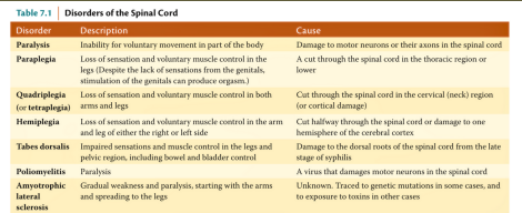
polio
a contagious viral disease that attacks the spinal motor neurons, which can lead to paralysis.
caused by poliovirus which spreads from person to person
symptoms of polio
muscle pain
fatigue
body ache
treatments for polio
There are no treatments for polio, only medications to relief symptoms.
vaccination against poliovirus
avoid consuming uncooked and unsanitary meals
Accidental spinal cord damage
damage to any part of the spinal cord
The spinal cord can be accidentally damaged when the protective vertebrae which surrounds the cord breaks and compresses in the cord itself.
Symptoms of accidental spinal cord damage
Loss of feeling or sensation
Loss of control of movement or paralysis (depends on the level of damage)
Cervical Damage: Quadriplegia or loss of movement in both arms and legs
Lumbar Region Damage: Paraplegia or loss of movement in the legs
Amyotrophic Lateral Sclerosis
where the motor neurons of the spinal cord and brainstem progressively deteriorate
also known as “Loy Gehrig’s Disease.”
The muscles served by these deteriorating motor neurons degenerate when their input ceases
Symptoms of Amyotrophic Lateral Sclerosis
Twitching and cramping of muscles
Loss of motor control
Impairment in the use of arms and legs
Tripping and falling
Essential Tremor
s a common motor disorder which is characterized by involuntary shaking or trembling.
typically affects the hands, arms or head when doing a voluntary movement.
symptoms of essential tremor
Trembling or shaking begin gradually, and it is more noticeable on one side of the body. → worsens with movements
treatment for essential tremor
not curable, it can be lessened through anti-seizure medications.
treatment of muscular dystrophy
no treatment
Muscle Proprioceptor
detects the stretch and tension of a muscle and send messages that enable the spinal cord to adjust its signals.
CONNECTIONS FROM THE BRAIN TO THE SPINAL CORD
messages from the brain must reach the medulla and spinal cord which control the muscles.
The path from the cerebral cortex to the spinal cord are called the corticospinal tracts:
2 KINDS OF CORTICOSPINAL TRACT
lateral corticospinal tract and medial corticospinal tract
Lack of acetylcholine or receptors = impaired movement Every muscle only makes one movement — a contraction Muscles relax when no message to contract is received.
ANTISACCADE TASK
task to look at the opposite direction of a visual stimuli
Requires sustained activity in parts of prefrontal cortex and basal ganglia in preparation for the task
Ability to perform this task gradually improves as prefrontal cortex matures