Electrical activity of the heart
1/15
There's no tags or description
Looks like no tags are added yet.
Name | Mastery | Learn | Test | Matching | Spaced |
|---|
No study sessions yet.
16 Terms
Describe the structure of cardiolomyoctes
elongated, cylindrical, striated cells
Have a single nucleus and many mitochondria
Have gap junctions: allow depolarising currents to from from one cell to another
Desmosomes + gap junctions form intercalated discs
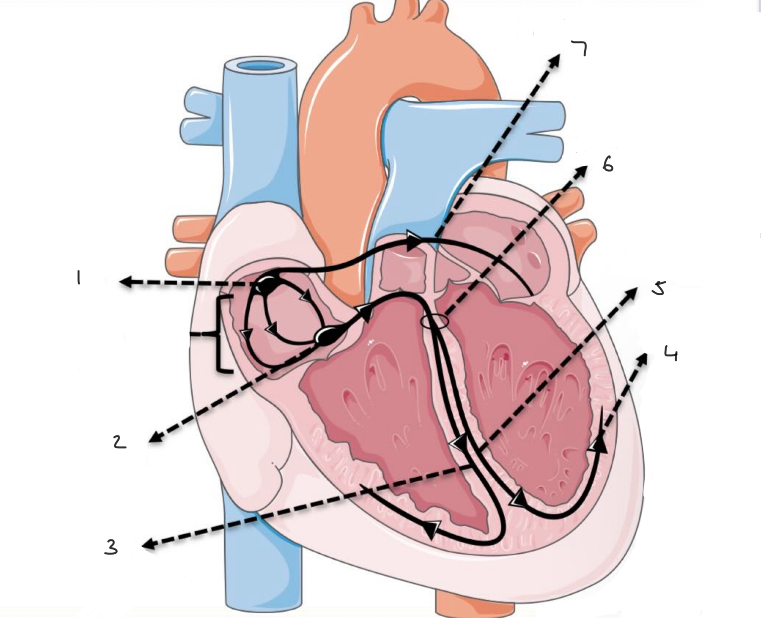
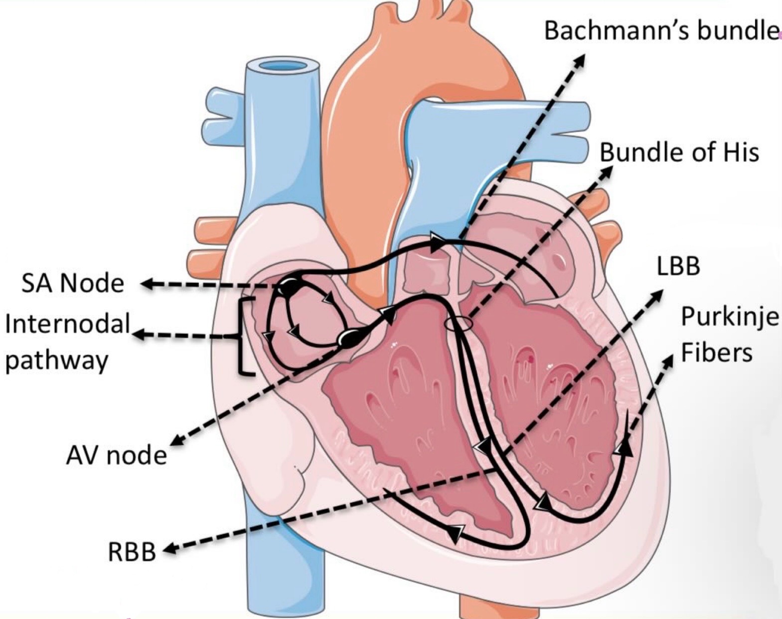
Name all the hearts nodal cells
SA node
AV node
Bundle of his
L + R bundle branches
Purkinje fibres
** generate spontaneous action potentials (slow response)
Structure of contractile cells
Have actin, myosin, troponin, tropomyosin
Have sarcoplasmic reticulum
Fast response action potentials
Where is the SA node located and what does it do?
Inferior to the superior vena cava
Pacemaker of the heart → sets sinus rhythm (60-80bpm)
What does the Bachmann’s bundle do?
Pass signals from the SA node to the L atrium
Where is the AV node located and what does it do?
Inferior to the pulmonary trunk
Gives time for atria to contract so the ventricles can fill
What does the bundle of His (AV bundle) do?
Carries action potentials from AV node to bundle branches
What does the R bundle branch do?
Carries action potential to the right myocardium
What does the L bundle branch do?
Carries action potentials to L myocardium
What does Purkinje fibres do?
Carry action potentials to ventricular muscle
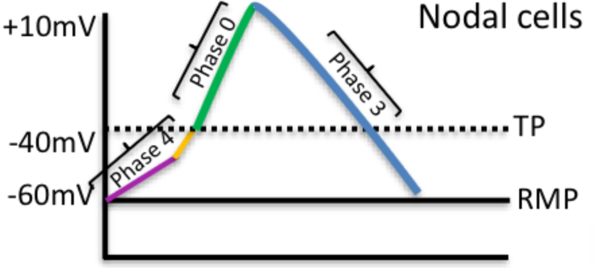
What happens in nodal cells?
Phase 4(diastole): funny Na+ channels open + T type Ca2+ channels open → reaches threshold (-40mV)
Phase 0 (upstroke): L-type Ca2+ channels open → +10mV → Ions move to contractile cells via gap junctions
Phase 3 (final repolarisation): L-type Ca2+ channels inactivate. K
+ channels activate and K+ leaves the cell → cell depolarises
Is the resting membrane potential of contractile cells?
-90mV
How do contractile cells generate an action potential?
In flow of positive ions: -90mV → -70mV
Phase O (upstroke): voltage gated Na+ channels open: -70 → +20
Phase 1 (initial repolarisation): Na+ channels close. K+ channels open → K+ moves out → 0mV
Phase 2 (plateau): K+ moves out and Ca2+ moves in (Sam’s amount of ions in and out)
Phase 3 (final repolarisation): L-type Ca2+ channels close. Ca2+ is taken back to SR by sodium-calcium exchanger and calcium proton ATPase pumps. K+ channels open and K+ exits the cell
Phase 4 (RMP): little movement of ions
Why do contractile cells have phase 1 and 2?
Excitation-contraction coupling
Ca2+ activate ryanidine receptors (RYR) in the SR
RYR opens up Ca2+ channels so more move out of the SR into the cytoplasm
Ca2+ bins to tropocollagen. → change shape of tropmyosin → moves tropmyosin away → myosin head interacts wirh actin → more cross bridges → more contraction
What is it called when the cells all contract at the same time?
Functional syncytium