Chapter 8: The Nervous System
1/184
There's no tags or description
Looks like no tags are added yet.
Name | Mastery | Learn | Test | Matching | Spaced | Call with Kai |
|---|
No analytics yet
Send a link to your students to track their progress
185 Terms
Functions of the nervous system
1. sensory input : sensory receptors respond to external & internal stimuli. Ex: t sensors in skin- to brain & spinal cord
2. integration : interpretation of sensory input. Ex: from t receptors- to hypothalamus. (controls body temp.)
3. motor output : response by muscles, glands, and organs. Ex: cold t- hypothalamus- shivering (skeletal m. Contraction) - produce heat- warm body.
Divisions of the Nervous System
1. Central Nervous System (CNS) - midline.
-brain
-spinal cord
2. Peripheral Nervous System (PNS) - project out from CNS, includes all the cranial & spinal nerves.
-Afferent (sensory) division: includes somatic and visceral divisions.
-Efferent (motor) division: includes somatic and autonomic divisions.
Organization of the Nervous System
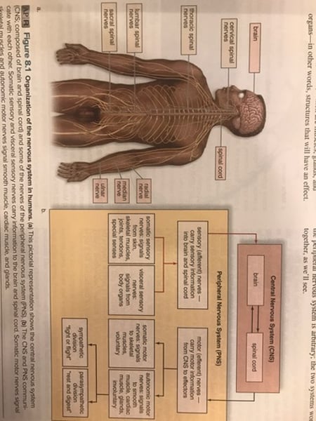
Afferent nerves
Come from 2 sources:
1. Somatic sensory nerves come from the surface of the body, skeletal muscles, tendons & joints.
2. Visceral sensory nerves come from internal organs.
Efferent nerves
Have 2 destinations :
1. Somatic efferent nerves innervate skeletal muscles.
2. Autonomic efferent nerves supply organs under involuntary control
-Ex: heart, digestive organs.
-There are 2 divisions of the autonomic nervous system: sympathetic & parasympathetic nervous systems.
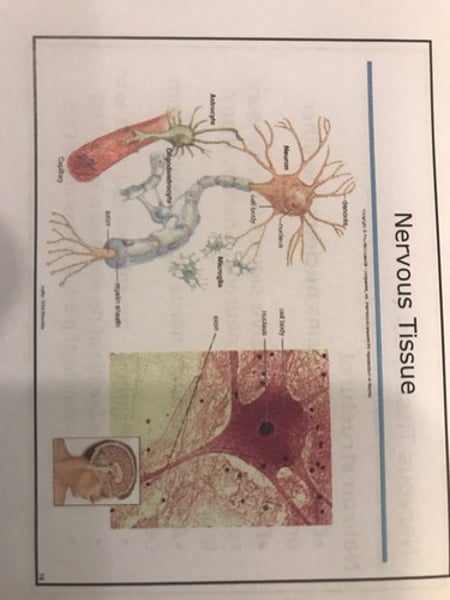
Nervous tissue Cells
1. Neurons (nerve cells): transmit impulses
2. Neuroglia: support & nourish neurons.
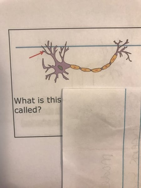
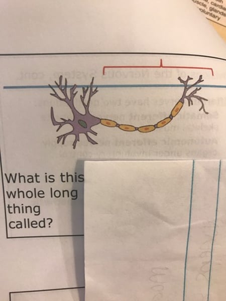
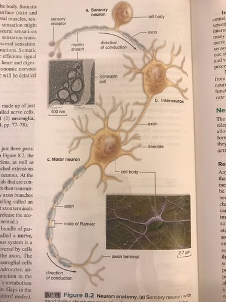
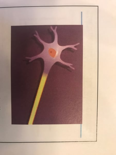
Neuron structure
-Dendrite: receive signal from sensory receptors and other neurons. Shorter, many branches.
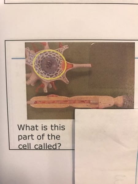
-Cell body: contains the nucleus and other organelles.
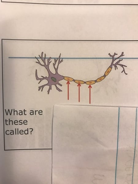
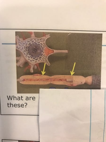
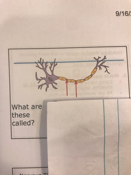
-Axon: conduct nerve signals away from cell body.
Parts of Axon
-Axon terminal: enlarged end of Axon
-Nerve: bundle of parallel axons in PNS
-Tract: bundle of parallel axons in CNS.
Cell body of neuron
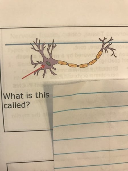
Cell body
Dendrite
Dendrites
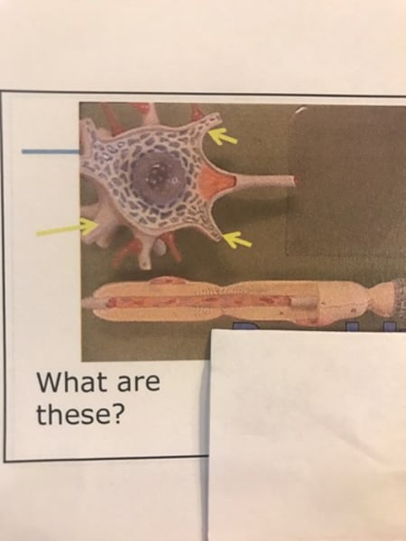
Axon
Neuron structure cont'd.
-Axon may be covered by a myelin sheath (lipid coating)
-formed by Schwann cells or neurolemmocytes in PNS.
-formed by oligodendrocytes in CNS
-provides insulation.
-increases speed of impulse conduction.
-Nodes of Ranvier: gaps in myelin sheath.
Nervous tissue
Schwann cells
Axon
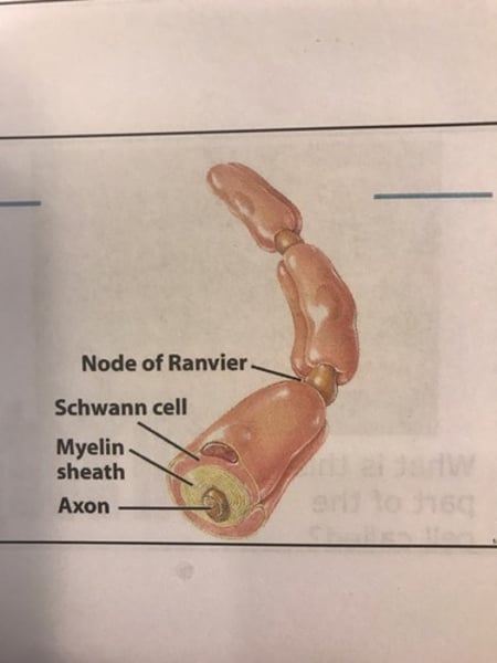
Schwann cells
Nodes of Ranvier
Nodes of Ranvier

Types of Neurons
1. sensory neurons
2. motor neurons
3. interneurons
Sensory neurons
-carry nerve impulses from sensory receipts to the CNS.
-almost all are unipolar- have one extension coming off of the cell body; it splits into 2 branches- one comes to the periphery & another goes to the CNS.
Interneurons
"Association neurons"
-all are in CNS
-the vast majority of neurons in the body are interneurons.
-typically multipolar- have many dendrites and a single axon.
-convey nerve impulses between various parts of the CNS (between sensory & motor neurons, form brain to cord & vice versa)
-form complex pathways for thinking, memory, and language.
Motor neurons
-carry nerve impulses from CNS to muscles, organs, or glands.
-all are multipolar neurons.
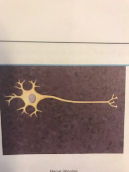
Neuron Anatomy
Nerve signal conduction
-neurons release neurotransmitters to communicate electrical signals between cells.
-axons carry electrical signals, called action potentials.
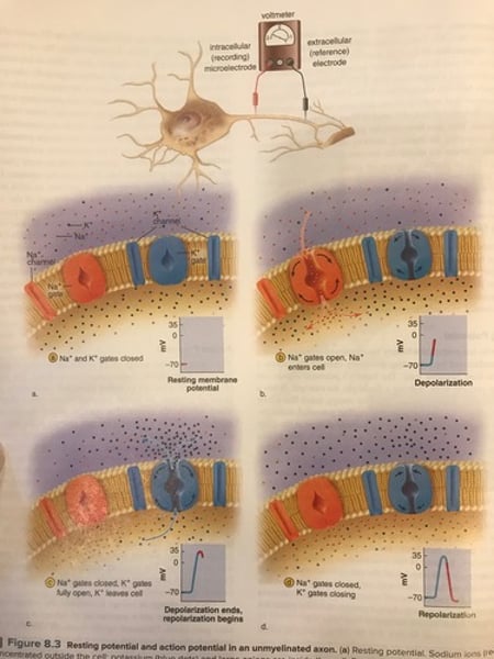
Resting potential
Potential energy of a neuron.
-The cell membrane is polarized.
-positively charged outside the cell due to sodium ions (Na+).
-negatively charge inside due to potassium ions (K+) and large (can't leave the cell) negatively charged proteins.
-At rest, the membrane is impermeable to Na+ But permeable to K+.
Resting potential cont'd.
-resting potential = 70mV!!
When the neuronal membrane is at rest, the resting potential is negative due to the accumulation of more sodium ions outside the cell than potassium ions inside the cell.
-the sodium/potassium pump moved ions to maintain resting potential. (Actively transports 3Na+ out of the neuron and 2K+ in).
Action potential and membrane potential
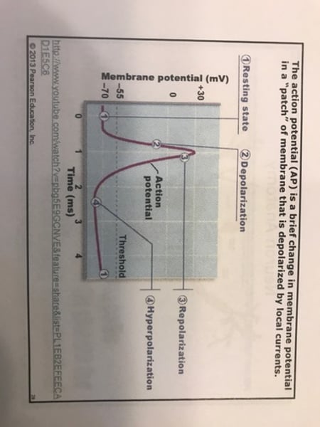
Action potential
-process of conduction of nerve signals.
-occurs in axons.
-begins with a stimulus, which activated voltage-regulated sodium gates (Na+ channels in the cell membrane)
-these gates are closed when the cell is at rest; the stimulus opens them & sodium ions rush into the cell (depolarization); the membrane becomes negative outside and positive inside.
Action potential cont'd.
-if enough Na+ enters the cell, the cell potentially may reach the cell's threshold.
-if threshold is reached, large numbers of voltage-regulated sodium gates will open, & the cell potential will rise abruptly.
-this sharp depolarization continues until the fell potential is +35mV; at this point, the voltage-regulated sodium gates close and voltage-regulated potassium gates open.
Repolarization
Cell potential becomes negative again!
Action potential cont'd.
-after the action potential is complete, the Na+/K+ pump moves Na+ out & K+ into the cell (reach resting potential) to get ready for another action potential.
-action potentials are conducted, or propagated, down the axon away from the cell body!
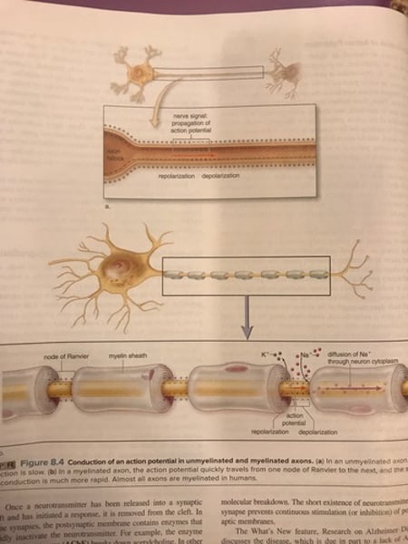
Resting potential & action potential in an unmyelinated axon
nerve impulse
Conduction of Action potentials
-as the axon depolarizes, Na+ will diffuse into the next section of the axon, causing depolarization to threshold at that location.
-continues down the axon, followed by repolarization.
-it is an all-or-none event; action potentials don't vary in size - the intensity of message is determined by the number of action potentials, not their size.
Refractory period
-period of time after an action potential, during which an Axon cannot conduct another action potential.
-the refraction period is the time needed for
Na+/K+ pump to restore the original Na+ and K+ concentrations.
-ensures one-way direction of an impulse.
Action potential in unmyelinated axon
Conduction of action potentials cont'd.
In unmyelinated axons:
-slow (~1 meter/second)
- each section of Axon must be stimulated.
In myelinated axons:
-An AP at one node of ranvier can "jump" over myelinated portion of Axon to the next node.
-called saltatory conduction.
-much faster (>100 meters/second)
Conduction of an action potential
Transmission across a synapse.
-Axon terminal: small swelling at tips of branched end of Axon.
-Synapse:
-region of close proximity between 2 neurons.
-presynaptic membrane: membrane of first neuron.
-postsynaptic membrane: membrane of the next neuron.
-synaptic cleft: small gap between the presynaptic and the postsynaptic neuron.
-the neuromuscular junction is a type of synapse.
Synapse
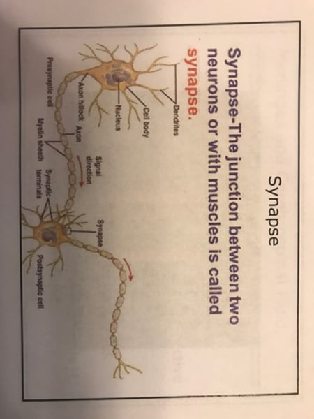
synaptic structure and function
Synaptic structure & function
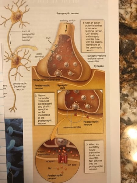
Transmission across synapse cont'd.
-Neurotransmitters: molecules stored in synaptic vesicles in the axon terminal that transmit a nerve impulse across a synapse.
-nerve impulse reaches Axon terminal
-calcium channels are opened and Ca2+ enters the terminal.
-causes synaptic vesicles to fuse with the presynaptic membrane and release the neurotransmitter by exocytosis into the synaptic cleft.
Transmission across synapse cont'd.
-neurotransmitter then diffuses across the synaptic cleft to the postsynaptic membrane & binds to its specific receptor on ligand-regulates gates; they then open.
-the gates are closed when the cell is at rest; the NT acts as a ligand that opens the gates.
-removal is the ligand (the NT) closes the gates.
-depending on the NT, the postsynaptic membrane will be exited or inhibited.
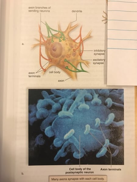
Neurotransmitter molecules
-at least 100 neurotransmitters have been identified.
-2 well known neurotransmitters : Acetylcholine (ACh); Norepinephrine (NE) = adrenaline.
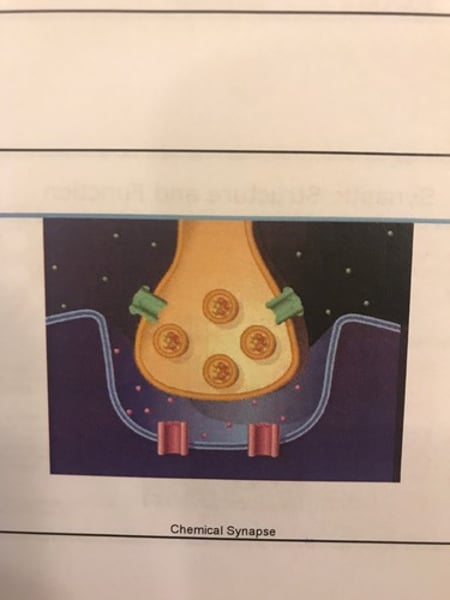
Chemical synapse
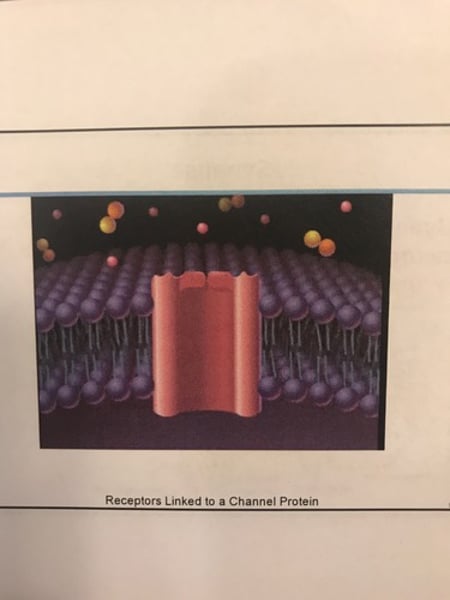
Receptors linked to a channel protein
Neurotransmitter molecules cont'd.
After a neurotransmitter has initiated a response, it is removed from the synaptic cleft to prevent continuous stimulation.
-enzymes may inactivate the neurotransmitter (Ex: ACh inactivated by acetylcholinesterase -AChE)
-it may be reabsorbed by presynaptic membrane.
Many drugs block or enhance the effect of neurotransmitters in the body.
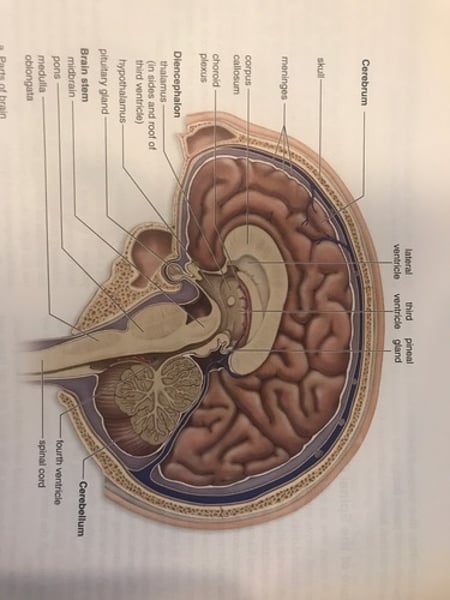
Central Nervous System (CNS)
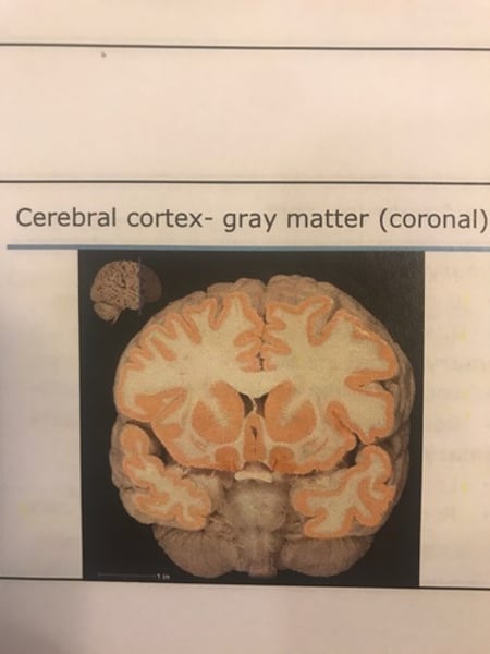
-consists of the brain and spinal cord
-gray matrer: contains cell bodies & unmyelinated fbers.
-white matter: contains myelinated axons; myelin appears white.
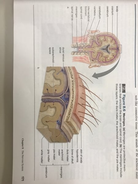
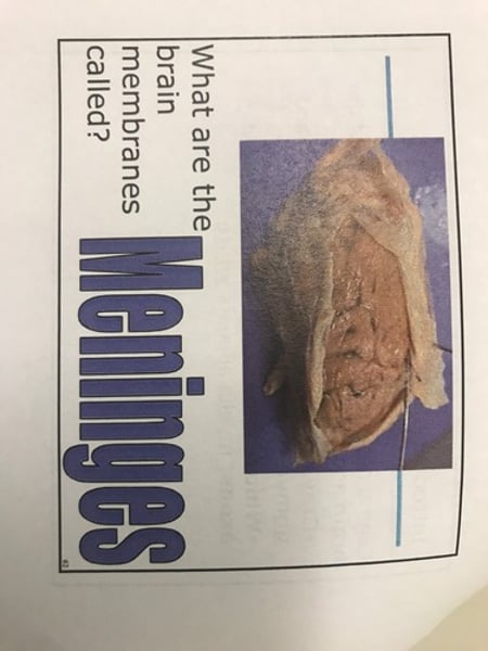
Meninges
-protective membranes of the CNS.
1. Dura Mater
2. Arachnoid matter
3. Pia matter
Dura mater
Outer menix composed of 2 layers of tough, fibrous connective tissue.
-epidural space: fat-filled space between the
Dura mater & the skull or vertebrae.
-rural venous sinuses: spaces between the dura
Mater layers containing venous blood.
Arachnoid Mater
Middle menix composed of spider web like connective tissue.
Pia Mater
Deepest menix that adheres to the brain & spinal cord. Capillaries from pia Mater called choroid plexus (ependymal cells) secrete cerebrospinal fluid.
Meninges
Meninges
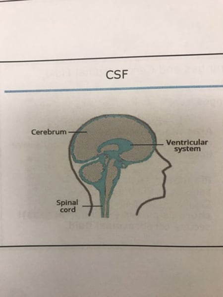
Cerebrospinal fluid (CSF)
Produced from blood by choroid plexus into the ventricles (protective cushion, nutrition).
-found in the:
-subarachnoid space: space between the
arachnoid Mater & pia Mater.
-ventricles: hollow, interconnecting cavities
Of the brain (choroid plexus).
-central canal: hollow, space of spinal cord.
-reabsorbed back into dural venous sinuses.
-blockages can occur that can cause hydrocephalus.
CSF
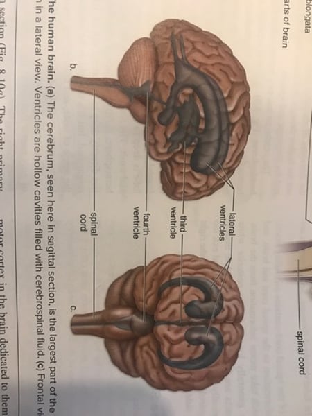
Ventricles of the brain
Ventricles of the brain

The spinal cord
Extends from foreman magnum to first lumbar vertebrae.
-protected by vertebral column.
-occupies vertebral canal, which contains the
spinal cord.
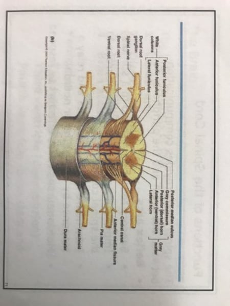
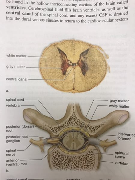
Structure of spinal cord
-spinal nerves extend from spinal cord in between vertebrae.
-intervertebral disks: (fibrocartilage) separates each vertebrae.
-herniated disk: disk torn open; may press on
spinal nerves & cause pain & loss of function.
-central canal and subarachnoid space contain CSF.
Structure of spinal cord cont'd.
-centrally located gray matter.
-H shaped
-contains interneurons & portions of sensory &
motor neurons.
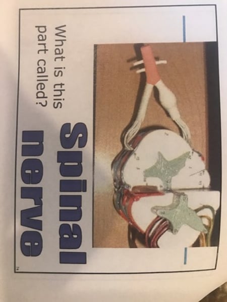
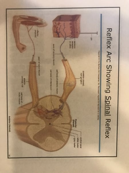
-Spinal nerve:
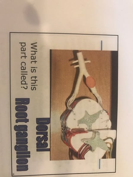
-Posterior (dorsal) roots: contain sensory fibers
That are entering the grey matter of the spinal
cord.
-posterior root ganglion: enlarged area of
posterior root; contains cell bodies of
sensory fibers.
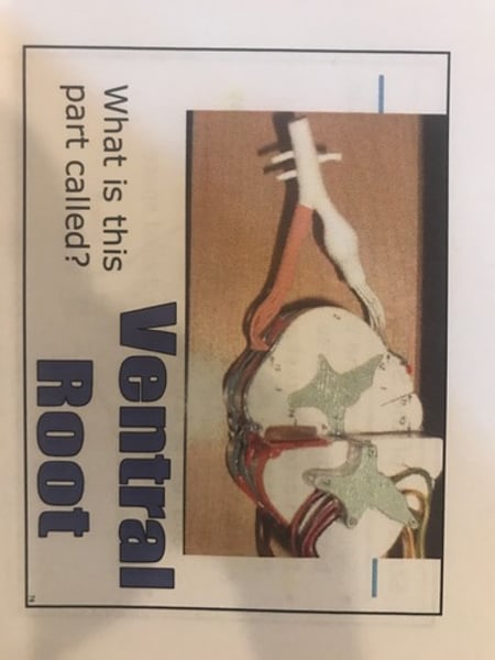
-Anterior (ventral) roots: contain motor fibers exiting the grey matter.
Spinal cord
Spinal cord
Spinal cord
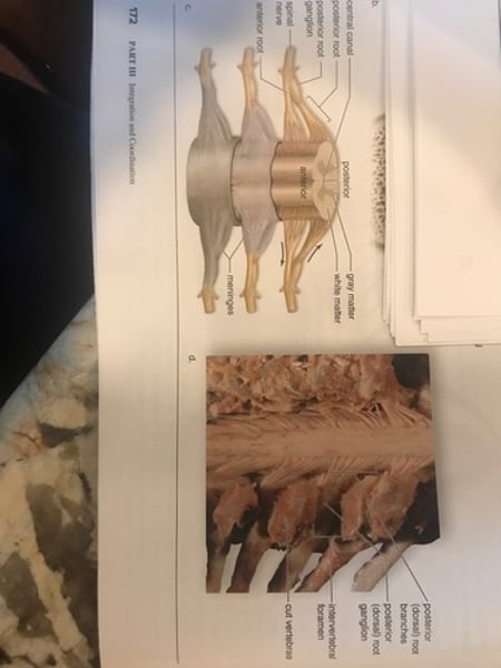
Spinal nerve
Dorsal root ganglion
Ventral root
White matter
-Posterior: white matter composed of ascending tracts carrying sensory information to the brain.
-Anterior: white matter composed of descending tracks carrying information from the brain.
-Tracts generally cross from one side of the spinal cord to the other.
Spinal cord
Provides communication between brain & peripheral nerves.
Reflex arcs
-sensory receptors respond to a stimulus.
-impulse travels over sensory neurons to the spinal cord.
-interneurons integrate data and relay a response by way of motor neurons.
-motor neurons cause effectors to respond.
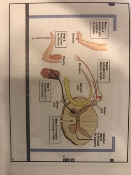
Reflexes
-Automatic involuntary responses to change inside & outside the body.
-cranial reflexes involve the brain.
-spinal reflexes involve only the spinal cord.
Reflex arc showing spinal reflex
Reflexes cont'd.
-reflexes can be used to determine if the nervous system is reacting properly, can help avoid injury, and help maintain balance.
-2 examples of reflexes used to test the function of nervous system:
1. Knee-jerk reflex.
2. Ankle-jerk reflex.
These 2 reflexes are used to maintain balance.
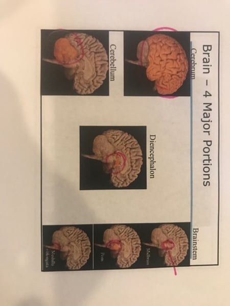
Brains 4 major structures
1. Cerebrum
2. Diencephalon
3. Cerebellum
4. Brain stem
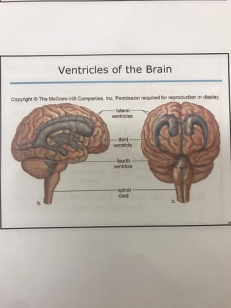
ventricles of the brain
-2 lateral ventricles (cerebrum)
-third ventricle (diencephalon)
-fourth ventricle (brain stem & cerebellum)
4 major portions of the brain
The human brain
Ventricles of the brain
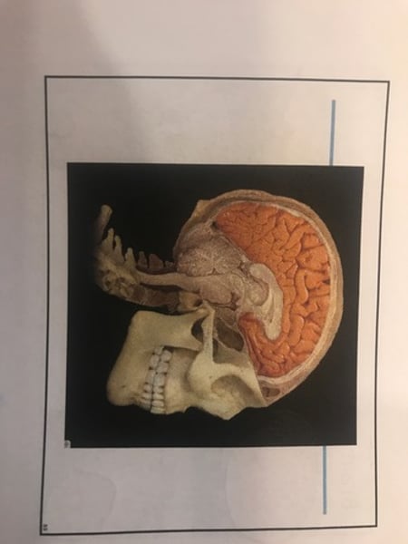
Cerebrum
Largest portion of the brain.
-receives sensory input, carries out integration & initiates voluntary motor responses.
-coordinates the activities of the other parts of the brain.
-involved in higher thought processes.
Cerebrum
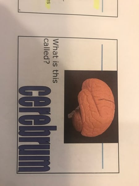
Cerebrum
Cerebrum cont'd.
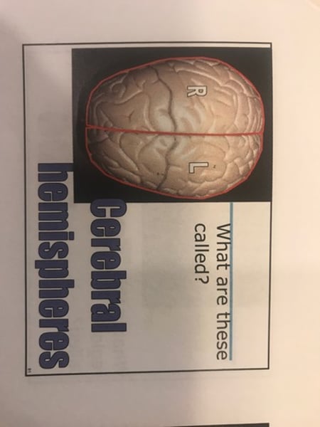
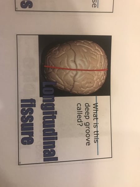
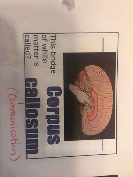
Divided into 2 cerebral hemispheres:
-longitudinal fissure: divides them.
-connected internally by the corpus callosum (white matter)
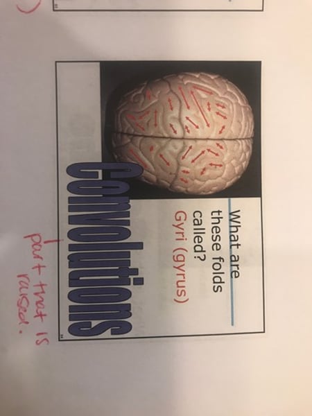
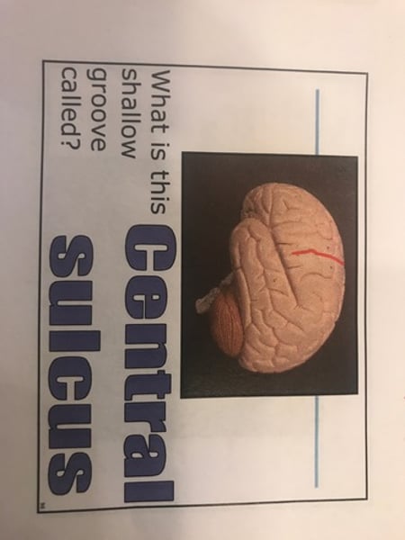
-Gyri: (ridges =convolutions) are separated by sulci (shallow grooves).
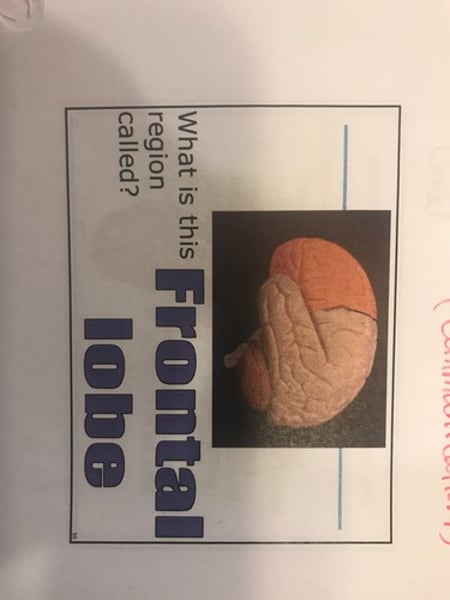
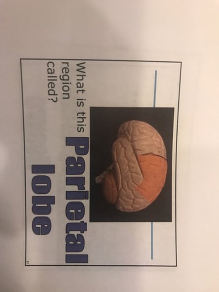
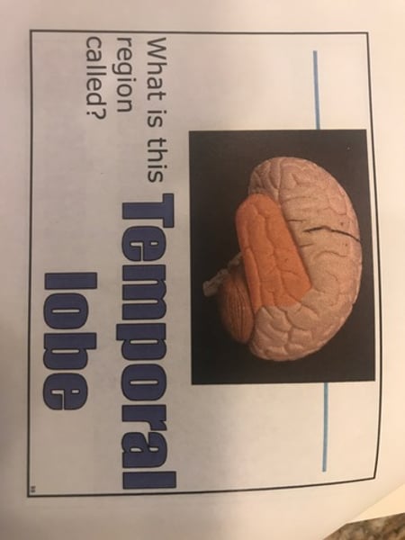
Five lobes:
1. Frontal lobe
2. Parietal lobes
3. Temporal lobes
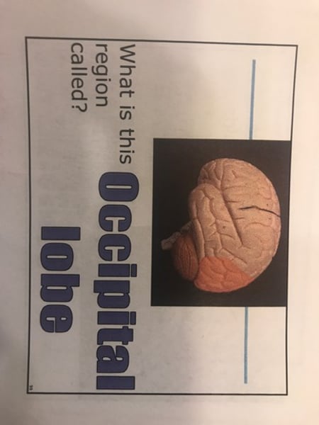
4. Occipital lobe
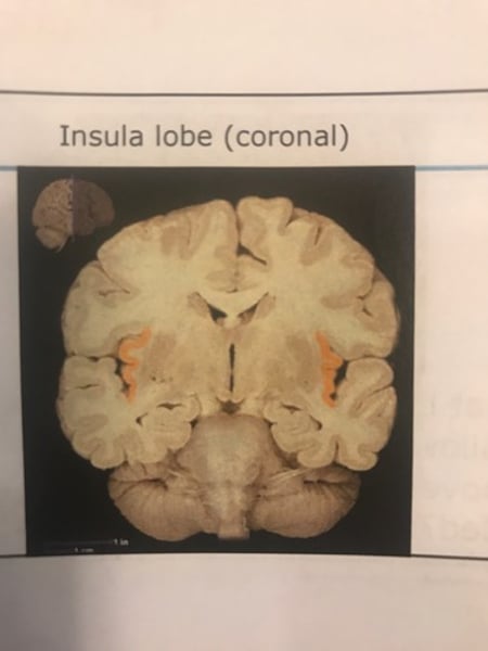
5. Insula
Cerebral hemispheres
Longitudinal fissure
Corpus callosum
Convolutions (gyri)
Frontal lobe
Central sulcus
separates frontal and parietal lobes
Parietal lobe
Temporal lobe
Occipital lobe
Insula
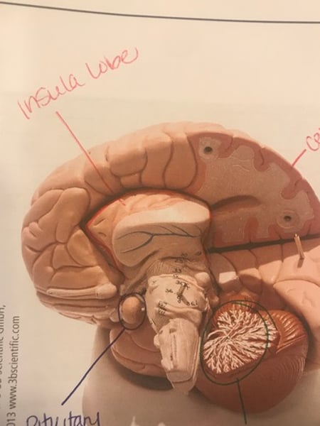
Insula lobe
lobes of cerebral hemisphere
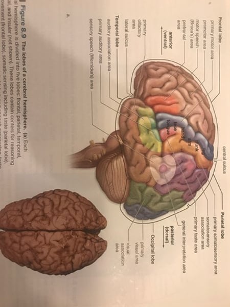
Cerebral cortex
-outer layer of cerebrum; made of gray matter.
-accounts for sensation, voluntary movement, information processing, consciousness.
Cerebral cortex

Cerebral cortex - gray matter