CLRS 205 - Nuclear Medicine & PET
1/55
There's no tags or description
Looks like no tags are added yet.
Name | Mastery | Learn | Test | Matching | Spaced |
|---|
No study sessions yet.
56 Terms
What is the primary purpose of nuclear medicine?
To diagnose and sometimes treat disease processes using radiopharmaceuticals.
What are radiopharmaceuticals?
Radioactive materials (radionuclides) combined/tagged with a pharmaceutical agent (element/compound).
How are radiopharmaceuticals usually administered to patients?
By injection, but can also be given by ingestion or inhalation.

What type of imaging does nuclear medicine use?
Planar (2D) imaging and SPECT (3D; Single Photon Emission Computed Tomography).
What does SPECT produce?
Slices of images from the emitted gamma radiation.
What happens once the radiopharmaceutical reaches the target organ or tissue?
The emitted gamma rays are detected, and the data is analyzed to image and evaluate function.

What does a radiopharmaceutical contain?
A gamma-emitting radionuclide and a pharmaceutical metabolized by a specific organ or system.
What intake pattern is characteristic of a normal organ?
Characteristic uptake pattern
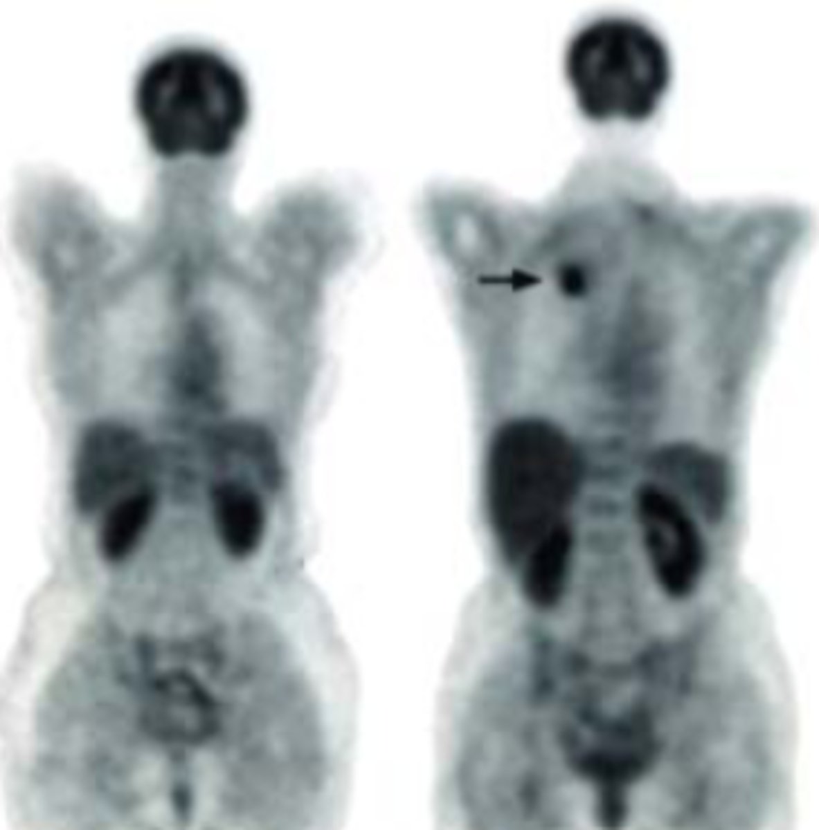
What indicates abnormal organ function in nuclear medicine imaging?
Variations from the normal uptake pattern, such as increased uptake (hot spot) or decreased uptake (cold spot).
What additional information can nuclear medicine studies provide?
Quantitative or numerical data about the functioning of the organ.
What is clearance time in nuclear medicine?
It measures the time an organ takes to eliminate the radiopharmaceutical, indicating its functional status.
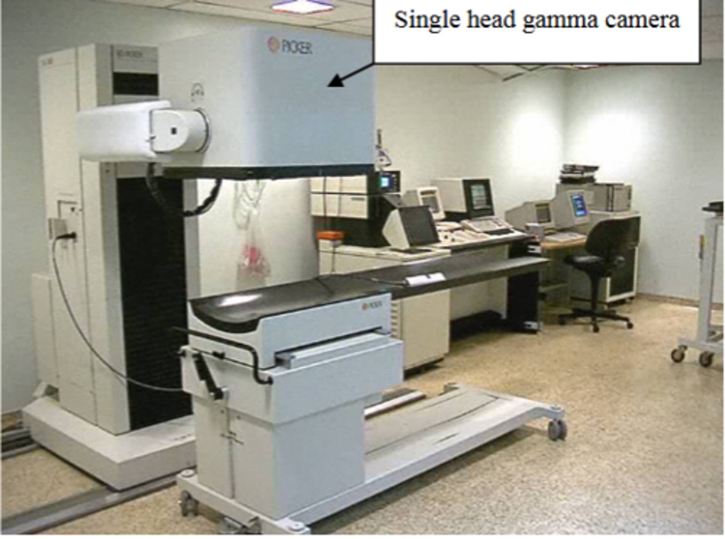
Equipment of nuclear medicine
Radiopharmaceutical, gamma camera, and computer
What is a radionuclide?
unstable atomic nucleus that decays to stability and emits gamma rays
What is a pharmaceutical?
element or compound that alters the biodistribution of the radionuclide; controls the final destination of the radionuclide within the body.
What is a radiopharmaceutical?
A combination of a radionuclide and a pharmaceutical that alters the biodistribution of the radionuclide, suitable for administration to humans.
What is the most common nuclear medicine radiopharmaceutical?
Technetium-99m (99mTc)
Why is Technetium-99m widely used in nuclear medicine?
Due to it being widely available and its favorable physical properties: a half-life of 6 hours, emits 140 keV photon energy, and does not undergo beta decay.
What types of radionuclides are used in nuclear medicine besides Technetium-99m?
Isotopes of thallium, gallium, indium, iodine, and xenon gas.
Define half-life (t 1/2).
The time it takes for the quantity of radiation to be reduced to half its original value.
What is used for bone imaging in nuclear medicine?
A phosphate derivative, which is normally metabolized by the bone.
What is the purpose of MAA (microaggregate albumin) in lung perfusion scans?
Trap radionuclides in the arterioles of the lungs for imaging.
What is the significance of gamma rays in radiopharmaceuticals?
Gamma rays are emitted during the decay of radionuclides, allowing for imaging in nuclear medicine.
What is the primary function of the gamma camera?
Image receptor in nuclear medicine that absorbs gamma radiation emitted from the patient and convert it into usable information for the computer.
From top to bottom, what makes up the gamma camera?
1. Photomultiplier tubes
2. Scintillation crystals
3. Collimator
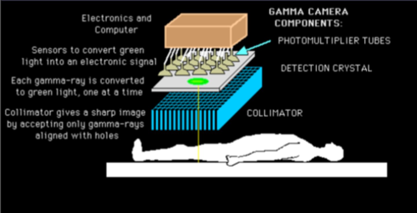
Purpose of scintillation crystals
Absorbs gamma rays emitted from patient and produces a flash of light
What material is commonly used for the scintillation crystal in gamma cameras?
Sodium iodide (NaI).
How thick and wide is the scintillation crystal typically in gamma cameras?
¼ to ½ inch thick and about 15 inches in width.
What is the function of a collimator in a gamma camera?
The lead collimator, shaped with holes, allows only photons traveling in the correct direction to interact with the scintillation crystal.
What is the role of photomultiplier tubes in scintillation imaging?
They convert light photons from the crystal into electrical signals and amplify them.
What type of information does the computer collect from the scintillation camera?
Both spatial and time information, providing quantitative information and data for image manipulation.
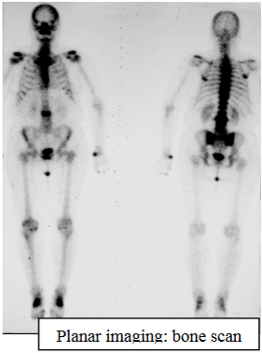
What is planar imaging?
Using a single gamma camera to absorb gamma rays from an area of interest and produce a two-dimensional image.
What does SPECT stand for?
Single Photon Emission Computed Tomography.
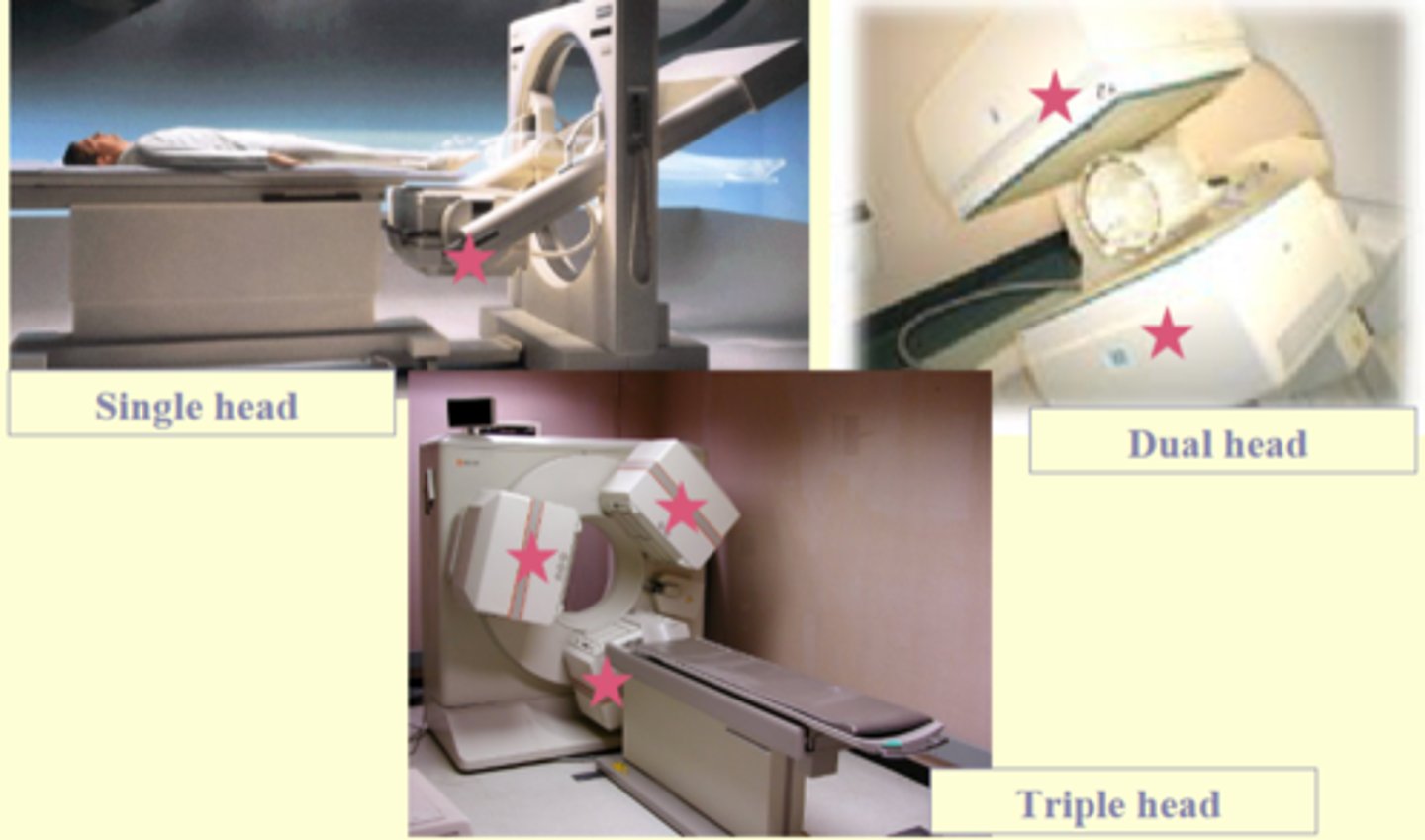
How does SPECT imaging differ from planar imaging?
SPECT creates 3D images by using a single photon that is absorbed by a rotating single head or 2/3 head gamma camera to collect data from multiple angles, producing a series of anatomical slices.
What is a common application of planar imaging?
Bone scan.
What is a common application of SPECT imaging?
Brain scan.

What types of organs and systems can be studied with nuclear medicine exams?
Essentially all organs and systems of the human body.
What are blood pool imaging agents?
Red blood cells (RBCs) combined with technetium (99mTc).
Which gas is used for lung imaging in nuclear medicine?
Xenon gas.
What can white blood cells (WBCs) combined with a radionuclide evaluate?
Inflammatory processes in the body, such as abscesses and tumors.
What are the most common types of nuclear medicine exams?
Cardiovascular studies, skeletal studies, and tumor imaging.
What conditions can nuclear medicine treat?
Thyroid disease and painful bone metastases using higher doses of radionuclide to destroy affected cells.
Who can benefit from nuclear medicine studies?
All groups of patients.
Role of the Nuclear Medicine Technologist (planar, SPECT, PET)
• Prepare, measure and administer radiopharmaceuticals in hotlab
• Adjust imaging variables
• Position patient and manipulate imaging equipment
• Perform computer processing of studies
• Dispose of radioactive waste
• Maintain records
• Practice radiation safety
• Perform quality control testing
• Provide professional patient care
What is a key advantage of nuclear medicine procedures?
Very early identification of disease progression, allowing for earlier treatment and a better prognosis.
How does the radiation exposure from nuclear medicine procedures compare to diagnostic x-rays?
NM radiation exposure is similar to x-ray exposure.
What does PET stand for in medical imaging?
Positron Emission Tomography
What type of particle do PET radionuclides emit?
A positron (a positively charged electron)
What happens when a positron collides with a free electron in PET imaging?
The two particles combine and annihilate each other, producing two 511 keV photons that travel in opposite directions.

What is the principle behind detecting annihilation events in PET imaging?
The annihilation event, observed by the detectors at the same time (coincidence), produces the image.
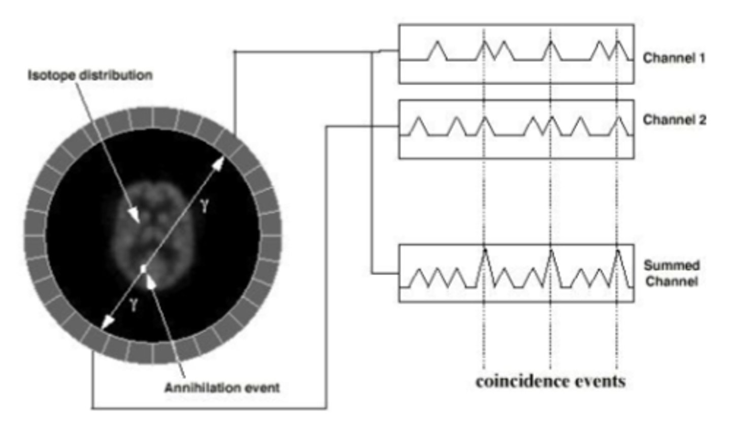
How does the patient movement affect data collection in PET imaging?
As the patient moves through the ring of detectors, more data is collected, allowing for the production of multiple image slices.
What are the most commonly used radionuclides in PET imaging?
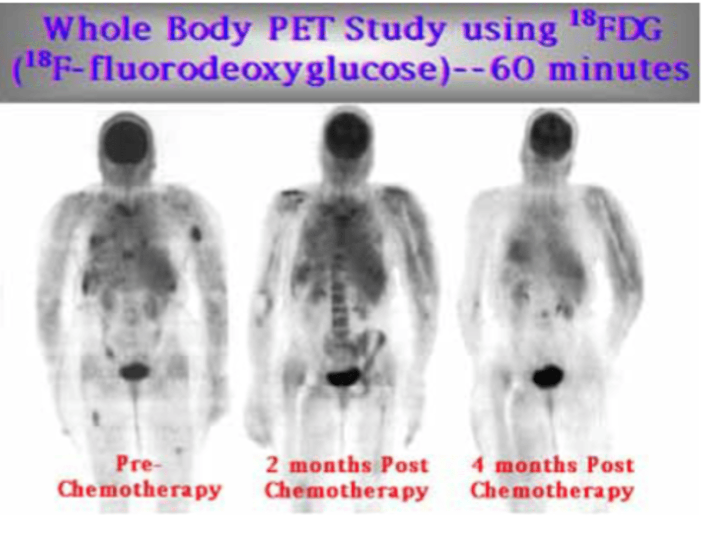
Isotopes of Oxygen, Carbon, Nitrogen, and Fluorine, which can be easily synthesized within the body.
What physiological processes can be measured and imaged using PET?
Blood flow, oxygen levels, and glucose metabolism.
What technology does PET utilize for greater resolution and quantitative accuracy?
Coincidence detection and a ring of detectors
Why are PET scanners considered expensive?
Due to the cost of the technology and the necessity of a cyclotron to produce PET radionuclides
What was the status of reimbursement for PET exams prior to recent changes?
They were considered experimental and not covered by insurance; currently, reimbursement is available for some PET scans
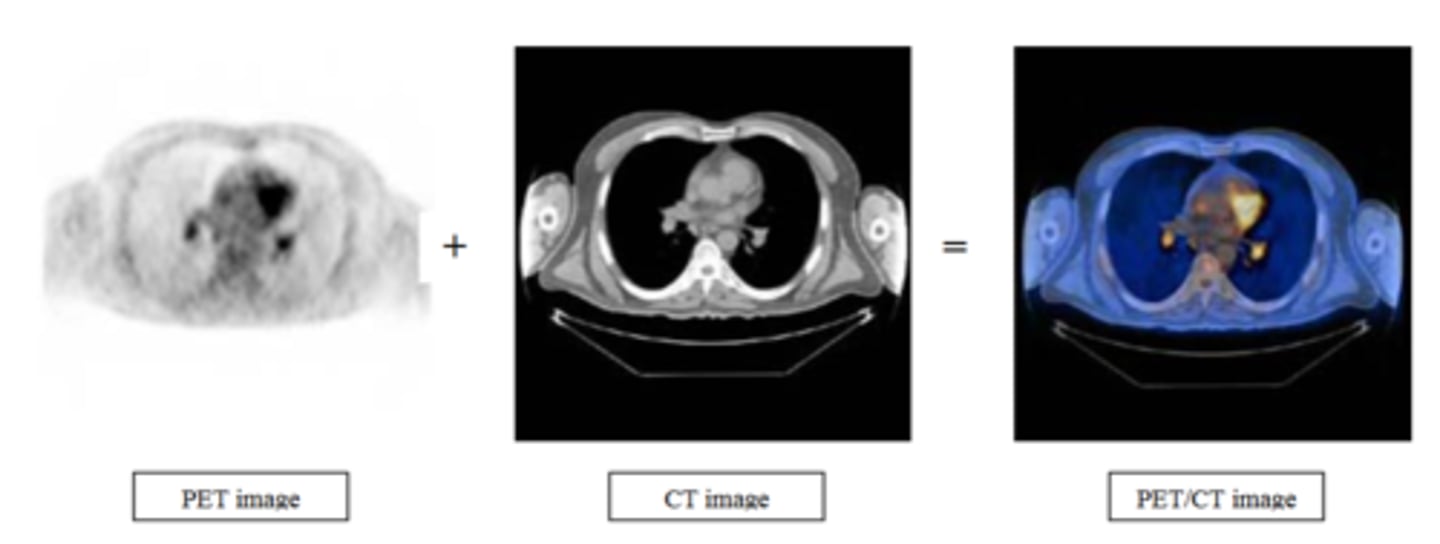
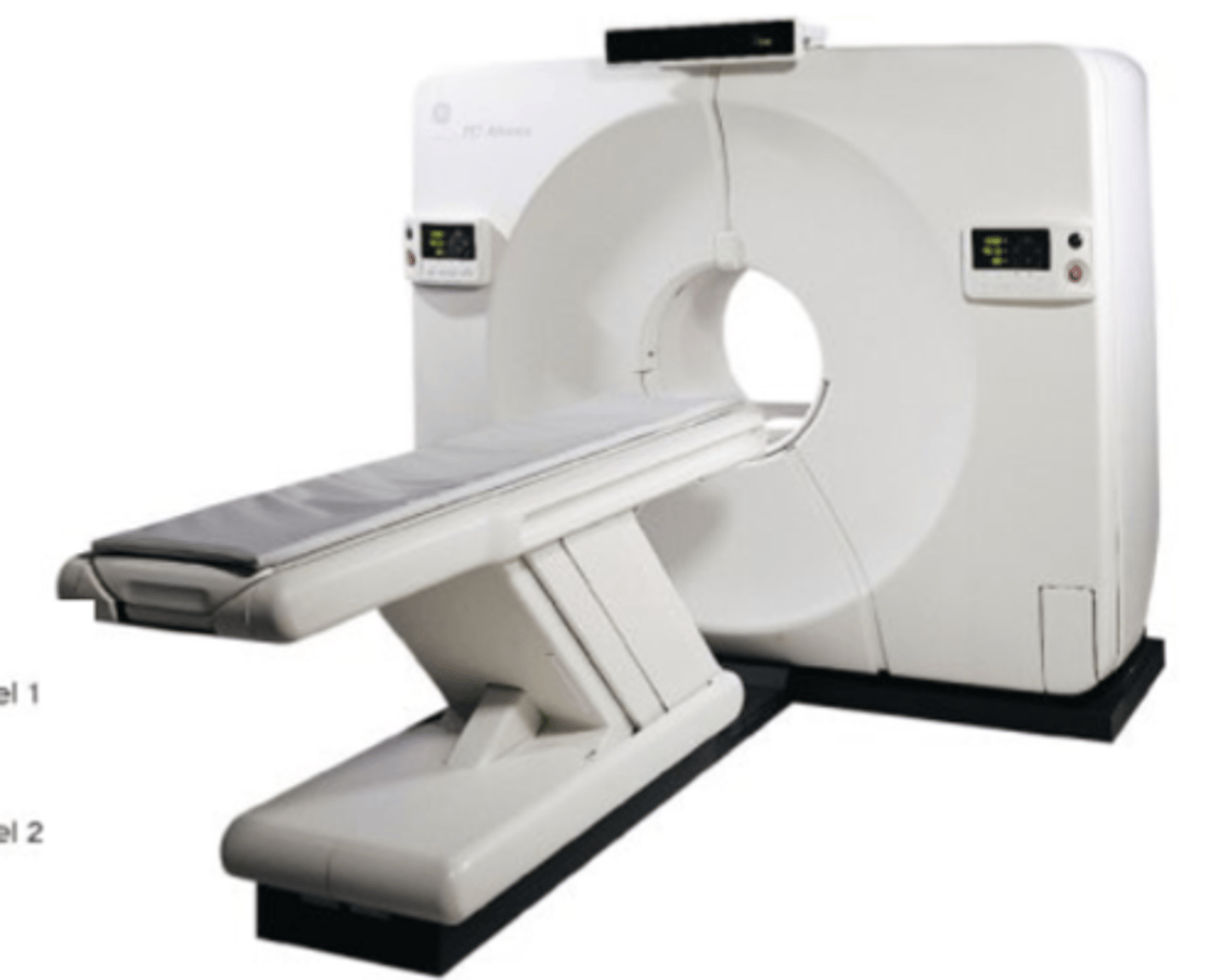
How does the combination of CT and PET imaging benefit medical imaging?
Combines CT's anatomic detail and PET's physiologic detail for high quality images