Adv EP Lesson 4 - AVNRT/AVRT/Atrial Flutter/Atrial Tachy/Atrial Fibrillation
 Call Kai
 Learn
Learn Practice Test
 Spaced Repetition
 Match
Match Flashcards
 Knowt Play
1/70

There's no tags or description
Looks like no tags are added yet.
Name | Mastery | Learn | Test | Matching | Spaced | Call with Kai | Chat |
|---|
No analytics yet
Send a link to your students to track their progress
71 Terms
AVNRT
localized in the AV node
slow antegrade pathway (alpha) with fast retrograde pathway
block in fast pathway because it is refractory
AVNRT EKG and Intracardiac ECG characterisitc
p wave not visible because they happen at the same time
short RP tachycardia, narrow QRS
Retrograde P wave after QRS or hidden within QRS
CS intracardiac electrograms appear to be “stacked”
short VA interval time
typical AVNRT
down slow pathway antegrade and up fast retrograde
slow fast AVNRT
atypical AVNRT
down fast pathway anterograde and up the slow retrograde
fast-slow AVNRT
AVNRT pacing maneuver
atrial extra stimulus; drive train of 6-8 beats at a given rate S1 followed by a premature beat (S2) and then a pause in pacing. The next drive train (S1) will be at the same pacing rate but the premature beat (S2) will be introduced 10 ms earlier than the previous S2. This series of pacing maneuvers will continue until either the atria becomes refractory or dual AV nodal physiology is identified.
dual AV nodal physiology indicator
a jump or sudden change in AH interval by greater than 50ms with only a 10ms decrease in prematurity of the S2
As and Vs on top of each other indicating AVNRT

Entrainment in AVNRT
used to rule out presence of a bypass tract
PPI - TCL > 115ms then it’s AVNRT in RV cath
SA-VA > 85ms in V1 cath

slow fast AVNRT

slow slow AVNRT

fast slow AVNRT

left variant AVNRT
triangle of koch
septal leaflet of the tricuspid valve, CS ostium and the tendon of Todaro
location of AVNRT
triangle of koch
advantage to focal cryo
allows for ablation during tachy
what to be careful of during cryo
AV block because it can occur during it with the vicinity of the cath to the AV node

which one is cryo which one is RF
A=RF
B=Cryo
RF ablation success indicators
junctional beats means ablating in the correct location
cryoablation success indicators
noninducibility or antergrade termination over the slow path
freeze thaw freeze
increaes ice formation of cryo lesion
AVNRT procedural end points
The use of Isoproterenol during post procedure testing phase may uncover dormant conduction.
Complete slow pathway elimination (non-inducibility of echo beats and A-H jump) may contribute to higher rates of long term efficacy.
Procedural end-points are usually assessed 30 min post ablation and vary by physician. Typically, end points include elimination of the slow pathway, or non-inducibility of tachycardia with max. 1 echo beat, with or without Isoproteranol administration.
During cryoablation, an AV ratio of 1:4-1:10 is acceptable T/F
F because targeted a small atrial and large ventricular signal, often between 1:4 to 1:2
WPW ECG indicator
short pr inteval <120ms
long QRS complex >110ms
delta wave
AVRT
bundle of kent is present and acts as an accessory pathway where a reentry circuit can occur

AVRT with antidromic conduction
initiated by premature contractions

label these as AVRT WPW, orthodromic, or antidromic
a WPW
b orthodromix reentrant tachy
c antidromic reentrant tachy
Orthodromic accessory pathway manifests on an ECG as a delta wave T/F
F
location of accessory pathways
right lateral, aneroseptal or mid septal, posteroseptal, or left lateral
manifest pathway
Manifests on ECG as a delta wave at baseline (pre-excitation, WPW)
May conduct both retrograde and antegrade
concealed pathway
Normal ECG at baseline (no delta wave)
Retrograde conduction only
AVRT extra stimlus pacing maneuver
ventricular extra stimulus drive train of 6 to 8 beats paced at a given rate (S1) followed by a premature beat (S2) and then a pause in pacing. The next drive train (S1) will be at the same pacing rate but the premature beat (S2) will be introduced 10 ms earlier than the previous S2. In the example below, retrograde atrial signal travels from the proximal to distal CS electrodes (right to left), displaying concentric activation pattern. There is also a V-A prolongation suggesting that the signal traversed the AV node. This would rule out the presence of an AP.
AVRT pacing maneuvers
atrial decremental pacing and ventricular extra stimilus
how to know if it is left sided accessory path/eccentric
During ventricular extra-stimulus pacing, if the retrograde atrial signal travels from the distal to proximal CS electrodes (left to right),
AVRT decremental pacing maneuver
premature atrial pacing leads to an A-H jump (conduction is occuring over slow pathway) and tachycardia is induced
In a patient with an accessory pathway, VA interval will _______ on decremental ventricular pacing.
remain unchanged
what will reveal the location of the AP
when mapping an AP, the part of the chamber that is activated first

ablation endpoint for AVRT
Delta wave disappears, return to normal ECG
Tachycardia is terminated & non-inducible
Wait period (usually 30 min)
Adenosine given to see if return of pathway occurs
atrial flutter
atria contract at high rates like 300bpm
sinus node signal propagates to AV node then ventricles but there’s a reentrant rhtyhm in the left or right atria; loop of depolarization happens on a piece of the tissue
Type 1 typical or Type 2 atypical
type 1 atrial flutter
typical
right sided
moves around tricuspid valve counterclockwise
cavotricuspid isthmus propagates signal more slowly
uses crista terminalis as the electrical barrier

type 1 atrial flutter ECG characteristics
concentric
negative p waves in inferior leads (2,3,aVf)
positive p waves in inferior leads if clockwise

type 2 atypical flutter
reentrant circuit in the right or left atrium
can be left or right sided
scar related
wavefront not travelling around the tricuspid annulus

how is the atrial flutter reentrant circuit initiated?
premature atrial contraction; electrical impulse sent out early in the atrium

atrial flutter side effects
prolonged tachy, atria not contracting properly causing blood clots, heart failure, stroke
treatment for atrial flutter
anticoagulants, beta blockers or calcium channel blockers, RF ablation of cavotricuspid isthmus
electrophysiologic prop of atrial flutter
Increase in intra-atrial conduction delay
•
No rate dependent shortening of AERP
•
Usually the faster you pace atrial tissue the faster it conducts and recovers
•
These changes increase the potential for a reentrant rhythm in the atria
what kind of tachycardia is an atrial flutter
macroreentrant atrial tachy
macroreentrant atrial tachy properties
Large circuit
Area of slow conduction and
Zone of unidirectional conduction block
Rate is seen between 220-350 beats/min
Usually requires rapid atrial pacing at 20-50% faster CL than TCL at several seconds to terminate
Atrial flutter if left unresolved can degenerate to AFIB over time
macroreentrant atrial tachy ECG properties
Sawtooth P waves in leads 2 and 3
Regular R-R intervals
Typically, 2:1 or 3:1 atrial to ventricular conduction blocks
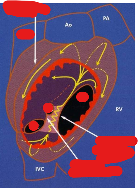
top to bottom
line of crista terminalis
TV
CS
IVC
isthmus and zone of slow conduction
eustachian valve and ridge

left to right
duodecapolar cath placed in the RA anterior to the crista terminalis
CTI line > path of ablation cath from the tricuspid valve to the IVC
ablation cath on CTI line
RV cath for vent pacing
CS cath for atrial pacing
atrial flutter on an intracardiogram
The first atrial signal is at the proximal CS catheter (not shown), followed by the HIS catheter. The atrial signal is next seen in the duo-decapolar catheter beginning in the HRA and ending in the lower right atrium, following a counterclockwise pattern. This type of typical flutter is called a counterclockwise flutter.

It is important to confirm a _______ blockfor a successful CTI ablation.
bidirectional
entrainment in atrial flutter
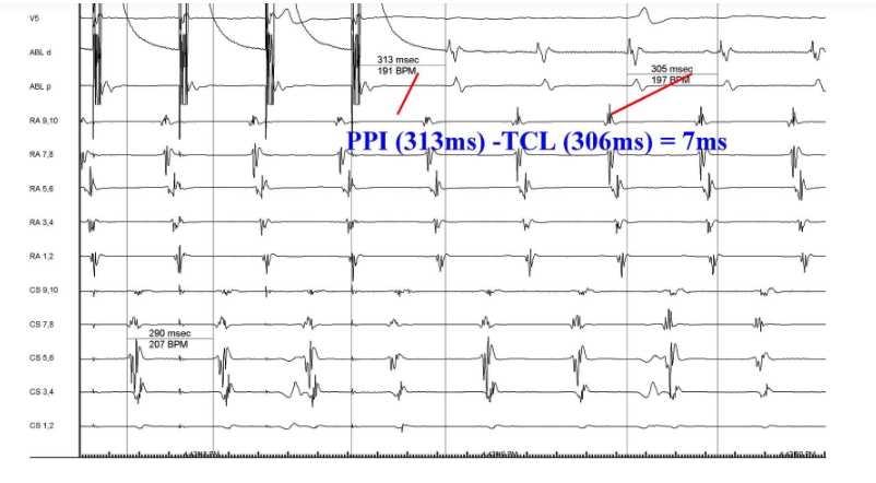
Pace at a rate 10-20 msec faster than TCL
Confirm capture
Off pacing, tachycardia must resume
If PPI – TCL < 30 msec, then we are within circuit of flutter; calculate on pacing lead
purpose for entrainment in atrial flutter
The ability to accelerate the tachycardia to the pacing cycle length without changing the tachycardia wave pattern on surface ECG.
If the tachycardia appears different or the post pacing interval PPI is longer than tachycardial cycle length during entrainment you are not within the circuit.
PPI (post pacing interval)
Once pacing is stopped, the tachycardia returns to the baseline tachycardia cycle length.
The time it takes to make one revolution around the flutter circuit is considered the PPI. If you are within the circuit, the PPI is equal to or less than 30 ms in reference to the Tachycardia Cycle Length (TCL).
right atrial non-isthmus dependent flutter (location and reentry loop type)
Lesional Right Atrial Macroreentrant Tachycardia
Upper Loop Reentry
left atrial macroreentrant tachycardia (location, circuit structures)
Mitral Annulus (Perimitral) Atrial Flutter
Circuits Involving the Pulmonary Vein(s) with or Without Left Atrial Scar
Left Septal Circuits
Lesional Right Atrial Macroreentrant Tachycardia (location and cause)
These macroreentrant circuits arise around a low-voltage area, incision, patch or scar in the lateral or posterolateral RA.
These areas usually develop after atriotomy and surgery for congenital disease. 3D activation mapping is an excellent modality to identify this type of AFL circuits.
wave morphology for left sided atrial flutter
A positive flutter deflection in the anterior precordial leads (V1, V2, V3, V4)suggest a left-side flutter
atrial tachycardia
an arrhythmia which can be caused due to both Focal and reentrant triggers
atrial tachycardia characterisitcs
Triggered, automatic, or micro-reentry
Common sites: Crista terminalis, tricuspid valve, CS Os, and pulmonary veins
P waves visible preceding QRS, but can be hidden within T wave if 1:1
P wave morphology differs from sinus morphology depending on where it’s originating from
AV block does not affect tachycardia
Can be multifocal AT with different P wave morphologies
how is atrial tachycardia induced?
atrial extra stimulus or incremental pacing
what does the VA-AV response to entrainment in atrial tachycardia mean?
The circuit is entirely within the atrium & does not involve the ventricle at all, so AV block does not affect it either.

left is sinus rhtyhm and right is atrial tachycardia
ablation strat for atrial tachy
Target earliest site of activation
Might speed up during Rf delivery & then terminate
ENDPOINT:
Termination of tachycardia
Non-inducible with pacing & drugs


basics of entrainment
patient must be in tachy to use entrainment
pace 10-20ms faster than tachycardia cycle length
stop pacing, ensure patient is still in tachy, then do PPI and TCL measurements
slow fast AVNRT morphology
P wave is usually not visible. This is due to the simultaneous activation of the atria and ventricles.
QRS morphology during AVNRT is usually the same as in NSR.
slow slo AVNRT morph
Retrograde P wave will appear inverted in the inferior leads and occur after the QRS complex. This is due to retrograde conduction
fast slow AVNRT morph
retrograde P wave is inverted in the inferior leads and will appear before the QRS complex.
CTI is bounded by
tricuspid annulus, CS, eustachian ridge, and IVC