apk2100c chapter 4 - tissues
 Call Kai
 Learn
Learn Practice Test
 Spaced Repetition
 Match
Match Flashcards
 Knowt Play
1/158

There's no tags or description
Looks like no tags are added yet.
Name | Mastery | Learn | Test | Matching | Spaced | Call with Kai | Chat |
|---|
No analytics yet
Send a link to your students to track their progress
159 Terms
Basic tissue components
Cells
Extracellular matrix
Four basic tissue types
Epithelial (ET)
Connective (CT)
Muscular (MT)
Nervous (NT)
Epithelial tissue
cover body surfaces and lines hollow organs, body cavities, duct, and form glands
Connective tissue
Protects, supports, and binds organs
stores energy as fat
provides immunity
Muscular tissue
generates physical force needed to make body structures move and generate body heat
Nervous tissue
detects changes in body and responded by generating nerve impulses/electrical signals (control)
Epithelial tissue arrangement
continuous sheets (single or multiple layers)
cells tightly packed and held together (little to no extracellular matrix)
Epithelial tissue location
Found at the boundary between two different environments
Skin (boundary between outside vs inside)
Always have free surface (one side facing free open area like cavity)
Types of Epithelium
Covering and lining
Glandular
Covering and lining (ET)
covering outer surfaces → skin
lines inner surfaces → stomach
Glandular (ET)
forms most glands of the body
sweat glands, oil glands, milk glands, pituitary gland, etc.
ex. produce sweat which goes to surface of skin
Epithelium tissue is what type of tissue?
Interface tissue: shared boundary/connection point
forms boundary between two diff areas
ET functions
Protects underlying tissue
Secretion - release products onto free surface; excretion of wastes
Absorption (Selective barrier)
Diffusion (SB)
Filtration (SB)
Sensory reception (has nerve endings)
High cellularity
ET
tons of cells
lots of cells lined up, no extracellular matrix
Specialized contacts
ET
in order for cells to line up need these to attach to one another in continual sheets
Polarity
ET
apical surface = faces the body surface, body cavity, lumen, duct
basal surface = opposite of apical surface, adheres to the basement membrane ( anchored to thin sheet of connective tissue)
Apical surface (ET)
faces the body surface, body cavity, lumen, duct
Basal surface (ET)
opposite of apical surface, adheres to the basement membrane ( anchored to thin sheet of connective tissue)
Avascular
ET
no blood supply directly into ET
No blood vessels into ET
Nervous innervation
ET
Nerve endings go into the ET
ex. sense being pinched or something cold
Regeneration
ET
high regenerative capacity
high mitotic activity
undergo cell division quickly
ex. skin paper cut heals quickly
Basal lamina
basement membrane layer
basal surface of epithelial cells will attach to directly
secreted by epithelium

Reticular lamina
deep to the basal lamina
part of underlying connective tissue

Cell junctions (ET)
Specialized contacts between epithelial cells
tight junctions
adhesive belt
desmosome
gap junction
How ET are classified
Arrangement of cell layers
Shapes of cells
Simple arrangement (ET)
One single layer of E cells

Stratified arrangement (ET)
Multiple layers (more than one)

Squamous cell shape (ET)
flat cell shape
tile like cells
nucleus bulges up while rest of cell flat

Cuboidal cell shape (ET)
square like in nature
round nuclear centrally located

Columnar cell shape (ET)
column shape
longer than it is wide
nucleus oval-like shape
nucleus more towards basal surface

How to classify when there are two cell shapes (ET)?
Look at what cell shape there is on apical surface
simple squamous ET description
single layer
flattened cells with disc shaped central nuclei and sparse cytoplasm
simplest
simple squamous ET function
Allows passage of materials by diffusion/readily
Filtration in sites where protection is not important
Produces lubricating fluid in serosae
simple squamous ET location
kidney glomeruli (site of filtration for blood to get made into urine)
air sacs of lungs (alveoli) = air needs to cross walls for o2 to get into bloodstream
lining inside of heart
ling of blood vessels = nutrient diffusion and gases
lymphatic vessels
lining of ventral body cavity (serosae)
visceral and parietal layers of serous membranes (made up of this)
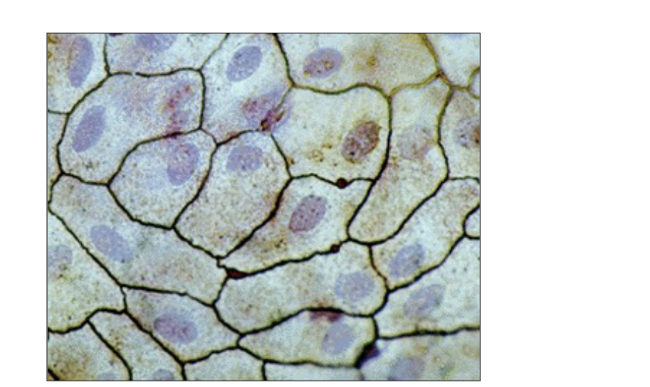
simple cuboidal ET description
single layer
cubelike cells with large spherical central nuclei
simple cuboidal ET function
thicker cells allow for
secretion
absorption
transport processes can happen
simple cuboidal ET location
kidney tubules
ducts and secretory portions of small glands
ovary surface
Lumen
hollow space in middle of cells
simple columnar ET description
single layer of tall cells
round to oval nuclei - closer to the basal surface
some cells have cilia
some cells can have microvilli
layer may contain mucus-secreting unicellular glands (goblet cells)
simple columnar ET function
absorption
secretion of mucus, enzymes, and other substances
ciliated type propels mucus (or reproductive cells) by ciliary action
simple columnar ET location
Nonciliated type
lines most of the digestive tract (stomach to anal canal)
gallbladder
excretory ducts of some glands
Ciliated variety lines
small bronchi (mucus when sick to move up to throat)
uterine/fallopian tubes (move egg)
some regions of uterus
Pseudo stratified columnar ET description
pseudo = fake
looks like multiple layers, but only one
all cells bound to the basement membrane, no multiple layers
single later of cells of differing heights
some do not reach free surface
nuclei seen at different levels
may contain mucus-secreting goblet cells and bear cilia
Pseudo stratified columnar ET function
secretion of mucus (goblet cells)
propulsion of mucus by ciliary action
Pseudo stratified columnar ET location
Nonciliated
male’s sperm-carrying ducts
ducts of large glands
Ciliated
lines trachea
most of upper respiratory tract
Stratified squamous ET description
thick membrane with several layers
basal cells are cuboidal or columnar → metabolically active
surface cells → squamous
keratinized type
surface cells are full of keratin and dead
basal cells are active in mitosis and produce cells of more superficial layers
Stratified squamous ET function
Protects underlying tissues in areas subjected to abrasion
even if top get scraped away bottom is still intact and there
Stratified squamous ET location
Nonkeratinized type
forms moist linings of the esophagus, mouth, and vagina (if layers are removed, that’s okay because there are more underneath)
places exposed to friction
Keratinized
forms epidermis of skin, a dry membrane
hardened layer of dead cells - initial dead keratinized cells
external skin

Stratified cuboidal ET description
generally two layers of cubelike cells
quite rare to find
large lumen and two rows of nuclei
Stratified cuboidal ET function
protection
layer along apical surface is cuboidal
Stratified cuboidal ET location
largest ducts of sweat glands, mammary glands, and salivary glands
Stratified columnar ET description
rare in body
multiple layers
basal cells usually cuboidal
superficial cells elongated and columnar
Stratified columnar ET function
protection
secretion
Stratified columnar ET location
rare in the body
small amounts in the male urethra (urinary system)
large ducts of some glands
Tight junction
specialized contact
lateral surface of cell (away from midline of cell)
sides of cell
interlocking junctional proteins = fuse membranes of two side by side cells
nothing can squeeze between the intracellular space
impermeable barrier/junction
Desmosomes
aka (Anchoring junctions)
formed by linker glycoproteins (cadherins)
tons of space between two cells
form internal tension-reducing network of fibers
allows for movement but not detachment
Gap junctions
channels or tunnels connecting two cells (proteins forming these channels = connexons)
communicating junctions allow ions and small molecules to pass from cell to cell
intraceullar communication
Basal lamina is part of…
basement membrane
(but can be used interchangeably)
Microvilli
found on apical surface
facing free surface
non-motile
increase surface area
microfilaments with actin
Cilia
can be found in apical surface
microtubules
move things along surface of cell (motile)
Glands
ET that make and secrete a product
Secretions are aqueous fluids that usually contain proteins → exocytosed
What can glands secrete?
Hormones
Mucous
Sweat and oils
Bile and digestive enzymes
Milk
ET that make and secrete a product form…
glands
Endocrine glands
secretion = hormones
hormones released directly into ECF and diffuse into blood stream w/o duct
effector organs (target) are far away
Exocrine glands
secretion flow onto body surfaces or into cavities (onto free surface only)
NEVER bloodstream
secretions act locally → effector organ is nearby
Multicellular = multiple cells form gland that secrete product via duct (sweat glands)
Unicellular = one-celled gland (goblet cell)
Goblet cell
unicellular exocrine gland
assoicated with columnar epithelial cells
has vesicles that has mucin (glycoprotein)
once mucin combines with water = mucus

multicellular exocrine gland
Has a duct(s) and secretory unit(s)
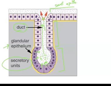
Simple tubular
multicellular exocrine gland
simple duct structure
tubular secretory structure
ex. intestinal glands

Simple branched tubular
multicellular exocrine gland
simple one duct
multiple tubular secretory structures

Compound tubular
multicellular exocrine gland
Compound duct structure (multiple)
Multiple tubular structures
ex. duodenal glands of small intestine

Simple alveolar
multicellular exocrine gland
Simple duct structure
Alveolar (circular) secretory structure = 1

Simple branched alveolar
multicellular exocrine gland
simple duct structure
multiple alveolar structures
ex. sebaceous (oil) glands

Compound alveolar
multicellular exocrine gland
Compound duct structure
Multiple alveolar secretory structure
ex. mammary glands

Compound tubuloalveolar
multicellular exocrine gland
compound duct structure/branches
multiple alveolar secretory structures
ex. salivary glands

Cutaneous membrane
Reference to skin
Mucous membranes
line open cavities and organs
tubes of respiratory, digestive, reproductive, urinary systems
Serous membranes
line closed cavities
thoracic and abdominal cavities (ventral body cavity)
Connective Tissue (CT)
most diverse and abundant type of tissue
CT main categories
CT proper
Cartilage
Bone
Blood
CT general characteristics
few cells
lots of extracellular matrix

Where do ALL CT derive or originate from?
Mesenchyme
Immature cell
-blast = to bud, sprout, produce
Retain capacity for mitosis and secrete matrix
Mature cell
-cyte
reduced capacity for cell division
most involved in matrix maintenance
doe not help with producing ground substance
Messenchyme cellular descendants
Fibroblast
Chondroblast
Osteoblast
Hematopoietic stem cell
Fibroblast
Becomes a fibrocyte
CT class: connective tissue proper
CT proper subclasses
Loose connective tissue
types: areolar, adipose, reticular
Dense connective tissue
types: regular, irregular, elastic
Chondroblast
Becomes chondrocyte
CT class: cartilage
Hyaline cartilage
fibrocartilage
elastic cartilage
Osteoblast
Becomes osteocyte
CT class: Osseous (bone)
Compact bone
Spongy (cancellous) bone
Hematopoletic stem cell
Becomes blood cells (and macrophages)
CT class: blood
CT Matrix (extracellular matrix)
contains protein fibers embedded in fluid, gel, or solid ground substance
ground substance = gel-like environment that everything is sitting in
bone tissue = ground substance gets calcified
fibers = collagen, elastic, and reticular (some CT will have more than another type of fiber)
ground substance (matrix)
material filling space between cells and fibers
fluid, semifluid, gelatinous, or calcified
functions to support and bind cells
store water
medium for exchange between blood and cells
combo of proteins and polysaccharides
fibers (matrix)
Synthesized by fibroblasts
provides strength and support for tissues
collagen, elastic, and reticular fibers
collagen fiber
tough
flexible
resists stretching
tensile strength
largest (rope like)
elastic fiber
allows for stretch and recoil
branches form networks
long, thin fibers
reticular fiber
form spongelike framework for support
smallest
special collagen fibrils (very thick)
cluster into networks
Other CT cells
fibroblasts
adipocytes (fat cells)
mast cells
white blood cells
macrophages
plasma cells
fibroblasts
secrete fibers and components of ground substance
CT
adipocytes (fat)
store triglycerides (fat) to be used later/energy
CT
mast cells
produce histamine (increase blood flow) and heparin (prevents blood clotting)
CT
white blood cells
immune response
neutrophils and esinophils
CT
macrophages
Engulf bacteria and cellular debris by phagocytosis
CT
Plasma cells
secrete antibodies
CT