Image Formation & Differential Absorption (RAD 1102)
 Call Kai
 Learn
Learn Practice Test
 Spaced Repetition
 Match
Match Flashcards
 Knowt Play
1/136

There's no tags or description
Looks like no tags are added yet.
Name | Mastery | Learn | Test | Matching | Spaced | Call with Kai | Chat |
|---|
No analytics yet
Send a link to your students to track their progress
137 Terms
What are the two primary parts of the X-ray tube?
cathode and anode

What is the cathode composed of?
filament and focusing cup
What is the filament made of?
tungsten
What is the focusing cup made of?
molybdenum
What charge is the cathode?
negative charge
What charge is the anode?
positive charge
The anode is made up of what?
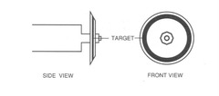
target
The target in the anode is made of what?
tungsten
The anode type that is used in everyday x-ray is known as....
rotating
thermionic emission
when filament is heated and electrons are boiled off
Heating of filament is controlled by what?
mA (milliamperes)
When mA is increased, what happens to the filament, the number of electrons boiled off, and the number of radiation?
gets hotter; increase; increase
Electrons are driven over to the anode at high speed by the ________ ________ of the negative charge placed on the ______ and a positive charge on the _________
*potential difference*; cathode; anode
When KV is increased, what happens to the energy of the x-ray, penetration, and the amount of radiation?
all will increase
quantity or exposure (intensity or output)
amount of radiation passing per second through a unit area of the surface perpendicular to the direction of the beam at the designated area
mA (milliamperes) controls what of the X-ray beam?
quantity or exposure (intensity or output)
quality
ability of the X-ray beam to penetrate matter
KV (kilovolts) control what of the X-ray beam?
quality of the X-ray beam
Direct transmission
radiation gets to the IR without interacting with tissue
Radiolucient - areas with low brightness
photoelectric interaction
*total absorption* of radiation by the body and is responsible for subject contrast

image receptor (IR)
devices that receives remaining radiation leaving the patient
differential absorption
image formation is result of this as x-ray beam passes through the body as variations of absorption and transmission of the exiting X-ray beam structurally represent the anatomic area of interest
The combination of ________ and ________ of the X-ray beam provides an image that represents the anatomic part
absorption and transmission
brightness
amount of light seen on the monitor
direct transmission
radiation gets to IR with *no interaction* of any tissues and passes through anatomic part unaffected (known as radiolucent material) which shows area with low brightness

radiolucent material
tissues that have low tissue density/opacity allowing X-ray to pass through it/easy to penetrate (i.e. air) and is shown as black/low brightness in a radiographic image
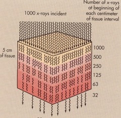
attenuation
total reduction in number of x-ray photon intensity as the photon passes through material (i.e. patient body) and occurs through absorption and scatter radiation
absorption
radiation is absorbed by the tissue through photoelectric effect and somewhat by Compton's effect
photoelectric effect
represents area with high X-ray absorption or is radio-opaque showing an increase in brightness
radio-opaque
tissues that have high density/opacity making it hard to penetrate or blocks radiation (i.e. heart and bone) and is shown as white in a radiographic image
What factors determine the probability of a photoelectric (PE) interaction?
energy of the radiation and the atomic number of the tissue
Compton interaction/scatter
*partial absorption* of radiation and gives fog on image (aka indirect transmission)

scatter radiation
some incoming photons are not totally absorbed but lose energy during the interaction and changes its original direction
The probability of scatter radiation depends on the energy of the _________ __________
incoming photon
What direction can scatter radiation occur?
any direction
Technologists experience the most occupational exposure because of...
scatter radiation
Intensity of scatter is affected by 3 things. What are those things?
- KV
- collimation (control of field size)
- patient factors such as thickness and density of tissue (i.e. fat creates more scatter than bone)
What are things that can control scatter radiation?
- grids
- beam restriction
- air gap
- compensating filters
- compression
Scatter photon ________ the quality of an image
decreases
The _______ & ______ of radiation will control the interactions that occur in the various tissues of the body
quantity (mA) & quality (KV)
What are factors that affects differential absorption?
- energy of beam
- tissue thickness
- atomic number of structure
- mass density or compactness
If there is a change in thickness of the body part, the technologist should change what?
mAS (milliamperes)
If there is a change in the atomic number or mass density (opacity) of a structure, the technologist should change what?
KVs (kilovolts)
i.e. using barium for upper GI
Tissue that has high tissue opacity will appear _____ on the radiographic image
light
Tissue that has low tissue opacity will appear _____ on the radiographic image
dark
What is the least dense tissue?
gas
What is the most dense tissue?
tooth enamel
primary photons
radiation produced in X-ray tube but occurs before the patient
Reduction of the X-ray beam occurs through which interactions?
photoelectric and Compton's/scatter
exit or image radiation
radiation which transmitted through or scattered in the body
low optical density or radiographic density
the overall degree of light or clear areas (like heart) on an X-ray image because the body's tissue blocked (attenuated) the X-rays
high optical density or radiographic density
the overall degree of black or dark areas (like lungs) on an X-ray image because a high amount of X-ray radiation passed through the body part to reach that area
high tissue density or opacity
tissue in which the cells are packed very close together (or are dense) like bone
low tissue density or opacity
tissue in which the cells are not packed close together (or are not dense) like air
Since the heart or cortex bone has high tissue density/opacity, it will have a ______ optical/radiographic density on a radiographic image
low
Since the lungs or cancellous bone has low tissue density/opacity, it will have a _______ optical/radiographic density on a radiographic image
high
Give examples of structures with a high tissue opacity and low optical/radiographic density. How does this show up on an image?
bones or heart; white
Give examples of structures with a low tissue opacity and high optical/radiographic density. How does this show up on an image?
air; black/dark
How does an image look when structures attenuate (absorb) more x-rays?
white/light
How does an image look when structures are easily penetrated?
black/dark
If tissue thickness increases, absorption will...
increase
If tissue thickness increases, attenuation will...
increase
If tissue thickness increases, transmission will...
decrease
If photoelectric effect increases, absorption will...
increase
If atomic number of tissue increases, absorption will...
increase
If atomic number of tissue increases, transmission will...
decrease
If tissue density increases, attenuation will...
increase
If mA increases, heat of filament will...
increase
If mA increases, number of electrons will...
increase
If mA increases, amount of radiation will...
increase
If kV increases, potential difference will...
increase
If kV increases, speed of electrons will...
increase
If kV increases, energy of x-rays will...
increase
If kV increases, penetration of x-rays will...
increase
If kV increases, transmission of radiation will...
increase
What is the effective atomic number of bone?
14
What is the tissue density (in g/cm³) of bone?
1.87 g/cm³
What interaction does bone have with X-rays?
photoelectric (total absorption)
What does bone look like on a radiograph?
white
What is the effective atomic number of muscle?
7.46
What is the tissue density (in g/cm³) of muscle?
1.0 g/cm³
What interactions does muscle have with X-rays?
photoelectric and Compton scatter
What does muscle look like on a radiograph?
grey
What is the effective atomic number of fat?
5.92
What is the tissue density (in g/cm³) of fat?
0.91 g/cm³
What interaction does fat have with X-rays?
Compton scatter
What does fat look like on a radiograph?
dark grey as it is porous
What is the effective atomic number of air?
7.64
Why is air's atomic number so high?
Because it is a gas
What is the tissue density (in g/cm³) of air?
0.00129 g/cm³
What interaction does air have with X-rays?
transmission (goes straight through)
What does air look like on a radiograph?
very dark/black
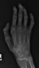
Alterations of Tissue Density
additive pathology
disease made harder to penetrate

destructive pathology
easier to penetrate i.e. osteoporosis
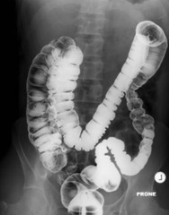
positive contrast media
barium or iodine added to see better structure better (i.e. during upper GIs)
negative contrast media
air
medical prosthesis
harder to penetrate but does not change technique i.e. hip replacements

Does additive pathology have a higher or lower atomic number/tissue density?
higher
Does additive pathology have an increase or decrease in transmission of x-rays?
decrease