OMSI- FAMM Exam 2
 Call Kai
 Learn
Learn Practice Test
 Spaced Repetition
 Match
Match Flashcards
 Knowt Play
1/175

There's no tags or description
Looks like no tags are added yet.
Name | Mastery | Learn | Test | Matching | Spaced | Call with Kai | Chat |
|---|
No analytics yet
Send a link to your students to track their progress
176 Terms
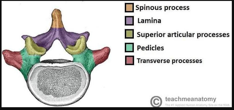
parts of the vertebra labled- top down
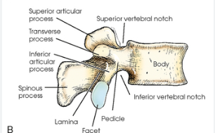
Parts of the vertebra- side view (thoracic)
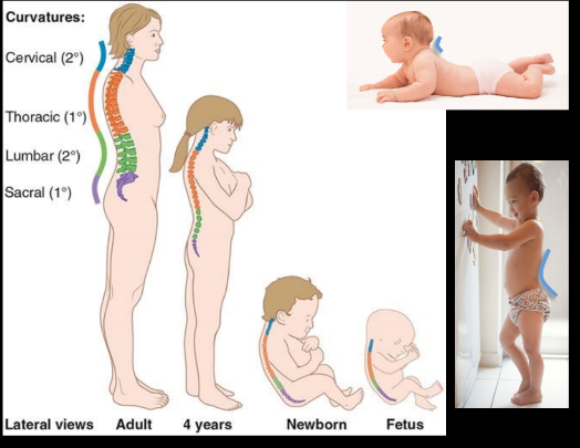
How many vertebra in each section
Cervical: 7
Throacic: 12
Lumbar: 5
Sacral: 5, fused
Coccygeal: 4, fused
A total of 33, 24 mobile and 9 immobile
Spinal curves
Primary curves- before birth
Thoracic
Sacral
Secondary curves- with mile stones, aka lordoses
Cervical- when baby lifts head
Lumbar- when toddler stands
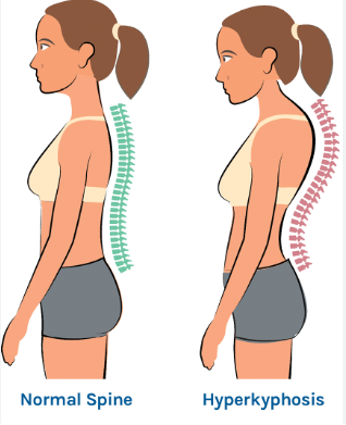
Hyperkophosis
hunchback, exagerated rounding of upper back
Hyperlordosis
swayback, exagerated inward curve of lumbar spine
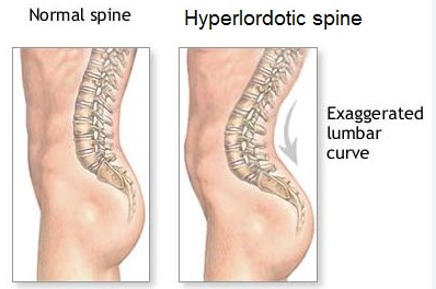
Lumbar kyphosis
flattening of the lumbar spine
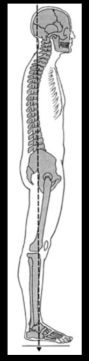
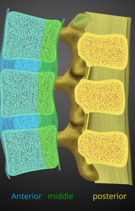
Denis Spinal Columns
A way of looking at a spinal cords section as 3 columns: Anterior, middle, and posterior
What is the precursor to the vertebra
scerotomes, on the medical sides of the somite during development
Klippel-Feil syndrome
partial or full fusion of the cervical vertebra, causes a shorted neck with little movement
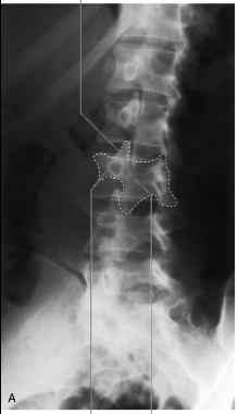
Lumbar region x-ray characteristic
The scottie dog formed by the body and posterior formations.
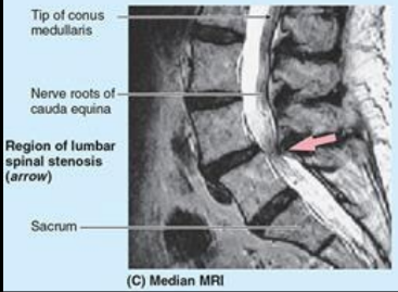
Lumbar spinal stenosis
narrowing of the spinal canal in the lumbar region, puts pressure on the nerves below it.
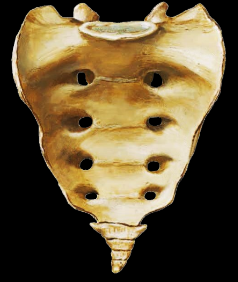
What are the anterial sacral foramen used for
sites for nerves for the glutear and lower limbs to pass through.
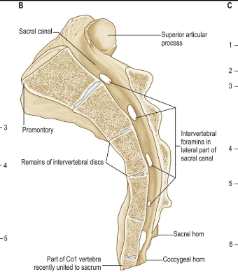
Sacral Canal
houses spinal nerve roots
*articular process of sacrum articulates with pelvis
lumbosacral angle
angle between L5 and S1
Too big: pain on lumbar back
Too small: stiffness of lower back
supernumerary ribs
rare extra rib- like protrusions on lumbar or cervical spine
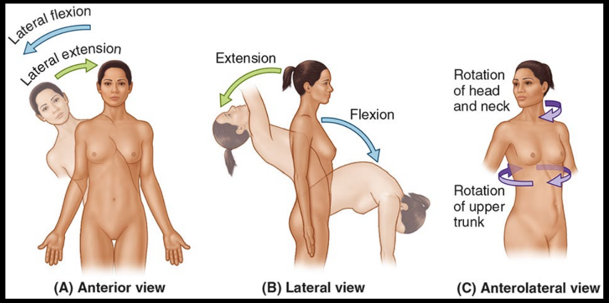
Spinal movements
herniated discs
When the soft center of the disk is pushed outward through the cord outer layer, causes radiating arm and leg pain
Motor neuron
Efferent, info from CNS to limbs
Cell body is in brain, spinal cord, or ganglion
Sensory neurons
Afferent, info goes from limbs to CNS
Cell body is located in brain or ganglion
ganglia
collection of cell bodies in the PNS
Nucleus
collection of cell bodies in CNS. Also called:
Cortical layer
body
center
tract
bundles of axons in CNS. Also called:
fasiculus
funiculus
lemiscus
peduncle
Epidural space
in the spine, a small cavity between the spinal sac and the dura mater. Contains fat, veins, and nerve roots. In all sections except sacrum, but epidurals are delivered between lumbar vertebra
connective tissue layers of a nerve
surrounding axon: endoneurium
surrounding fasicle: perineurium
Surrounding nerve: Epineurium
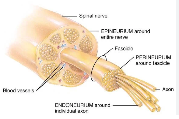
All nerves are
spinal or cranial, or derivatives of them
Spinal nerves
31 pairs
8 cervical
12 thoracic
5 lumbar
5 sacral
1 coccygeal
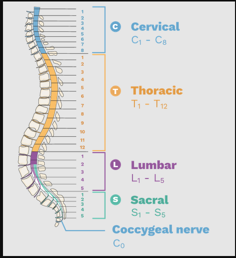
end of spinal cord
tapers off between L1 and L2, the end is called the conus medullaris. It is achored to the coccyx through the filum terminale. At this point, it turns into several branching nerves called the cauda equina
Spinal nerve structure
ventral root are motor neurons, somas in ventral horn of spinal cord
dorsal root are sensory neurons, somas in a ganglion close to the spine while fibers enter dorsal horn.
They will come together to form spinal nerve, then split into Rami
Dorsal Ramus: supplies mortor and sensory innervation to the skin/muscles of the back
ventral ramus: supplies motor and sensory innervation of the ventral and lateral skin and muscles of the trunk and limbs
How many cranial nerves are there?
12, all arise from brain accept CN XI, that originates from superior spinal cord
Which glucose form is more common
97% of glucose is cyclic, since that’s the more stable conformation. When linearized, it has a highly reactive aldehyde group.
Where is most glucose metabolized in the body?
60% in the liver
uses GLUT-2 transporters that have low affinity, a way of making sure only excess glucose is taken in
once in the liver, glucose will be phosphorylated (glucokinase) so it can’t escape the cell.
What two enzymes primarily phosphorylate glucose
Glucokinase: low affinity (high km) but high capacity (high Vmax), good for storeing excess of glucose
hexokinase: high affinity (low km) but low capacity (low Vmax), keeps cells that don’t store glucose from taking in too much
phosphofructokinase-1
catalyzes fuctose 6-P to fructose 1,6 bis
irriversable
promoted allosterically by AMP and Fructose 2,6 bisphosphate (most potent)
inhibited allosterically by ATP and citrate
Fructose 2,6 bisphosphate affect on PFK-1
F 2,6 -BP is stimulated by insulin
in turn, stimulates glycolysis and ATP formation
PEP to Pyruvate
irreversable step
stimulated by fructose 1,6 BP
pyruvate kinase
produces ATP
glycolysis energy yeild
2 ATP
2 NADH
2 pyruvate
*anerobic glycolysis also produces lactate by reducing pyruvate- regenerating the NAD+ needed for glycolysis
how is glucose stored for multi-day fasting
through gluconeogenesis- the formation of glucose molecules through building up hydrocarbons
critical steps of gluconeogenesis
Pyruvate to oxaloacetate (pyruvate carboxylase)
Oxaloacetate to PEP (PEP Carboxykinase)
F 1,6 B-P to F 6-P (Fructose 1-6 Bisphosphatase)
Glucose 6-P to Glucose (Glucose 6 phosphatase)
pyruvate carboxylase
converts pyruvate to oxaloacetate, kicking off gluconeogenesis
PEP Carboxykinase
Turns OAA to PEP
OAA is in mitochondria, must leave through malate (reduced OAA) -aspartate shuttle
removes a carbon, making a 3 carbon molecule
Fructose 1-6 Bisphosphatase
F 1,6 B-P to F 6-P
regulated by insulin(inhibits)/glucagon(promotes)
AMP inhibits
Glucose 6 phosphatase
Glucose 6-P to Glucose
needed to release glucose into blood
Glycogen in composed of
alpha 1,4 glucose linkages
branch points at alpha 1,6-glycosidic bonds (roughly every 18-12 glucose)
forms a spherical shape around progenetory protein- glycogenin (a glycosyltransferase)
Where is glycogen mainly found
liver
skeletal muscles (the most by weight)
however, unlike hepatocytes, glucose cannot be released into the bloodstream since skeletal muscle does not have glucose-6-phosphatase
at low levels: kidney, heart, brain
Glycogenesis
addition of glucose onto glycogen molecule
primarily controlled by insulin
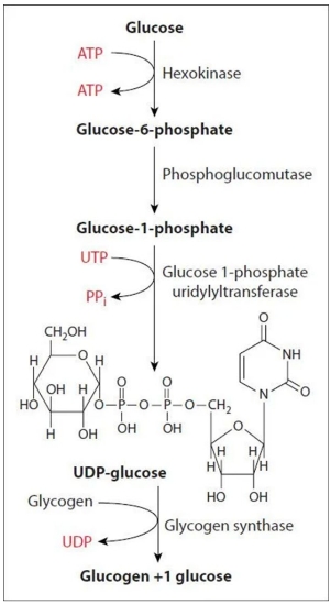
glycogenlysis
breaks glycogen down into glucose (liver) or glucose-1-phosphate (skeletal muscle)
rapidly releases glucose into body
glycogen phosporylase
removes 1 glucose from glycogen through phosphorylytic cleavage (PLP coenzyme)
debranching enzyme
removes branched glucose (alpha 1,6 bonds), then transfers most of the branch to the main chain. Then hydralizes the remaining alpha 1,6 glucose to create free glucose
phosphoglucomutase
converts released glucose-1-phosphates from glycogenlysis to glucose-6-phosphates for glycolysis and reverse
glycogen storage disease
insufficent or excessive glycogen is produced, can affect various organs.
pentose phosphate pathway
occures in cytosol
oxidative phase: generates nucleotide intermediates and NADPH
non oxidative: generates intermediates for glycolysis
what cells have a lot of PPP activity
cells that rapidly devide- nucleotide synthesis and NADPH to turn RNA to DNA
cells that have a lot of oxidative phosphorylation- NADPH as a antioxidant and cofactor to several antioxidant enzymes.
Glutathione (GSH)
a major cellular antioxidant. It’s oxidized (GSSG) form is reduced by NADPH to restore GSH afterwards.
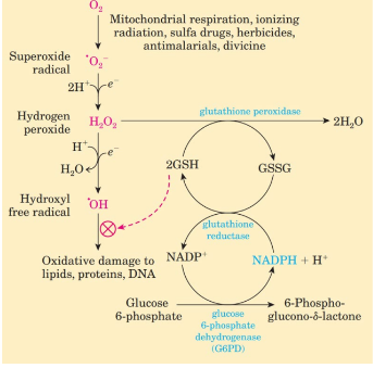
glucose-6-phosphate dehydrogenase
fist and rate limiting reaction in the Pentose phosphate pathway, it converts glucose-6-phosphate into 6-phosphogluconolactone (a short lived intermediate to ribulose) while producing NADPH.
Glucose-6-phosphate dehydrogenase deficiency
restricts the Pentose phosphate pathway, especially seen in red blood cells since their only way of replenishing NADPH is PPP. Causing hemolytic anemia (RBC are destroyed faster than made.
X-linked recessive, especially in middle Easters, tropical African, Mediterranean, and asian population.
Is exacerbated if diet is high in foods or drugs that create reactive oxygen species, such as fava beans
Lattisimus dorsi
A: Adduction, extension, Internal rotation, horizontal abduction of humerus. turnkey support, scapula depression, respiration aid.
O: posterior iliac crest to T7 spinous process
I: Intertubercular grove of humerus
N: thoracodorsal (C6-8)
Trapezius
A: covers upper back, shoulders, neck
Elevation of scapula, extension/rotation of head and neck to opposite side
elevation, upward rotation and adduction of scapula
depression, adduction, and upward rotation of scapula
O: External occipital protuberance, superior nuchal line, nuchal ligament, and spinal process C7-T12
I: lateral 1.3 clavicle, acromion and spine of scapula
N: CNXI and C 2/3-4 ventral rami
What muscles move the shoulder girdle
pectoralis minor: abduction, downward rotation, depression
Serratus anterior: Abduction, upward rotation, depression, stabilize
Trapezius: elevation, adduction, upward rotation, depression
Levator Scapulae: elevation, downward rotation
Rhomboids: adduction, downward rotation
Levator Scapulae
A:
elevate and downward rotate scapula
laterally flex head and neck to same side
extend head and neck
O: Tansverse process’ of C1-C4
I: medial border of scapula below superior angle
N: dorsal scapular nerve and C3-C4 nerve
Rhomboids (major + minor)
A: elevate, adduct, and downward rotate scapula
O:
Minor: spinal process of C7-T1
Major: spinal process of T2-T5
I: medial boarder of scapula
N: dorsal scapular nerve
Serratus posterior superior
A: elevates ribs 2-5
O: C7-T3 spinal process
I: Ribs 2-5
N: anterior Rami T2-T5
Serratus posterior inferior
A: depresses ribs 9-12
O: T11-L3
I: Ribs 9-12
N: anterior rami T9-T12
Errector Spinae family
Deep to serratus posterior, flexes spine to same side and extends vertebral column. Innervated by dorsal rami.
spinalis- connects to spinous processes to spinous processes
longissimus- connects common tendon (deep portion of thoracocolumbar fascia) and transverse processes to ribs, then transverse processes and mastoid
Illiocostalis- connects common tendon and ribs to transverse processes and ribs
Transversospinalis family
Rotates spine to opposite side, extends vertebral column. Innervated by dorsal rami
Rotatores- transverse process to spinal processes, creates a V shape
Multifidi- Sacrum and transverse processes connect to spinous processes 2-4 vertebra below
Semispinalis- transverse process’ of C4 to T12 to spinal processes 4/6 segments inferior.
semispinalis capitis: transverse processes to between superior and inferior nuchal lines
Minor deep layer
stabilize vertebra, aid in movement and respiration. innervated by dorsal rami (intertransversarii also innervated by anterior Rami.
interspinales: between spinal processes, extension and rotation
intertransversarii: between transverse presses, flexion
Levatores costarum: C7 to T11 transverse presses to the ribs 1-2 vertebra below. Elevates ribs and lateral flexion
posteiror neck muscles
splenius capitis
A: bilateral (both sides of body) and unilateral (one side of body) extension of head and neck
O: SP C7-T4 and ligamentum nuchae
I: mastoid process and superial nucchal line
N: Posterior Rami C2-3
Splenius cervicis (aka colli)
A : bilateral and unilateral extension of neck, lateral flexion and rotation on the same side
O: SP of T3-T6
I: TVPs C1-C2 or C3
N: C4-6
semispinalis capitis
most superior portion of the semispinalis that connects to occipital protrusion
A: extension of the head and neck
O: anterior protrusion of C4-7 and the transverse processes of C7 to T6
I: between nuchal lines (four curved ridges of bone on the back of the skull's occipital bone)
N: posterior Rami
suboccipital region muscles- rectus capitis posterior minor and major
A: extend head and ipsilateral (turns head to same side as agonist muscle) rotation
O: spinal process of C2 (major) and posterior tubercle of C1 (minor)
I: Inferiror nuchal lines
N: suboccipital nerve
suboccipital region muscles- obliques capitis superior and inferior
A: extend head (superior) and ipsilateral rotation (inferior)
O: spinal process of C2 (inferior) and Transverse process of C1 (superior)
I: between nuchal lines laterally (superior) and transverse process of C1 (inferior)
N: suboccipital nerve
suboccipital triangle
Composed of rectus capitis posterior major, obliquus capitis superior, and obliquus capitis inferior that protect:
vertebral artery
suboccipital nerve
suboccipital vernous plexus
Triangle of auscultation
Where vitals are taken in the back. Framed by trapezius and latrissumus dorsi on two sides
joints of the vertebral column
atlanto-occipital: between Atlas and occipital bone
atlantoaxial: between Atlas and axis, where most cervical rotation occured.
Intervertebral: cartilage joints between disks and vertebra
zygapophysial: facet joints between vertebra protrusions not connected by disk
costovertebral: joins connecting ribs and spine
IV disk structure
outer, fiberious layer for abosrbing shock and support
pulpy inner nucleus, houses spinal cord
Glycolysis steps that produce energetic molecules
For anerobic respiration, lactate dehydrogenase will reduce pyruvate into lactic acid- oxidizing NADH to NAD+ to replinish stores.
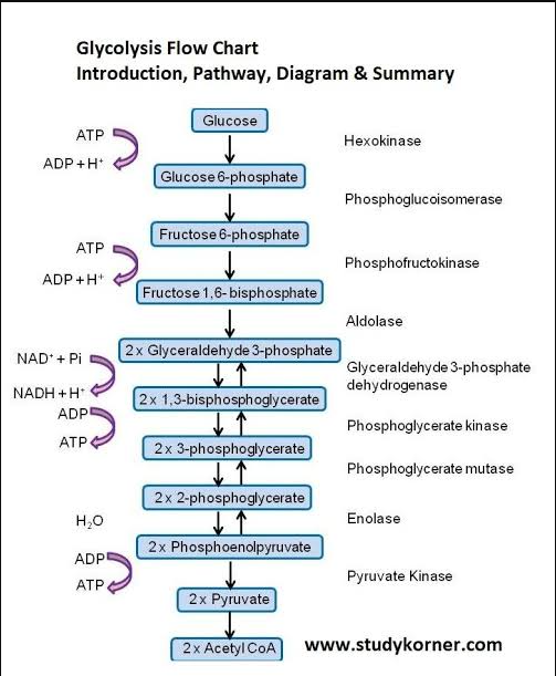
when is pyruvate converted to acetyl coa
when cell’s every level is low- Pyruvate Dehydrogenase complex (PDHC) is driven by mass action ratio
how does PDHC work
will ultimately transfer the pyruvate acetyl group to coenzyme A, forming a CO2 and a NADH.
Located in the mitochondria, so pyruvate must be trafficked inside
E1 complex has TPP as a cofactor
E2 complex has lipoic acid and coA as cofactors
E3 complex has FAD and NAD+ as cofactors to facilitate high energy electron movement
How does arsenic kill?
It binds to the thiol group in the lipoic acid (that allows it to oxidize) of PDHC, hauling conversion of pyruvate to acetyl coa
wernicke-korsakoff syndrome
a thymine deficiency, which is needed for TPP. This means pyruvate would not be converted, and if a patient with this is given glucose their cells would instead undergo anerobic respiration (which would produce acid in the brain)
therefore, any patient with extremely low blood sugar in the ER is given a dose of thymine just in case
PDHC regulator enzymes
PDH kinase inactivates E1 (Adds phosphate)
promoted by high energy molecules like ATP, Aceytl Coa, and NADH
PDH phosphatase activates E1 (removes phosphate)
promoted by high pyruvate levels
promoted by high calcium levels, since in skeletal muscle high intercellular calcium means the cross bridge cycle is activated
Citric Acid Cycle
remeber what steps produce high energy molecules. Key steps are the oxidations:
isocitrate dehydrogenase - produces a-ketoglutartate, produces NADH and CO2. stimulated by ADP and Ca, inhibited by high energy molecules
a-ketoglutartate dehydrogenase complex- produces succinyl coA, NADH and CO2. same inhibitors/promoters. Has the same cofactors as PDHC
Succinate dehydrogenase: produces Fumarate and FADH.
Malate dehydrogenase: produces oxaloacetate, generated NADH. Not significantly allosterically regulated.
Succinyl Coa to succinate produces a GTP as the thoester bond in CoA is broken
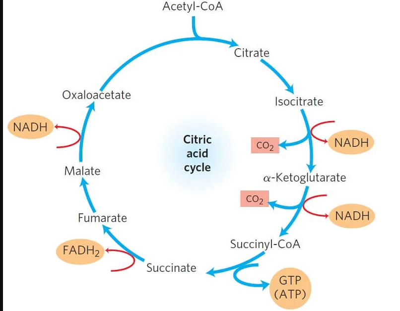
Total citric acid cycle yeilds
3 NADH
1 FADH2
1 GTP
Electron Transport Chain inhibitors
inhibit electron flow at complex 4
cyanide
carbon monoxide
inhibits ATP synthase
oligomycin (from streptomyces)
Thermoginin (UCP1 protein)
Found in brown fat of newborns, this uncoupling protein acts as a more favorable rout for protons to enter the mictochondrial matrix. As protons pass through, heat is generated.
General cell signaling steps
signaling cell
signal molecule
signal receptor
intracellular signal transcution with second messengers
cellular response
Neuroendocrine signaling
for long distance targets and cells with neuronal input. Triggers hormone release into blood
paracine signaling
short distance, works on neighboring cells
Juxtracrine signaling
cells must be in physical contact, ligand and recepotor are on cell membrane (no signal molecule)
Autocrine
cells produce a signal that affects themselves
nuclear vs membrane receptors
nuclear- steroidal signaling molecule comes into cell and find nuclear/cytosolic receptor. This will go to nucleus and effect gene transcription, slow response
cell surface receptor: ligand is hydrophlic and cannot enter cell, receptor is on outside of membrane and uses second messengers. Fast reponse
second messengers
Hydrophilic
cAMP- kinase activation (glycogen breakdown, HCL secretion, smell)
cGMP- kinase activation (smooth muscle relaxation, phototransduction)
Ca2+ - exocytosis, apoptosis, muscle contraction
IP3- Ca2+ release
Hydrophobic (membrane associated)
PIP2
PIP3
DAG- kinase activation (cell growth/metabolism, HCL, smooth muscle contraction)
Gases (diffuse across membranes)
NO
CO
Botulinum toxin
neurotoxin produced by botulism, interfears with neuromuscular junction communication, no neurotransmitter is released
loss of skeletal muscle function
blurred vision
drooping eyelids
difficulty speaking
muscle weakness
respiratory failure
GPCR
activates trimeric G protein by releasing GDP and allowing GTP to associate. Will inactivate once GTP is hydralized
Beta andrenergic receptor
muscarinic acetycholine receptor
glucagon receptor
G proteins
a type of gtpase, are on/off switches for a signal transduction.
monomeric: activated by other signaling proteins like Ras, Rab, Rho, Ran
Heterotrimeric: activated by GPCRs, membrane bound. when activated (associated to GTP) Alpha (binds GTP) and beta/gamma subuts will dissasociate and signal other molecules
Major trimeric g protein pathways
Gs- alpha subunit stimulates cAMP
Gi- inhibits cAMP
Gq- stimulates phospholipase C pathway
Protein Kinase A (PKA)
cAMP dependent protein Kinase, is regulated with a bound regulator protein.
cAMP present: regulator protein dissasociate, PKA is activated
pseudohypoparathyroidism (PHP)
a parathyroid hormone resistance, cannot regulate blood calcium and phosphorus levels. An example of a Gs and GPCR disfunction
Gs loss of function
elevated phosphorus, low calcium
short stature
short limbs (especially 4th metacarpal)
obesity
delayed development
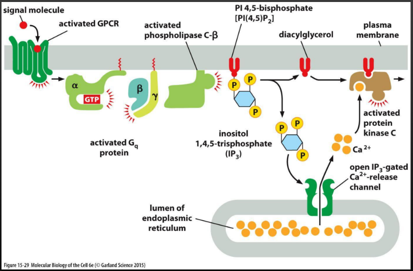
phospholipase C pathway
Gq protein activates phospholipase C, which triggers IP3 and DAG to dissasociate and signal the release of calcium, which activates protein Kinase C. Used in:
smooth muscle contraction
hormone secretion
glycogen breakdown
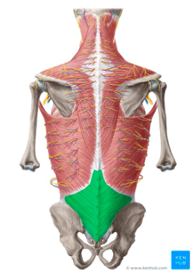
thoracocolumbar ligament
fascia covering the latissimus dorsi
ligamentum nuchae
a thick, triangular band of connective tissue at the back of the neck