Microbiology midterm
1/183
There's no tags or description
Looks like no tags are added yet.
Name | Mastery | Learn | Test | Matching | Spaced | Call with Kai |
|---|
No analytics yet
Send a link to your students to track their progress
184 Terms
Types of microorganisms
Acellular - viruses
Prokaryotes - bacteria and archaea
Eukaryotes - protists - algae, fungi, protozoans
Basic methods in microbiology
Microscopy
Cultivation
Sterility
Microorganisms in health
Pathogenic microbes - Harmful microorganisms that can cause diseases in their host organisms.
Pathobionts - Microbes that are normally harmless but can cause disease when certain conditions are met.
Symbionts - Mutually beneficial to their host, often providing advantages like improved nutrition or protection.
Microbiota - The entire community of microorganisms, including bacteria, viruses, fungi, and other microbes, residing in a specific environment, such as the human body or a particular ecosystem.
Antibiotics
Natural products of microbial metabolism - penicillin, tetracycline, chloramphenicol/chemically modified
Mostly produced by molds and a group of microbes called actinobacteria
Antibiotic resistance arises due to
Presence of natural antibiotics in the environment
Mutations and a process called horizontal gene transfer
Metagenomics
Discovering uncultivable microbes directly from the environment through next generation sequencing.
Other pharmaceuticals of microbial origin
Steroids
Prebiotics and probiotics
Therapeutetic enzymes
Bio pharmaceuticals
Vaccine production
Recombinant DNA technology
A method that combines genetic material from different sources to create new DNA sequences with desired traits or functions.
Uses of microbes in medical diagnosis
As assay organisms to determine concentrations of antibiotics, vitamins, and amino acids.
To determine mutagenic or carcinogenic activity.
Koch’s postulates
The organism must always be present in every case of the disease, but not in healthy individuals.
The organism must be isolated from a diseased individual and grown in pure culture.
The pure culture must cause the same disease when inoculated into health, susceptible individuals.
The fourth postulate was added by an American plant pathologist Erwin Frink Smith in 1905, and is stated as:
The same pathogen must be isolated from the experimentally infected individuals.
Viruses
Composed of nucleic acid and proteins.
Naked or enveloped with glycoproteins.
No organelles, no cells.
Intracellular parasites - rely on host enzymes to replicate.
Will parasite on bacteria, archaea, and eukaryotes.
Viroids
Viroids ONLY infect plants and are replicated at the expense of the host cell.
Prions
Infectious peptides
Do not replicate, instead induce a conformational change of a host protein leading to the development of disease.
Resemble brain proteins and cause neurodegenerative disease.
Prokaryotes
Single-celled organisms without proper nuclear envelope.
Ribosome as its only organelle - everything takes place in the membrane.
One circular chromosome and extrachromosomal elements (plasmids).
Haploid, reproduce asexually by binary fission.
Lack of recombination → mutations and horizontal gene transfer.
Archaea
Prokaryotic, with no nuclear membrane, but distinct biochemistry and RNA markers from bacteria.
Ancient and thrive in extreme environments - high temperatures, salinity, pH.
Fungi
— Non-photosynthetic eukaryotes
— Chitlin in cell walls
Yeasts - unicellular, reproduce by binary fission or budding
Molds - multicellular, cells known as hyphae, grow into mycelium
— Dimorphic fungi - can switch between yeast and mold lifecycle
Protozoans
— Single-celled eukaryotes
— No true photosynthesis
— Considered animals
— Some pathogenic
Methods of study
Classical methods
Microscopy
Cultivation
Aseptic techniques
Contemporary methods
Genomics
Metagenomics
Single-cell genomics
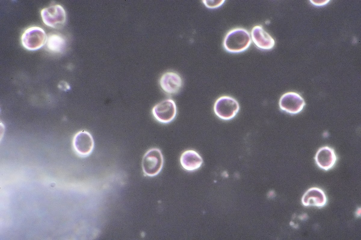
Dark-field microscopy
Type of electron microscope, excludes the unscattered beam from the image, resulting in a generally dark field around the specimen.
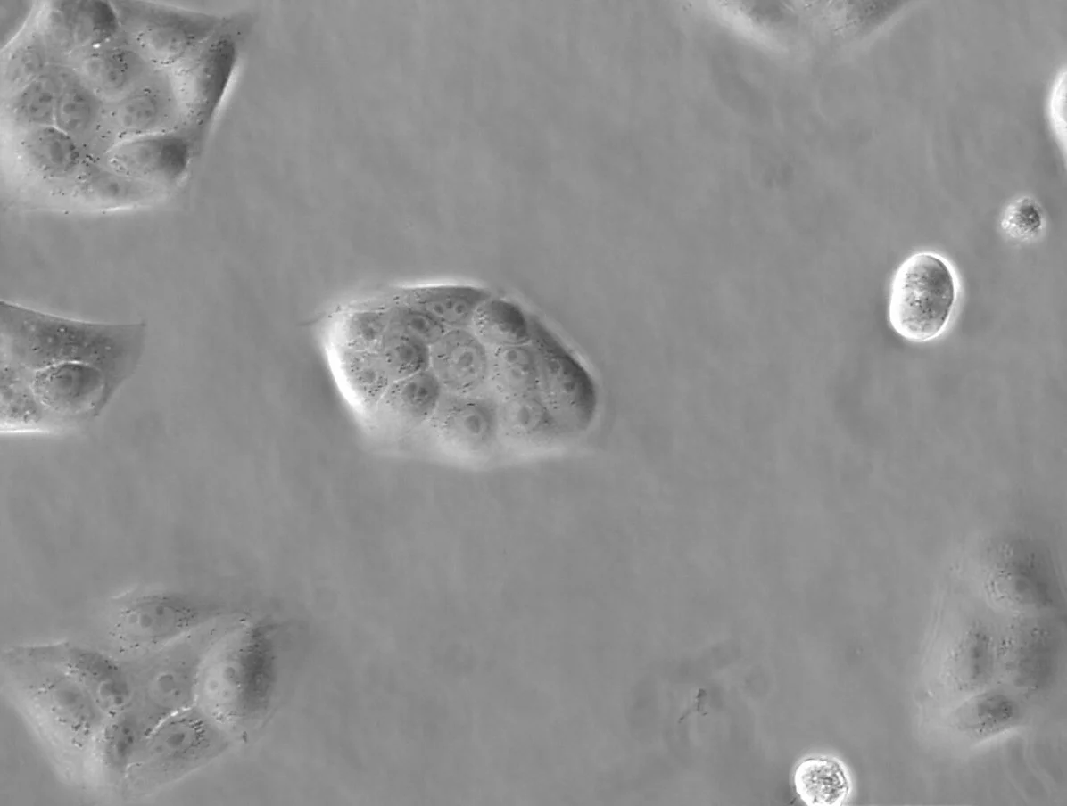
Phase-contract microscopy
Type of electron microscope, converts phase shifts in light passing through a transparent specimen to brightness changes in the image.
Pure culture
The study of colonies; large microbial populations which arose from a single initial cell.
Isolation of microbes in pure culture
Streak plate
Pour plate
Agar slants
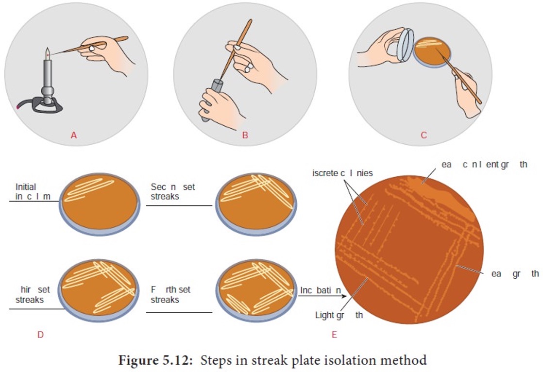
Streak plate
For the culture to be considered pure, a single colony must be transferred three times.
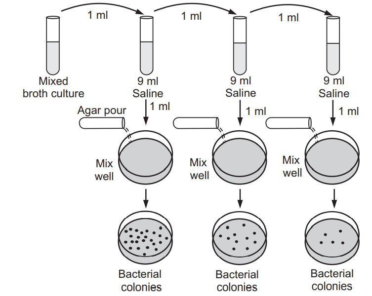
Pour plate
Microbes are serially diluted until single colonies are obtained and purified using streak plate.
Agar slants
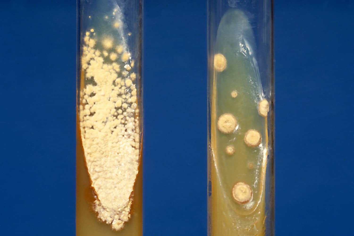
— Slanting the surface of the agar gives the bacteria a greater surface area on which to grow in a test tube.
— Slants are created in test tubes that can be capped, which minimises water loss.
— Cultures can be stored for a longer time.
Culture media nutrients necessary for growth
Sources of carbon, phosphorous, and nitrogen
Growth factors (vitamins, etc.)
Salts and minerals required in minuscule amounts
Culture media types (states of matter)
Solid media - contains agar (inedible by bacteria)
Liquid media - used to propagate large number of bacteria
Semisolid media - allows bacteria to be incorporated within the medium
Culture growth media types
Complex growth media - no exact composition (ex. Blood)
Synthetic (chemically defined) growth media - exact composition known
Selective growth media - promotes growth of certain species and represses the growth of others
Differential growth media - contains compound only certain species can eat and changes colour when digested
Most common growth media for medical use
Blood agar - complex, used to isolate general pathogens and bacteria that will eat anything
Chocolate agar - ‘cooked’ blood agar, used to release growth factors present in blood cells
Mueller-Hinton agar - standard growth medium for antibiotic testing
Saburaud-dexterose agar - standard growth medium for fungal growth
The great count plate anomaly
States that 99.9% of microbes present in the environment cannot be obtained in pure culture. Microbes that grow in culture are often not important for the community and are rare.
Cultivation independent techniques
Genomics - studies individual organisms (isolates)
Metagenomics - DNA isolated directly from the environment followed by next-generation sequencing. Allows study of individual genes (diversity) or reconstruction of complete genomes (MAGs - metagenomic assembled genomes)
Single-cell genomics - involves sorting individual cells microorganisms on a chip and sequencing the DNA present in the single cell.
Removal of microorganisms
Sterilisation - destruction of all microbes including spores
Disinfection - removal of pathogenic microorganisms but not spores
Antisepsis - a process in which bacteria is prevented from growing but not killed (ex. Iodine solution)
Methods of sterilisation
Dry heat
Moist heat
Filtration
Radiation (UV-light)
Dry heat sterilisation
Inoculation loops, needles - open flame
Laboratory glassware - hot oven (180-220C, 3h)
Moist heat sterilisation
Steam under pressure (121C, 20m)
Destroys spores
Autoclave - pressure cooker
Sterilises glassware and media
Denatures cellular proteins
Monitoring autoclaves
Physical method - measures temperature using thermometer
Chemical method - uses a heat sensitive chemical that changes colour at the right temperature and exposure time, autoclave tape.
Biological method - ensuring spore-bearing organisms are killed during sterilisation.
Filtration sterilisation
Sterilisation of fluids that would break down in heated - antibiotics, sera
Conducted by passing the liquid through a filter with pores small enough to keep the microbes on the filter
Radiation (UV-light) sterilisation
Used for enclosed areas - inoculation rooms
Used for sensitive materials
Damages DNA
High-level disinfectants
As effective as sterilisation
Glutaraldehyde, hydrogen peroxide, peracetic acid, chlorine compounds.
Disinfects surgical instruments, endoscopes
Intermediate-level disinfectants
May not be effective against spores
Alcohols, iodophors, phenolics
Disinfects body surfaces (a,i), medical devices (p)
70% alcohol more effective than 90%
Low-level disinfectants
Quaternary ammonium compounds, ex. contact solution
Which of the following chemicals may be as effective as sterilisation?
High-level disinfectants - glutaraldehyde, hydrogen peroxide, peracetic acid, and chlorine compounds
Prokaryotic cell shapes
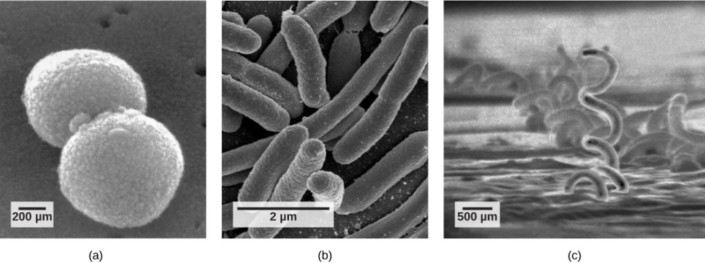
Coccus - sphere
Bacillus - rod
Spirillum - spiral
Pleomorphic - tend to change shape as they grow/combination of multiple shapes
Basic arrangements
Arise from the ability of the cell to divide across x, y, and z axes.
Diplo - two
Strepto - chain
Staphylo - cluster
Mechanisms that drive bacterial shape
Cell division and segregation
Attachment to surfaces
Passive dispersal
Active motility
The need to escape predators
The cytoplasm
Water (80%), nutrients, wastes, enzymes, gases, inorganic ions
Small (70S) ribosomes in archaea and bacteria
Dispersed throughout the cytoplasm
Features of plasmids
Extrachromosomal elements
Circulator or linear molecules capable of independent replication
Cells can increase its number of plasmids without dividing
Often contain genes that give advantage in a given environment.
Episomes - plasmids integrated into the genome
May be transferred among bacteria
Cell membrane
Selectively permeable barrier that encloses the cell
Lipid bilateral is fluid and elastic → fluid mosaic
All bacterial process occur in the membrane
Molecular structure of the cell membrane
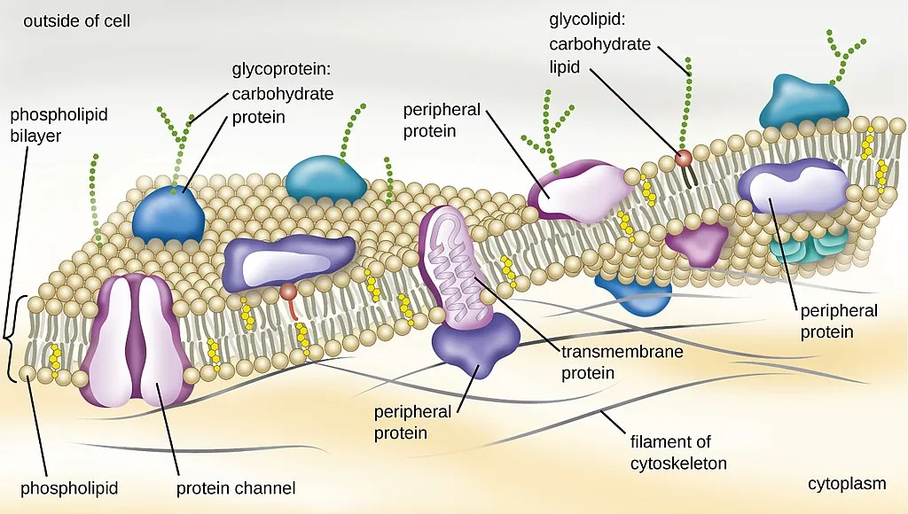
Hydrophilic polar head connected to hydrophobic fatty acid tails through glycerol linkage. Form a bilayer.
Other membrane components
Lipids - increase rigidity and flexibility
a. Sterols in eukaryotes
b. Cyclic hopanoids in prokaryotes
Proteins - 60% of the membrane
Types of membrane proteins with respect to location
Integral proteins - span the entire membrane, often act as receptors and transfer signals. Hydrophobic amino acid regions embedded in the membrane, hydrophlic regions located outside the membrane.
Peripheral proteins - No hydrophobic regions, hydrophilic amino acids on the surface prevent them from being sucked into the membrane. Can detach/reattach in response to a signal.
Main membrane functions
Permeability barrier
Protein anchor
Energy conservation
Membrane as a permeability barrier
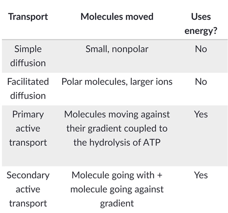
Strict control of transport is orchestrated by carrier proteins. Water and lipid-soluble small compounds are the only compounds that passes freely.
Simple diffusion
Facilitated diffusion
Primary active transport
Secondary active transport
Membrane as a protein anchor
Receptors relay signal between the cell’s exterior and interior
Enzymes have many activities
Adhesion molecules identify and interact with other cells
Transport proteins move molecules and ions across the membrane
Importance of carrier proteins
Uptake against the concentration gradient is necessary and mediated by carrier proteins.
The classes of membrane-transporting systems
Simple transporters - transport substances without chemical modification
Group translocation - requires phosphorylation of the transported substance
ABC system - couples the energy of ATP binding and hydrolysis to substance transport
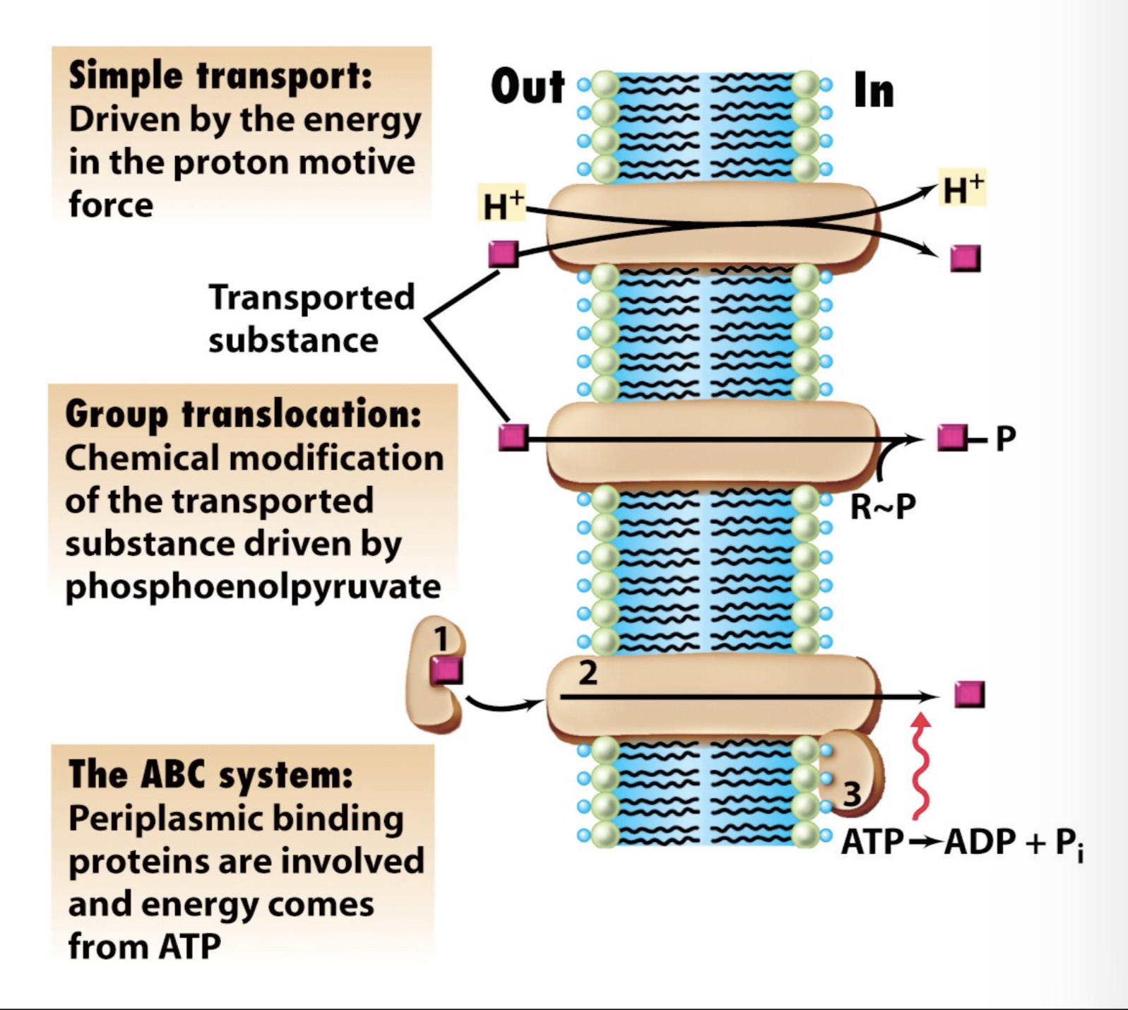
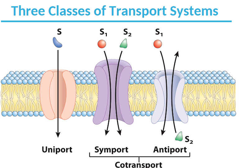
Types of transport events
Uniport - Unidirectional transport of a molecule
Symport - Transport of a substance along with another substance, frequently a proton
Antiport - Transport of two substances in opposite directions
Membrane potential
A difference in electric potential between the interior and the exterior of a biological cell, allows formation of proton motive force.
Proton motive force consists of
Ion transporters - actively pushes ions across the membrane and establishes concentration gradient, acts like a battery.
Ion channels - allows ions to move across the membrane down the concentration gradient, acts like a resistor.
Cell wall
The outermost boundary of bacterial cells, very important to prokaryotes.
Turgor pressure (balances the osmotic pressure difference between the interior and exterior): 2 atm in gram-negative cells.
Prokaryotes without cell wall: either intracellular parasites or posses a unique membrane (lipoglycan)
Unique structure:
One of the few bidimesional polymers in nature
Just one molecule can make it a rigid container.
Cell wall functions
Prevents osmotic lysis
Maintains cellular shape
Provides sufficient stability, but elastic enough to allow growt
Enables communication with the environment
Enables cellular of septum during cellular division
Permeability barrier (in gram-negative cells)
Provides motility (anchors flagella)
Complex peptidoglycan structure
Polymer backbone
Alternating residues of sugars NAG and NAM
NAG - N-acetyl-glucosamine
NAM - N-acetyl-muramic acid
A four-aminoacid peptide (both D- and L-aa) connected to carboxyl group of NAM
Many bacteria substitute diaminopimelic acid at third position with another (L-Lys)
Peptidoglycan synthesis
Inside - Soluble substrates are activated and peptidoglycan units are built
Membrane - Activated units are attached and assembled on the undecaprenol phosphate membrane pivot
Outside - The peptidoglycan units are attached to, and cross-linked into, the peptidoglycan polysaccharide
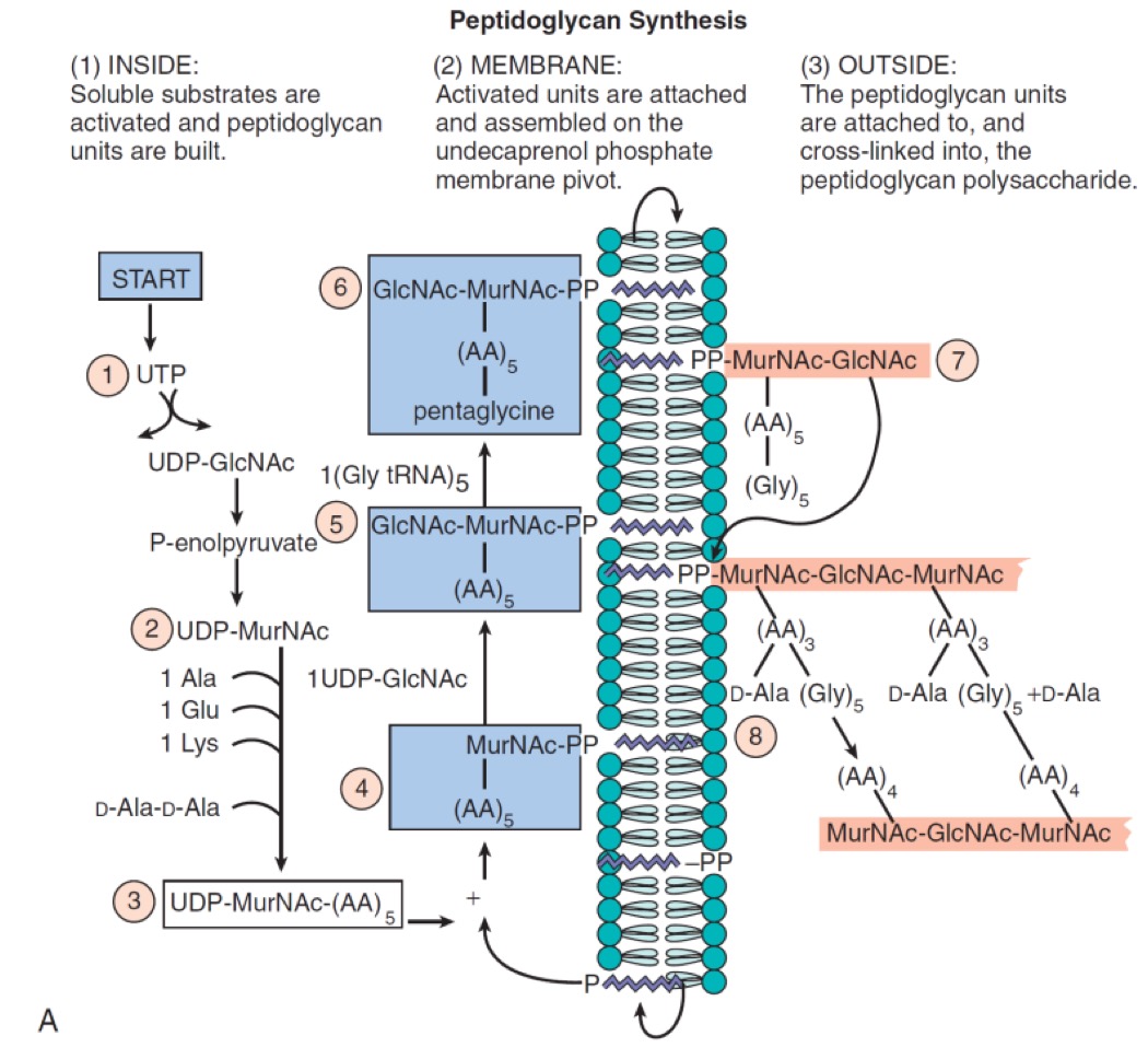
Problems and solutions to peptidoglycan synthesis
Fairly large precursors must be delivered across the membrane. The synthesis must take part on the outside of the membrane, where there is no ATP
Construct energetically rich units of peptidoglycane. Park nucleotide → UDP-N-acetylmuramyl pentapeptide. (3)
Use the conveyer belt (4-7)
- Undecaprenol (bactoprenol) is a membrane lipid carrier. Attach Park nucleotide to it, and it becomes a lipid.
- Assemble the unit on the conveyor belt.
- Flip it to the other side.
Insert into existing peptidogylcan (8)
- Use transglucosylases to insert and link new monomers into the breaks in peptidoglycan.
- Use transpeptidases to cross-link the peptidoglycan.
- Both reactions transfer bond energy, without the need for ATP.
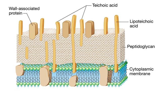
Gram-positive cell wall
Thick, multilayered, mainly peptidogylcan.
Trichroic acid - partially penetrates peptidoglycan
Lipoteichoic acid - lipid attached to it to anchor itself into the cytoplasmic membrane
Trichroic and lipoteichoic acids regulate autolytic cell-wall enzymes → maintain shape
Exoenzymes
Break down complex nutrients on the outside of gram-positive cell walls allowing them to diffuse through.
Gram-negative cell wall
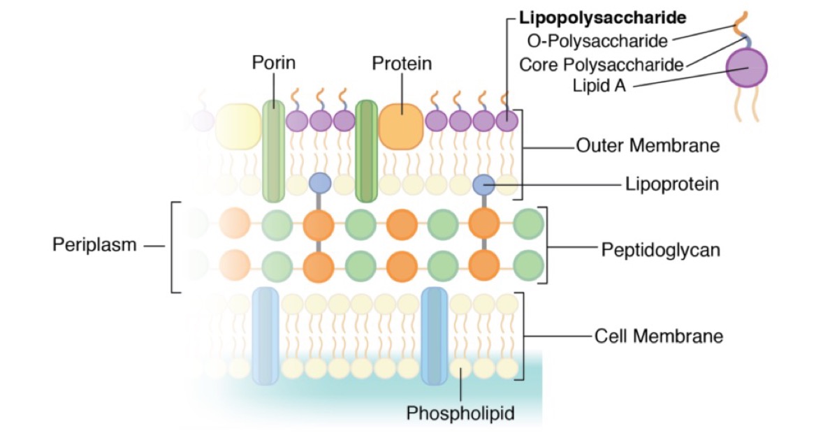
Peptidoglycan is thin
Embedded in periplasm
Contains an outer membrane
Gram-negative outer membrane
Inside - phospholipids
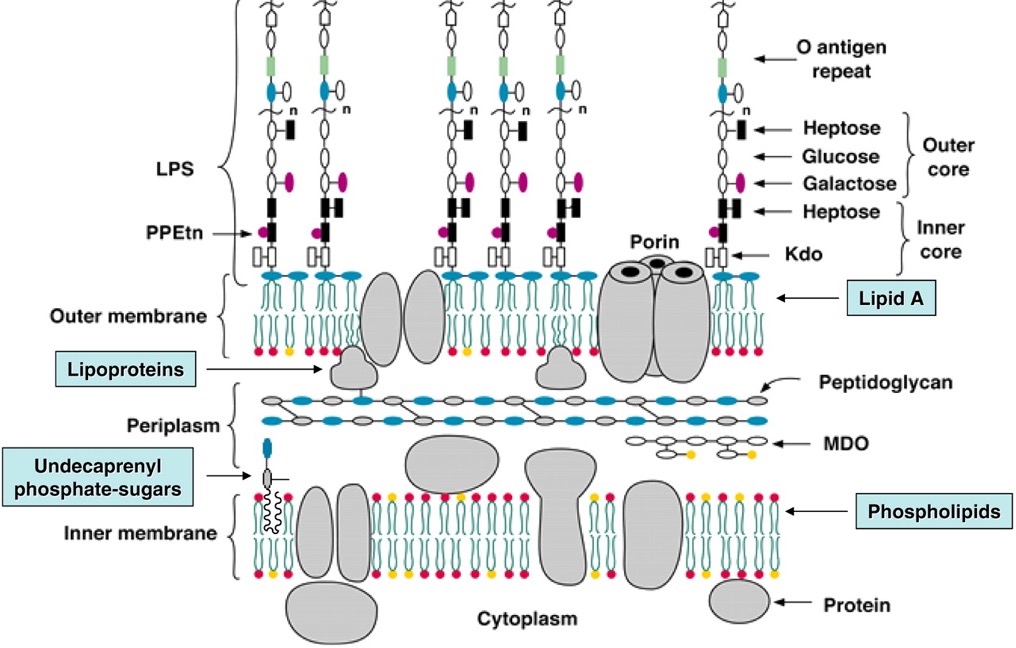
Outside- lipopolysaccharide LPS, attached to the peptidoglycan by lipoprotein
Lipopolysaccharide (LPS, endotoxin)
Contributes to the structural integrity of the cell
Protects the membrane from certain chemical attacks
Increases the negative membrane charge
Plays a role in surface adhesion, phage sensitivity.
Components of lipopolysaccharide
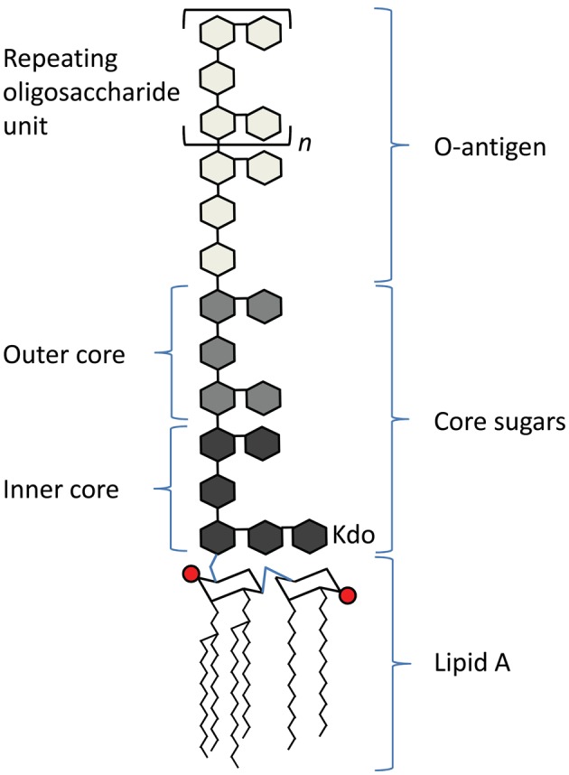
Lipid A
Inner core
Outer core
O-antigen
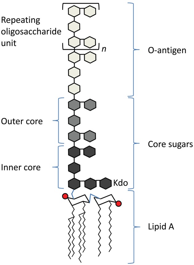
Lipid A
Toxic component of lipopolysaccharide
Responsible for endotoxin activity (arouses immune system)
Unique to gram-negative bacteria!!
Core polysaccharide
Branched polysaccharide of 9 to 12 sugars.
Essential for structure and viability.
Contains an unusual sugar, 2-keto-3-deoxy-octanoate (KDO) and is phosphorylated.
O-antigen
Long, linear polysaccharide attached to the core.
Species-specific - certain e.coli are nontoxic, other strains are and differ in the sequence of the o-antigen.
Nutrient transport in gram-negative bacteria
No exoenzymes
Periplasm contains transport systems for ions, protein, and sugars
These are degraded by periplasmatic enzymes
Porins traverse entire cell wall an allow diffusion of small hydrophobic molecules.
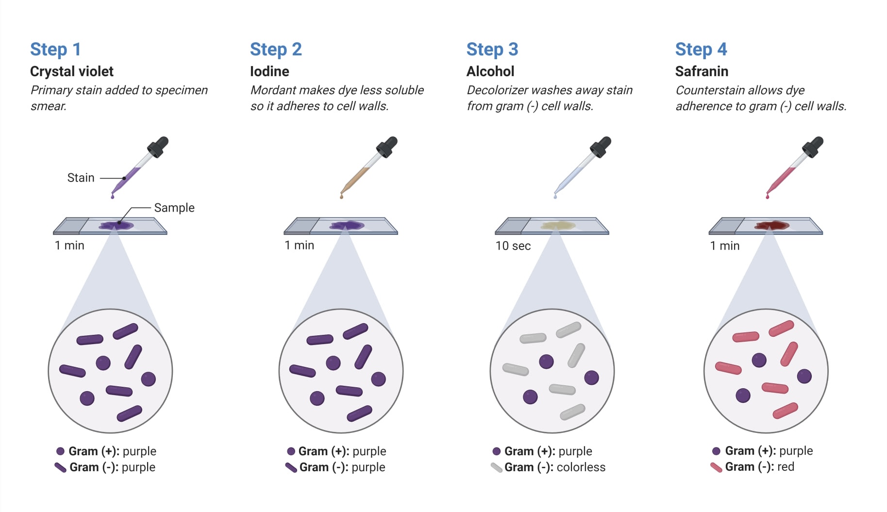
Gram stain
Alcohol decolorises gram-negative cell walls because of its lacks of peptidoglycan.
Gram-positive cells have thick cell walls and too much dye that cannot be removed easily.
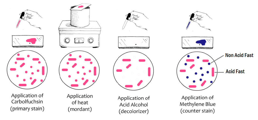
The Ziehl-Neelson/acid-fast stain
Used to stain mycobacteria (tuberculosis, leprosy), which have thick and waxy cell wall and will not stain using gram.
Alternative cell wall structures
Mycobacteria (tubercolosis)
- Peptidoglycan intertwined with arabinogalatan
- Surrounded by wax-like lipid Croat of mycolic acids
- Only stained using acid-fast stain
Mycoplasma (‘walking’ pneumonia, phylum Tenericutes)
- Have no cell wall, but incorporate host steroids into membrane to achieve rigidity
- The membrane is three-layered
- Evolved from gram-positive bacteria that lost their cell wall
Thermoplasma
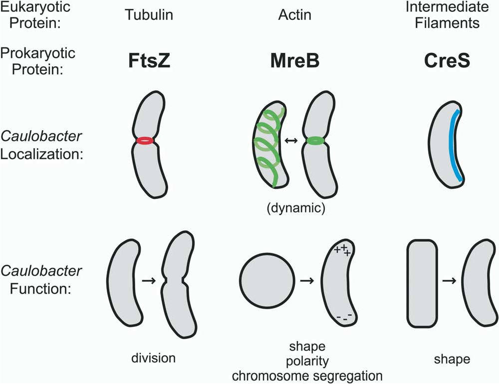
Bacterial cytoskeleton proteins
FTsZ - homologous to tubulin - forms a ring during cell division - formation of septum
MreB - homologous to actin - helical - determine rod shape
Crescentin - homologous to lamin and keratin - bends the cell
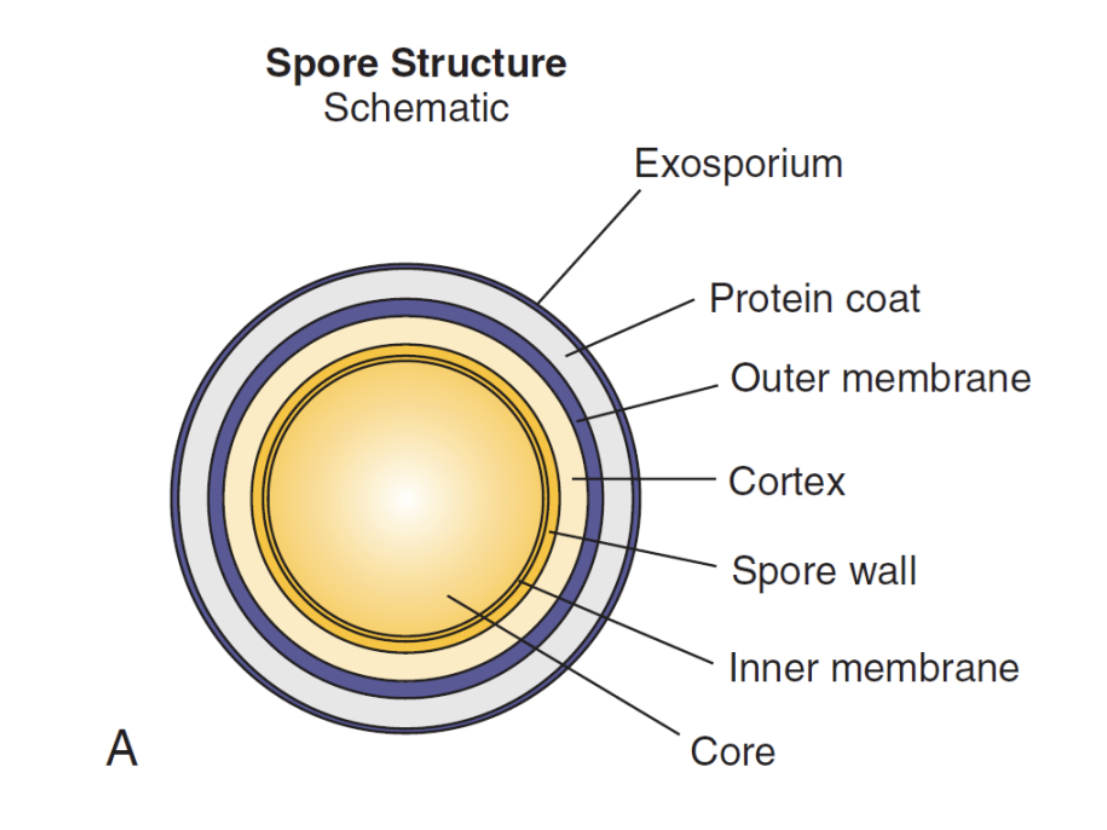
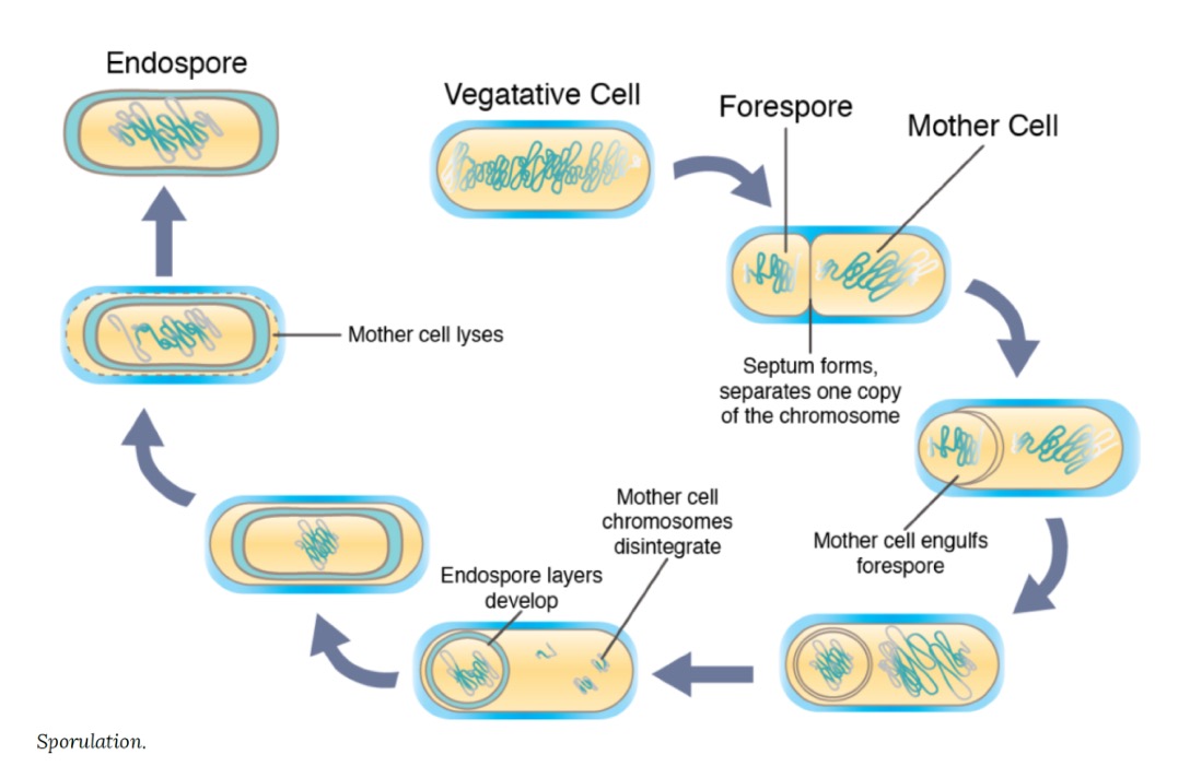
Endospores
Found only in Clostridium and Bacillus*, gram-positive bacteria
Produced under hostile conditions from vegetative cells
Extremely resistant to environmental conditions
High concentration of calcium bound to dipicolinic acid → increased DNA stabilisation
Outer coat made of keratin-like proteins protects the spore.
Formation of endospore
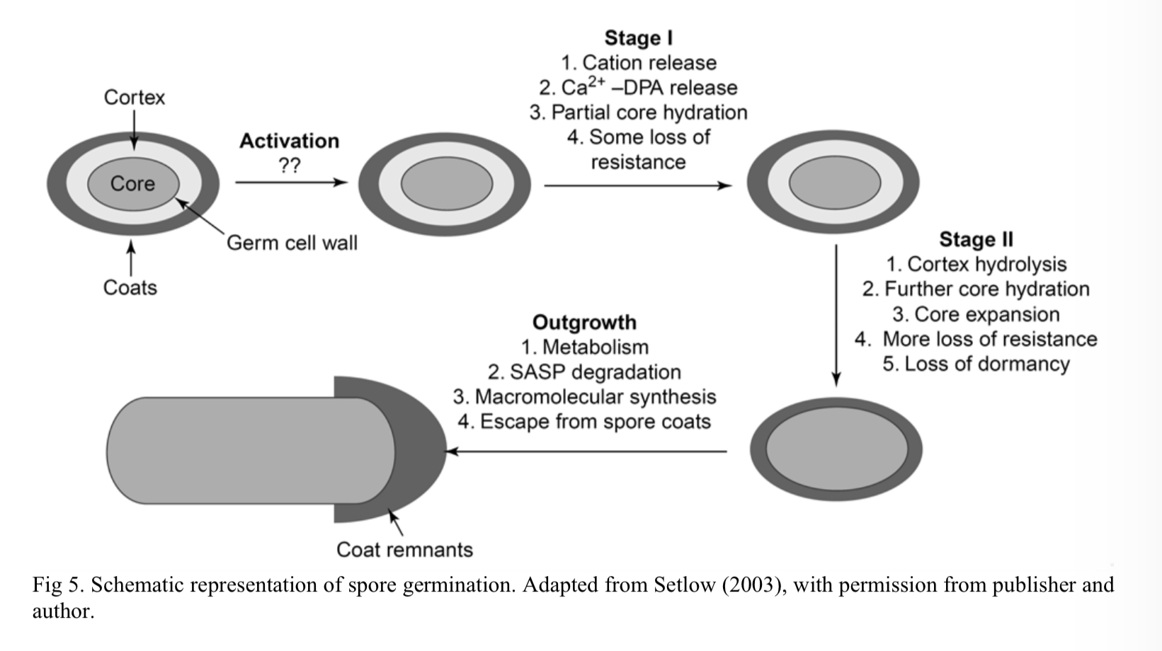
Germination of endospore
Triggered by an environmental change which initiates gene expression, completed in 6 hours.
Activation
Germination
Overgrowth
Surface structure of bacteria
Capsule
Slime layer - loosely packed polysaccharide layer, easily removed from the cell
S-layer - composed of proteins or glycoproteins, provides rigidity, cell shape, protection from environmental changes and predators
Capsule
Tightly backed polysaccharide or protein layer
Protects the bacterium from host immune response
Sticky, promotes adhesion to host surfaces
Promotes formation of biofilm - bacteria embedded in polysaccharide are protected from antibiotics and host defence.
Structures outside of the cell wall
Fimbriae
- Numerous extensions composed of protein pilin
- Help bacteria attach to surfaces and hosts
Pili
- Longer than fimbriae
- Conjugative pili participate in DNA exchange (sex)
- Type IV pili enable twitching motility
Bacterial flagellum
Turns clockwise or counterclockwise allowing bacteria to swim.
Hollow tube is made up of flagellin protein.
The hook allows its axis to point away from the cell.
A shaft runs between the hook and the basal body passing through a series of rings that embed it in the membrane and connect it with the motor.
The motor is powered by proton energy, H+ or Na+ flux.
Chemotaxis
Movement towards the attractant or away from the repellent using the flagellum
Orchestrated by bacterial chemoreceptors
Attachment of the molecule phosphorylates/methylates the receptor → activates the protein pathway.
Movement becomes biased towards counter clockwise rotation
Other types of locomotion in bacteria
Corkscrew motility - endoflagella put torsion on the entire cells
Gliding - does not require flagella, observed on surfaces, provided by surface proteins or slime
Biofilm formation
In a biofilm, bacterial cells are embedded in extracellular polysaccharide, making them particularly resistant to environmental conditions.
Pioneer cells attach to the surface through designs, fimbriae of extracellular polysaccharides.
Other cells are attracted to the biofilm.
Cells are kept at distance by polysaccharide molecules with small water channels allowing exchange of water and nutrients.
Protected from desiccation, antibiotics, immune system.
Bacterial classification
Genus name (capitalised) - species name (never capitalised)
Species are designated by biochemical and other phenotypic criteria and by DNA relatedness
Strains are category below species level and are classified below by serotyping, enzyme typing, identification of virulence factors, characterisation of plasmids, protein patters, or nucleic acids.
Identifying a microorganism
Colony morphology and staining
Metabolic examination
Use of bacterial viruses - bacteriophages
Use of serology - antibody - antigen reactions
Genetic differentiation - DNA hybridisation, PCR
Bacterial replication and division cycle
Occurs through binary fission - splitting into two cells.
Daughter cells must be capable of life
Have chromosomes
Have mRNAs, tRNAs, ribosomes, and cytochromes
Cellular division of gram-negative bacteria
Gram-negative cells have an outer membrane and divide by constriction, followed by membrane fusion.
The ring separating the cells is formed by a FtsZ protein.
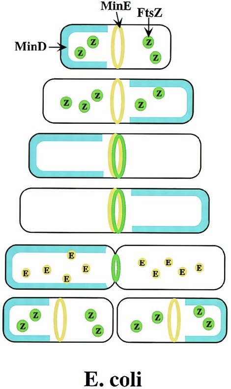
Cellular division of gram-positive bacteria
Gram-positive cells have a thick cell wall and must develop a cross-wall in order to divide.
In addition to FtsZ ring, the cross wall is made out of DivIVA protein.
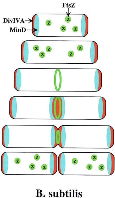
Timeline of E. coli cellular division
Replication: 45m
Constriction and cell wall formation: 15m
Cells can initiate several rounds of chromosomes replication and can segregate partially replicated chromosomes into new cells - can sometimes divide every 20 minutes with the right food and aeration.
Generation time
The interval between two cell divisions, the number of cells increases exponentially.
Batch culture
A closed-system microbial culture of fixed volume.
Four phases of bacterial growth curve
Lag phase
Exponential/logarithmic phase
Stationary phase - number of bacteria remains the same.
Death phase - no more nutrients present, some cells remain viable because they can cannibalise the others.
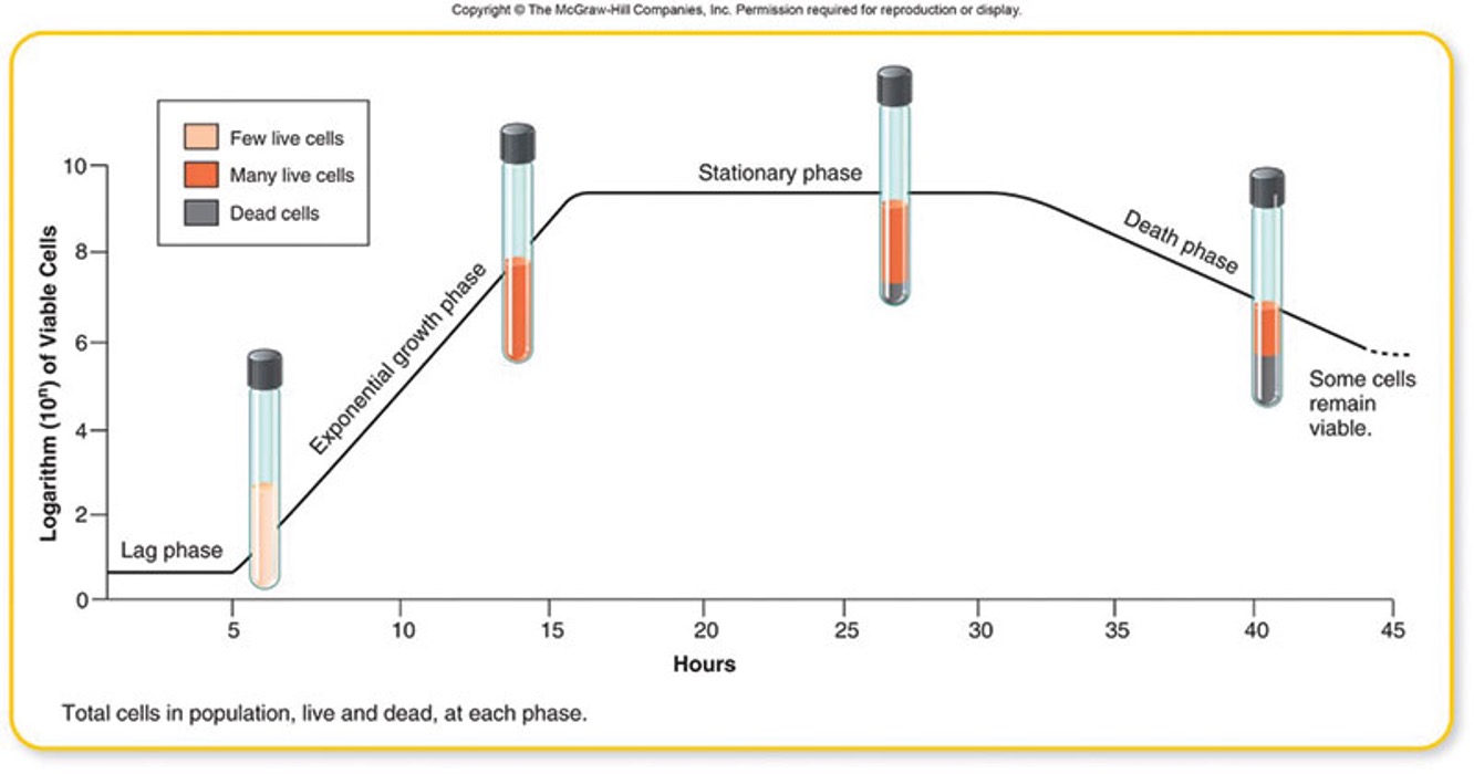
Open culture
Resembles environments of pathogens; constant supply of nutrients and removal of waste.
Physical factors that influence growth
Temperature
pH
Water activity/solutes
Availability of oxygen
Availability of nutrients
Physical factors that influence growth - Temperature
Pathogens grow at the temperature of its host.
- human pathogens - 37C
- avian (bird) pathogens - 42C
Optimal growth is often closer to maximum temperature than minimal temperature