ANAT 5010 - unit 6
1/72
There's no tags or description
Looks like no tags are added yet.
Name | Mastery | Learn | Test | Matching | Spaced | Call with Kai | Chat |
|---|
No analytics yet
Send a link to your students to track their progress
73 Terms
cubital fossa
a transition area between the arm and the forearm; a triangular depression on the anterior aspect of the elbow
- base: line between epicondyles of the humerus
- medial: pronator teres muscle
- lateral: brachioradialis muscle
- floor: brachialis muscle
- roof: deep fascia
what are the boundaries of the cubital fossa?
- tendons of biceps brachii
- arteries (brachial, radial, and ulnar)
- median nerve
what are the contents of the cubital fossa?
superficial branch of the radial nerve
descends under the brachioradialis muscle; innervates the dorsal-radial aspect of the hand and first 3.5 digits EXCEPT the DIP joints; sensory innervation
deep branch of the radial nerve
runs between the two heads of the supinator muscle to innervate the muscles in the posterior compartment of the forearm; motor innervation
styloid processes of radius and ulan
two palpable bony projections at the wrist; stabilize the carpal bones and anchor ligaments
distal skin crease
proximal border of flexor retinaculum
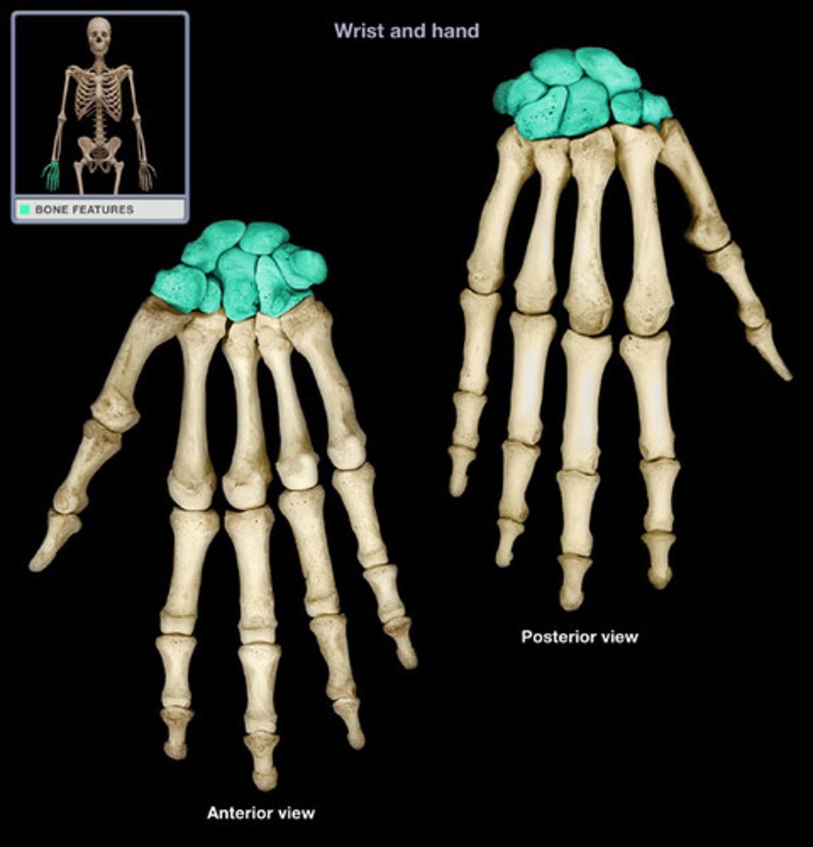
- proximal row: scaphoid, lunate, triangular, pisiform
- distal: trapezium, trapezoid, capitate, hamate (has a hook)
what are the carpal bones?
antebrachial fascia
dense connective tissue sheath enveloping the forearm muscles
palmar carpal ligament
thickening of antebrachial fascia on the anterior wrist
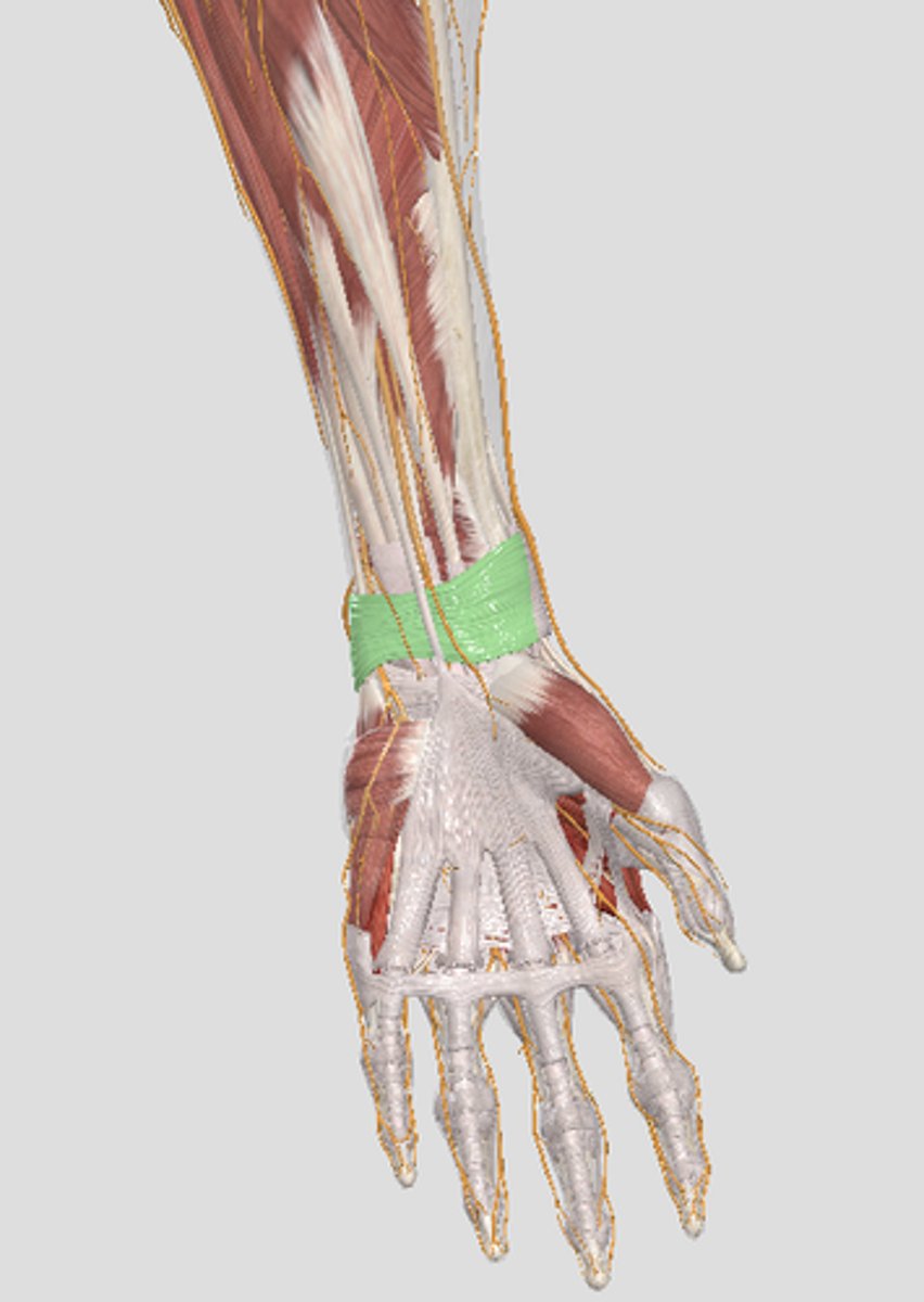
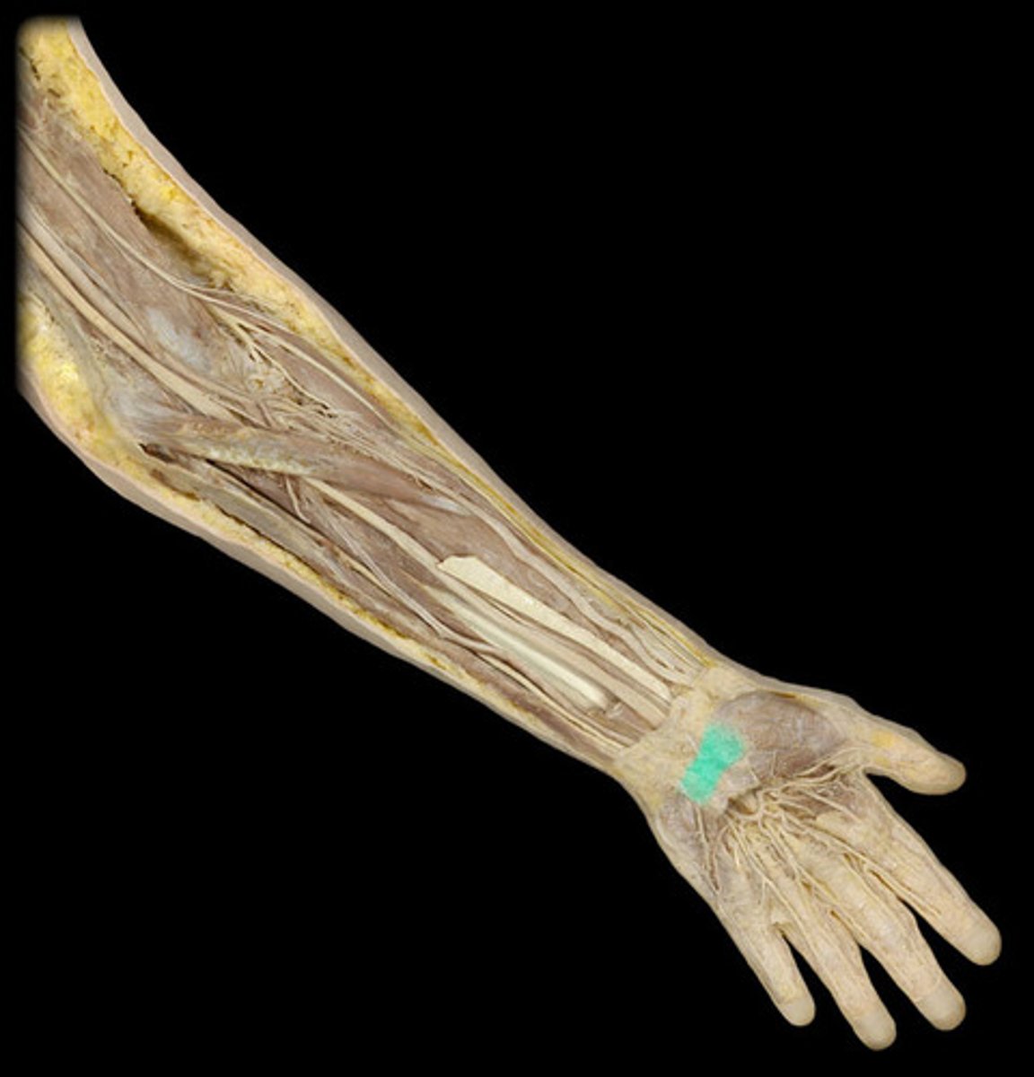
retinaculum
a thickened band of deep fascia that serves to hold tendons in place where they cross joints
flexor retinaculum (transverse carpal ligament)
connects the tubercles of scaphoid and trapezium and pisiform and the hook of the hamate; forms the roof of the carpal tunnel
thenar fascia
a thin layer of connective tissue covering the thenar muscle
hypothenar fascia
a thin layer of connective tissue covering the hypothenar muscle
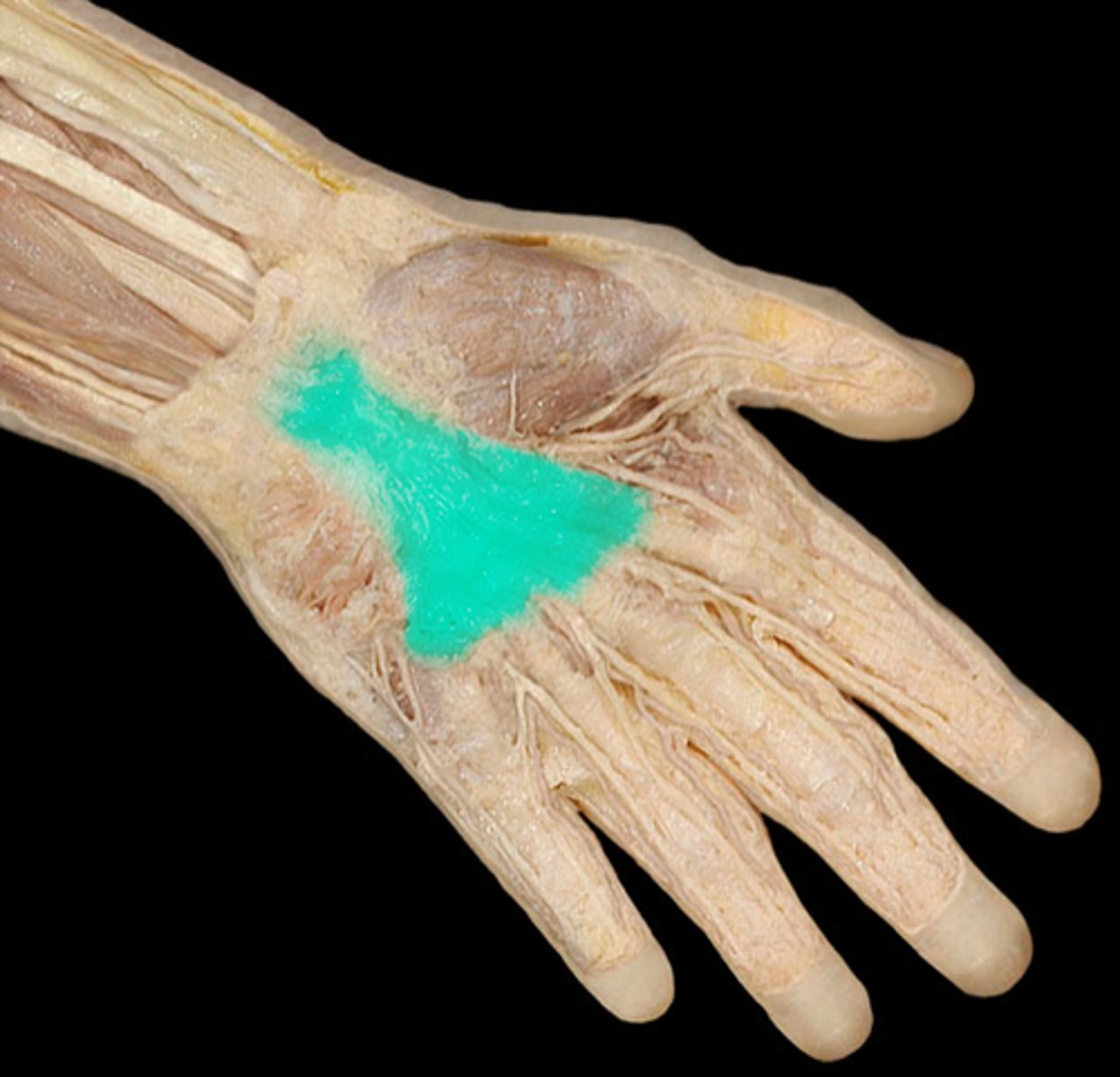
palmar aponeurosis
a thick, triangular sheet of deep fascia in the center of the palm, stretching from the wrist to the base of the fingers
- flex the wrist
- flex the digits
- pronate the hand
what are the actions of the muscles in the anterior forearm?
origin: medial epicondyle (superficial head); coronoid process of ulna (deep head)
insertion: midway of later surface of radius
what is the origin and insertion of the pronator teres muscle?
- pronator teres
- flexor carpi radialis
- palmaris longus
- flexor carpi ulnaris
what muscles are in the first layer of the anterior forearm?
action: pronates hand
innervation: median nerve (passes between the two heads)
what is the action and innervation of the pronator teres muscle?
origin: medial epicondyle
insertion: on the bases of the 2nd and 3rd metacarpal bones on the palmar surface
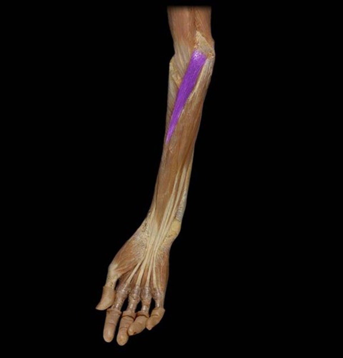
what is the origin and insertion of the flexor carpi radialis muscle?
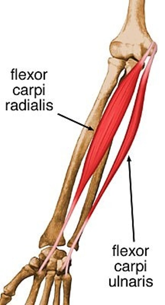
action: flex and abduct the wrist
innervation: median nerve
what is the action and innervation of the flexor carpi radialis?
origin: medial epicondyle
insertion: flexor retinaculum and palmar aponeurosis
what is the origin and insertion of the palmaris longus muscle?
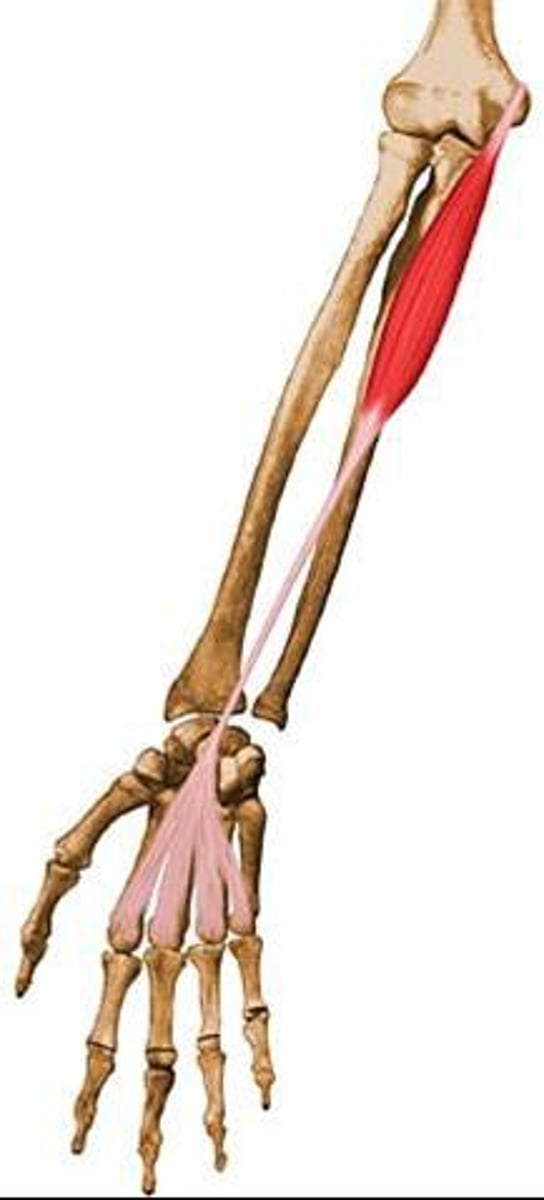
action: flex the hand
innervation: median nerve
what is the action and innervation of the palmaris longus?
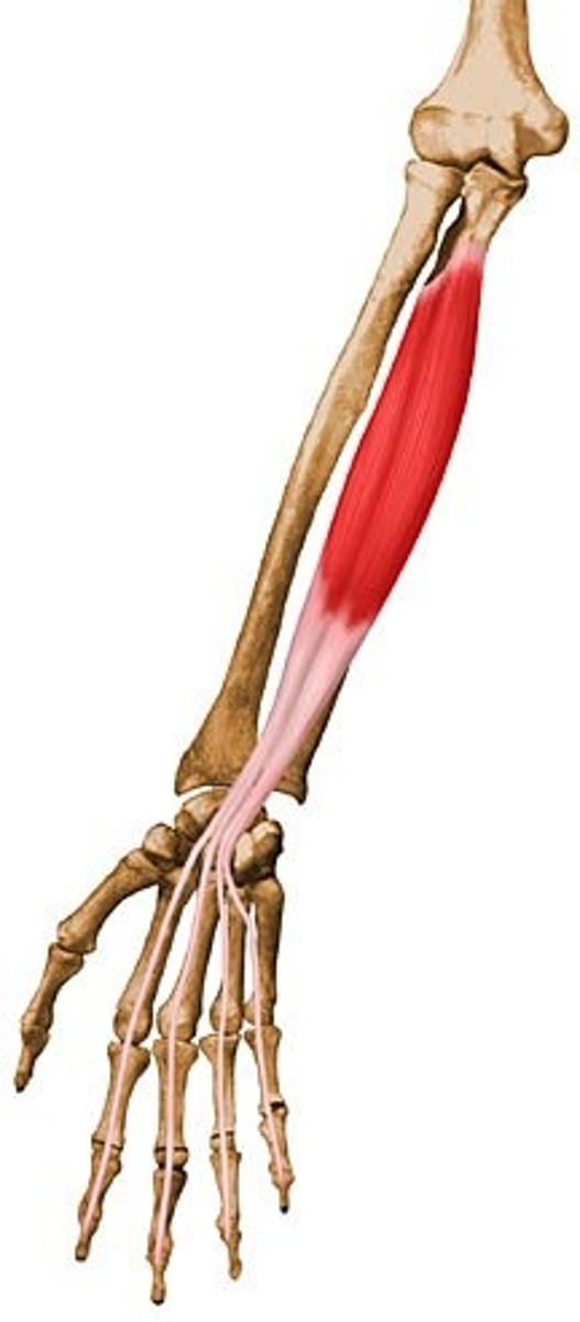
origin: medial epicondyle, olecranon and posterior border of ulna
insertion: pisiform (goes through the pisohamate and pisometacarpal ligament) and also inserts on the hamate and base of the fifth metacarpal bone
what is the origin and insertion of the flexor carpi ulnaris muscle?

action: flexes and adducts the wrist
innervation: ulnar nerve
what is the action and innervation of the flexor carpi ulnaris?
flexor digitorum superficialis muscle
what muscle is in the second layer of the anterior muscles of the forearm?
origin: humero-ulnar head (medial epicondyle and coronoid process of ulna), radial head (oblique line of radius)
insertion: bases of middle phalanges of medial four fingers
what is the origin and insertion of the flexor digitorum superficialis muscle?
action: flexes the wrist joint, the metacarpophalangeal joints, and the proximal interphalangeal joints
innervation: median nerve
what is the action and innervation of the flexor digitorum superficialis muscle?
- flexor digitorum profundus muscle
- flexor pollicis longus muscle
what muscles make up the third layer of the anterior muscles of the forearm?
origin: medial and anterior surfaces of the ulna, interosseous membrane
insertion: bases of distal phalanges of medial four fingers
what is the origin and insertion of the flexor digitorum profundus muscle?
action: flexes wrist, flexes MP, and flexes PIP and DIP
innervation: medial 2 tendons -> ulnar nerve; lateral 2 tendons -> anterior interosseous nerve
what is the action and innervation of the flexor digitorum profundus muscle?
origin: anterior surface of radius, interosseous membrane
insertion: base of distal phalanx of thumb
what is the origin and insertion of the flexor pollicis longus muscle?
action: flexes MP joint of first digit, flexes interphalangeal joint of first digit, flexes the wrist
innervation: anterior interosseous nerve
what is the action and innervation of the flexor pollicis longus muscle?
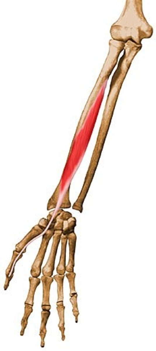
pronator quadratus muscle
what muscle is in the fourth layer of the anterior forearm muscles?
origin: distal anterior surface of ulna
insertion: distal anterior surface of radius
what is the origin and insertion of the pronator quadratus muscle?
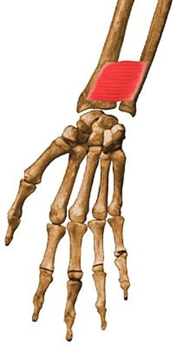
action: pronation
innervation: anterior interosseous nerve
what is the action and innervation of the pronator quadratus muscle?
brachial artery
ends at the neck of the radius and divides into the radial and ulnar arteries
- radial recurrent artery
- muscular branches
- palmar carpal branch
- superficial palmar branch
- deep palmar arch
what does the radial artery split into?
- anterior ulnar recurrent
- posterior ulnar recurrent
- common interosseous (then to anterior and posterior interosseous)(posterior becomes the interosseous recurrent)
- muscular branches
- palmar carpal branch
- superficial palmar arch
- deep palmar arch
what does the ulnar artery split into?
superior ulnar collateral artery and posterior ulnar recurrent artery
what anastomoses posterior to the medial epicondyle?
inferior ulnar collateral artery and anterior ulnar recurrent artery
what anastomoses anterior to the medial epicondyle?
radial collateral artery and radial recurrent artery
what anastomoses anterior to the lateral epicondyle?
middle collateral artery and interosseous recurrent artery
what anastomoses deep to the anconeus?
median nerve
runs between the two heads of the pronator teres, then deep to the FDS, is located between the palmaris longus and FCR at the wrist
median nerve
innervates all the anterior forearm muscles of the first and second layer besides the flexor carpi ulnaris
- anterior interosseous nerve (innervates the third and fourth layer muscles except for the medial half of the FDP)
- palmar branch (sensory innervation to the skin superficial to the flexor retinaculum
what does the median nerve give off in the anterior forearm?
ulnar nerve
travels behind the medial epicondyle of the humerus and runs between the two heads of the flexor carpi ulnaris, is on the FDP to the wrist and enters the hand through guyon's canal
ulnar nerve
innervates the flexor carpi ulnaris and the flexor digitorum profundus (4th and 5th digits)
- palmar branch (sensory innervation of the skin over the hypothenar eminence)
- dorsal branch (entire 5th and ulnar side of the 4th digit, ulnar side to the middle phalange of the 3rd digit, radial side to the middle phalange of the 4th digit)
what does the ulnar nerve give off?
- lateral side: tubercle of the scaphoid and tubercle of the trapezium
- medial side: pisiform and hook of hamate
what makes up the base of the carpal arch?
flexor retinaculum
connects the carpal arch
- flexor digitorum superficialis tendons (two layers)
- flexor digitorum profundus tendons (one layer)
- flexor pollicis longus tendon
- median nerve
what is in the carpal tunnel?
carpal tunnel syndrome
common condition that causes pain, numbness, and tingling in the hand and arm; occurs when the median nerve is squeezed or compressed as it travels through the wrist
- lateral: capitulum of humerus and head of radius
- medial: trochlea of humerus and trochlear notch of ulna
what are the articulations for the elbow joint?
ulnar collateral ligament
connects the medial epicondyle of the humerus and the ulna
anterior ulnar collateral ligament
connects the medial epicondyle of the humerus to the coronoid process of the ulna
posterior ulnar collateral ligament
connects the medial epicondyle of the humerus to the olecranon process of the ulna
intermediate ulnar collateral ligament
connects the olecranon process to the coronoid process
radial collateral ligament
connects the lateral epicondyle and the annular ligament
- head of the radius
- radial notch of the ulna
what is the articulation of the proximal radioulnar joint?
annular ligament
connects the radial notch of the ulna and the head and neck of the radius; provides stability
quadrate ligament
connects the inferior border of the radial notch of the ulna and the neck of the radius; closes the cavity between the radius and the ulna; allows rotation of the radius
interosseous membrane
connects the radius and ulna
head of ulna and ulnar notch of radius
what is the articulation of the distal radioulnar joint?
- palmar radioulnar ligament
- dorsal radioulnar ligament
what are the ligaments in the distal radioulnar joint?
colles' fracture
occurs to a fall on an outstretched hand with an extended wrist; fracture of the distal radius; dorsal displacement of the wrist and hand
flexion, extension, adduction, abduction, and circumduction
what does the wrist joint allow?
- radius and fibrocartilaginous disc
- proximal row of carpal bones (except pisiform)
what are the articulations of the wrist joint?
fibrocartilaginous disc
connects the radius and the styloid process of the ulna
palmar radiocarpal ligament
connects the distal end of the radius with the scaphoid, lunate, and triquetrum
dorsal radiocarpal ligament
connects the distal end of the radius with the scaphoid, lunate, and triquetrum on the dorsal side
radial carpal collateral ligament
connects the styloid process of the radius with the scaphoid and trapezium
ulnar carpal collateral ligament
connects the styloid process of the ulna with the pisiform and triquetrum