Optics of Vision, The Retina & the Retinal Visual Field
1/33
There's no tags or description
Looks like no tags are added yet.
Name | Mastery | Learn | Test | Matching | Spaced | Call with Kai |
|---|
No analytics yet
Send a link to your students to track their progress
34 Terms
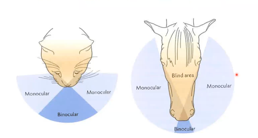
What anatomical feature determines the visual field?
The head morphology
Cat has larger binocular field due to more forward facing eyes
Horse have a very small binocular field of vision with extended monocular vision due to eyes being on the side of the head
What does vision require to be detected?
Vision requires detection of various light wavelengths
Humans, apes and Old world monkeys have enhanced visual acuity, distance perception and colour range compared to other mammals.
Birds, are superior and have the most advanced visual sense of all animal kingdom. Photons (light particles) contain more energy in lower (i.e. violet) range and less in the higher (i.e. red) spectrum range.
High energy is detrimental to sensory cells. Infrared waves are usually not detected by retinal photoreceptors, many animals absorb heat energy for body processes
What is the fovea centralis?
The fovea centralis is located in the center of the macula lutea, a small, flat spot located exactly in the center of the posterior portion of the retina. As the fovea is responsible for high-acuity vision it is densely saturated with cone photoreceptors.
What gland has the main role of keeping the lens clean within the eye?
The lacrimal gland
Lacrimal gland secrets saline tear onto the eye through ducts
Fluid cleans and lubricates the front of the eye during a blink
Prevents cornea from drying out and frost injury during cold weather
Tears are empty into the nasal cavity and function to maintain mucous membrane within the cavity
Fluid contains Iysozyme and immunoglobulin A to protect against infections
Describe the two axis of the eye involved in light detection.
These terms are about how light travels through the eye and how we perceive clear vision.
Optical Axis - Maximizes the visual field presented to the back of the eye, giving optically clear image (Crosses the cornea, lens dead center)
Visual Axis - visual axis to fovea gives the best color vision
Light traveling along this axis lands on the fovea, giving the best color vision and visual acuity.

How are images refracted through the eye or lens?
Light 'bends' (i.e. refraction) when passing from across the cornea, lens and vitreous humor, leads to an inverted image in the back of the eye
Refraction is the bending of light when it passes through a medium with a different optical density, like the cornea or lens.
In the eye, refraction focuses light onto the retina, a focal point in the back of the eye
Distance from the focal point behind the lens is dependent on the distance to the image
Divergent lens does not produce a focused image at a focal point; distributed leads to a distributed production of that image
What is the accommodation ability of the lens?
Ability of lens to change power - through ciliary muscles
Focal length is fixed but increased power increases ability of lens to refract divergent light (ie near objects) and achieve focus
Ciliary body relaxes = expansion of the lens, widening (Increase in power
Ciliary body contracts = decreased in elongated structure
Near objects → thicker lens → more refractive power
Far objects → flatter lens → less power
Describe the species variabilities in accomodation.
Horse lacks full accommodation ability. Shape of retina means it can see near and far by moving head/eyes
Distant objects - head depressed
Close objects - head raised
to maximize acuity
Describe the neural response to light.
Light enters - Binding to photoreceptors at the back of the retina (Either cones or rods)
Conveyed back to front via the bipolar cells and retinal ganglion cells which integrate information and send it along the optic nerve
Ganglion cells also express photoreceptors called melanopsins
At back of eye there is either dark pigment or tapetum
Retinal detachment is separation of retina from pigment epithelium
Describe how rods are able to detect light.
Rods
Rods are photoreceptor cells in the retina that detect low light (dim) conditions.
They have stacked discs inside their outer segment.
These discs are plasma membrane structures.
Rhodopsin, the photopigment, is embedded in these membranes.
Rhodopsin contains opsin (protein), and retinal (a light sensitive molecule) Light converts cis-retinal in rhodopsin to trans-retinal (isomerisation), generating a signal. Enzymes convert it back to cis-retinal so rods can respond to light again.
Describe the intracellular phototransduction of rhodopsin,
1. Photon hits rhodopsin (R)
2. Isomerisation of retinal
3. Activation of alpha subunit of transducin
4. Activation of phosphodiesterase (PDE)
5. Decreases GMP levels and closes ion channels
Hyperpolarises the receptor cell, leading to the release of NTs
In rods, the baseline is active in the dark (depolarized, releasing neurotransmitter).
Light reduces neurotransmitter release, which is interpreted by the downstream cells as a “signal.”
So, hyperpolarization = light detection → brain receives information about light.
How does the distribution of photoreceptors change across the retina?
Cones dominate the central retina (fovea) for color and detail. Rods dominate the peripheral retina for motion detection and low-light vision. Optic disc = (“blind spot”) - No photoreceptors here. This is where optic nerve exits the eye.
Fovea: 1 photoreceptor → 1 nerve → sharp vision
Periphery: Many photoreceptors → 1 nerve → sensitive, low-detail vision
Describe the light sensitivity between the rods and cones within the retina.
Rods are much more sensitive to light (irradiance).
• One rod responds to one photon
• There are multiple rods per bipolar cell
• Rods work in the scotopic region
“Scotopic” = night vision or low-light vision.
Relies almost entirely on rods.
Cones are not functional at very low light levels.
• Rods are poor for spatial detail
Cones provide more detailed information (including colour)
• But they need more light (photopic region), more photons required to activate one cone receptor
• The ratio of cones to ganglion cells is 1:1
What role do the ganglion cells have in their relationship to the photoreceptors?
Ganglion cells integrate multiple photoreceptor signals
Some ganglion show higher activation when light is shown in the center of the field
Some show higher activation when light is shown in the periphery
Depending on receptors, determines the frequency of action potentials
Ganglion cells show ____ ___________.
intrinsic photoresponsiveness
What is melanopsin?
A photoreceptor that responds to irradiance information, the signals control the daily rhythms
Melanopsin is very sensitive to light in blue light range
What is the clinical relevance of the pupillary light reflex?
Pupillary reflexes indicate functional state of the afferent and efferent that control the pupil
For example, if a light stimulus directed to the left eye elicits a consensual constriction in the right eye, but not a direct one in the left eye, then the afferent limb of the reflex (i.e. optic nerve is intact; but the efferent limb to the left eye is damaged)
Possible reason: damaged oculomotor nerve
If shining light and there is a response in both eyes, optic nerve is intact
If there is a lack of response, there is damage to ciliary ganglion or the preganglionic parasympathetic fiber
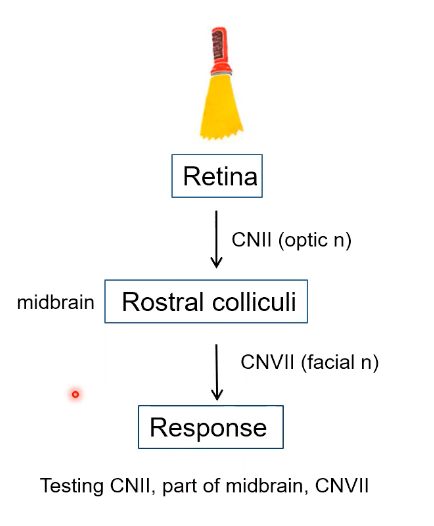
What is the dazzle reflex?
Subcortical mediated brainstem response
Palpebral fissure closes in response (e.g. blink, head movement) in response to sudden intense illumination of the eye
Ipsilateral response is greater than the contralateral response
Absence of a response indicates blindness
Retina recieves light (CN II(Optic Nerve)) into rostral colliculi (Midbrain) through CNVII (Facial Nerve) → Response = blink, sudden movement, blindness
Tests CNII and CNVII
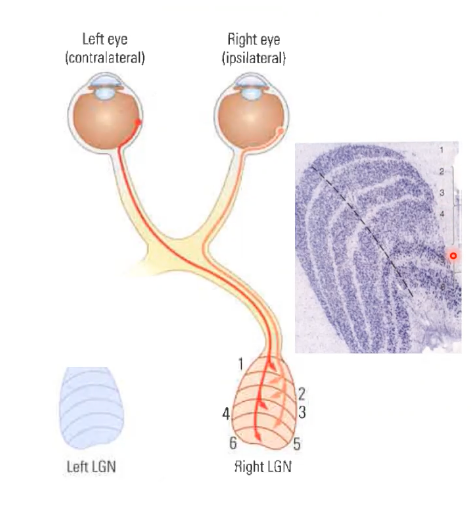
Describe the neural circuit in the eyes from the retina to the lateral geniculate nucleus.
Lateral geniculate nucleus located in the thalamus
LGN, maintains a retinotopic (i.e. topography)
6 layers of the 'retinal map' consists of parvocellular (1-4) and magnocellular (5,6) cells
The lateral geniculate nucleus (LGN) is called a retinal map because it preserves the spatial layout of the retina — neighboring points on the retina project to neighboring points in the LGN. This retinotopic organization keeps the visual scene’s structure intact as it’s relayed to the visual cortex.
Parvocellular cells are small, integrate signals from cones and necessary for colour and form
Magnocellular cells are large, integrate signals from rods, involved in movement, depth and Irradiance
From the LGN → where do the cells project?
The layers of cells project into the primary visual cortex or occipital lobe (Located in the back of the brain)
The primary visual cortex integrates multiple different LGN cells, in an attempt to formulate visual perception
What three cells are found in the primary visual cortex?
Simple cells: respond to stimuli/ edges or lines with a correct orientation.
Complex cells: respond to moving edges/stimulus of the correct orientation.
Hypercomplex (end-stopped) cells respond best when a stimulus (like a line or edge) is a certain length or size
Stop firing (“end-stopped”) if the stimulus extends too far beyond their preferred size
How do simple cells, complex cells and hypercomplex cells react to light?
LGN → dots
Simple cell → line detector
Complex cell → moving line detector
Hypercomplex cell → line ending / corner detector
Retina: dots → LGN: center-surround spots → V1: edges, orientation, motion → Higher areas: shapes, objects, faces
The primary visual cortex is a feature-detector factory.
It builds orientation, motion, and line-ending detectors from multiple LGN inputs.
These simple features are passed forward to higher visual areas where more complex perception (shapes, objects, faces) emerges.
Summarize the neural circuit for vision.
🟡 Photoreceptors (rods and cones) – in the retina, convert light into electrical signals.
🟢 Bipolar cells – relay signals from photoreceptors to ganglion cells.
🔵 Ganglion cells – their axons form the optic nerve, carrying visual information to the brain.
⚪ Optic chiasm – where fibers from the nasal half of each retina cross to the opposite side, ensuring that each hemisphere processes the opposite visual field.
🔴 Lateral Geniculate Nucleus (LGN) – in the thalamus; acts as a relay and maintains the retinotopic map.
🟣 Primary Visual Cortex (V1) – processes basic features like orientation, edges, and movement.
🔶 Higher visual areas (V2, V3, V4, MT, etc.) – integrate information for object recognition, color, depth, and motion.
Separation into dorsal and ventral streams:
Describe the dorsal stream and ventral streams.
From the occipital lobe (Primary center) → to the secondary centers, divided into:
Dorsal stream (Or the action stream) flows from V1 to V5/V3A - parietal visual areas
Guides movements such as the hand postures for grasping a mug or pen, how to reach, grasp, or interact with objects
damage to dorsal stream results in optic ataxia
Ventral Stream (Or the recognition stream), helps us recognize what is in the environment, temporal visual areas, object identification and recognition
Damage to the ventral stream prevents identification of objects
Reaching movements directed toward individual body (e.g. tactile or proprincentive feedhack) remains intact
What is the association cortex and grandmother cells?
The association cortex interprets and combines sensory information for recognition and meaning, while “grandmother cells” are a (mostly theoretical) idea of single neurons that respond to one very specific stimulus
Within the temporal cortex → direction of movement integrated at lower levels is integrated in temporal cortex, giving rise to species specific responses
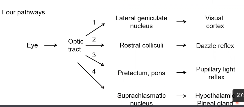
What are some of the non-image forming pathways responsible for biological rhythms?
Circadian Rhythm and the SCN
Melanopsin-containing retinal ganglion cells detect light levels (especially blue light) and send signals through the optic nerve to the suprachiasmatic nucleus (SCN) in the hypothalamus.
The SCN acts as the body’s master clock, regulating daily (circadian) rhythms like sleep, wakefulness, hormone release, immune function, and behavior.
🧬 Molecular Clock Mechanism (in SCN neurons)
During the day, the genes BMAL1 and CLOCK are active.
These genes produce proteins (BMAL1 and CLOCK) that form a dimer, which activates other genes — Period (Per1, Per2) and Cryptochrome (Cry1, Cry2).
PER and CRY proteins build up in the cytoplasm, pair up (dimerize), and move back into the nucleus.
There, they inhibit BMAL1 and CLOCK, stopping their own production.
As PER and CRY levels fall overnight, BMAL1 and CLOCK become active again — restarting the cycle.
⏰ Result
This feedback loop takes about 24 hours, creating a daily rhythm that synchronizes the body’s internal clocks with the external light–dark cycle.
In short:
Light → Melanopsin cells → SCN → activates BMAL1 & CLOCK → produce PER & CRY → inhibit BMAL1 & CLOCK → cycle repeats every 24 hours, setting the body’s circadian rhythm.
Summarize the four man visual and non-visual pathways in the brain.
Visual Field = 1
Blink/Head movements = 2
Pupillary Light reflex = 3
Control of rhythms = 4
What is visual adaptation or afteraffects?
Visual adaptation means your eyes and brain adjust to constant stimulation.
When you look at something for a while, the neurons responding to it get tired (their response decreases).
When you look away, you can get an aftereffect — you briefly see the opposite pattern or motion.
What are the main photoreceptors that underlie color vision?
The cones
3 Main types: red, green, blue
Each responds to a different wavelength
What are the three main theories of color vision?
Trichromatic theory - colour vision that is based on the coding of the three basic colours: red, green and blue
Opponent-process theory - colour vision that emphasizes the importance of the opposition of pairs of colours: red versus green and blue versus yellow.
Explain why color vision starts in the ganglion cells.
Colour opsins – These are proteins in your cone cells in the retina that detect different colours (like red, green, blue). Each type of cone has a different opsin sensitive to a certain wavelength of light.
Ganglion cells – These are cells in the retina that collect information from many cones. They “integrate” or combine the signals from the different types of cones to detect colour differences and patterns.
Transmit to LGN – After integrating the information, ganglion cells send it along their axons to the LGN (lateral geniculate nucleus) in the thalamus, which is a relay station in the brain.
Which secondary visual pathway is dependent on color vision?
Which is not?
dorsal, more action pathway, so - no color vision
ventral, more recognition pathway, so - color vision
Describe the organization of the secondary visual cortex.
Complex neurons in the visual temporal cortex (ie V4) respond to objects in the visual field that have color that is dependent on orientation, texture and shape.
Ocular dominance columns
Stripes of neurons in V1 that prefer input from one eye (left or right).
Help the brain combine signals from both eyes for depth perception.
Orientation columns / pathways
Neurons in V1 are arranged so that neighboring cells prefer similar line orientations (vertical, horizontal, diagonal).
This allows the brain to detect edges, shapes, and contours.
Blobs
Interspersed within these columns, blobs are color-sensitive neurons.
Mostly respond to wavelength (color) rather than orientation or motion.
In short:
V1 is organized into ocular dominance columns (eye preference), orientation columns (edge detection), and blobs (color), so different features of vision are processed in specialized, interwoven areas.
Why are mammals considered the exception, in terms of having light receptors outside the retina?
1. Non-retinal light detection
Most animals have light-sensing cells outside the eyes, in places like the brain, skin, or other organs.
These cells can detect light even if it doesn’t form an image.
Example: Many birds and reptiles can detect day length (photoperiod) directly in the brain to regulate hormones.
Mammals are the exception:
In mammals, almost all light detection is done through the retina in the eyes.
Mammals rely less on non-eye photoreceptors for physiological processes like seasonal reproduction.
2. Photoreceptors in multiple tissues
Some animals have opsins (light-sensitive proteins) in tissues like skin, brain, or pineal gland.
These opsins can sense light levels and day length, even if the animal’s eyes are closed or in the dark.
3. Guiding reproduction
Light detected outside the retina can influence hormones like melatonin.
Melatonin signals day length and seasons to the reproductive system.
Example: Birds or fish may start mating when days get longer in spring.
Simple analogy:
Photoreceptors in the eyes → “see the world”
Photoreceptors elsewhere → “sense the calendar” to know when to breed
So in short:
Many animals have light sensors outside the eyes that tell their bodies about the season, which helps time reproduction. Mammals mostly rely on the eyes for this, making them the exception.