Week 1 - Tissue Types
 Call Kai
 Learn
Learn Practice Test
 Spaced Repetition
 Match
Match Flashcards
 Knowt Play
1/35

There's no tags or description
Looks like no tags are added yet.
Name | Mastery | Learn | Test | Matching | Spaced | Call with Kai | Chat |
|---|
No analytics yet
Send a link to your students to track their progress
36 Terms
Tissue
A group of cells found together in the body
Types of Tissue
Epithelial Tissue
Connective Tissue
Muscle Tissue
Nervous Tissue
Epithelial tissue
Epi-: Greek prefix meaning “pond” or “on top of”
Sheets of cells found on the outer surfaces of the body (skin) or glands. Found on tops of, covering other tissues.
Joined by cell junctions - cells directly connected to one another
All substances must enter and exit through the epithelium
Apical-Basolateral Polarity - The cell’s top and bottom structure differ
Apical surface: ‘top’ surface facing the ‘outside’. Not attached to anything
Basolateral surface: ‘bottom’ that faces and attaches to the underlying tissue
All epithelial tissue is either in contact with blood or the external environment.
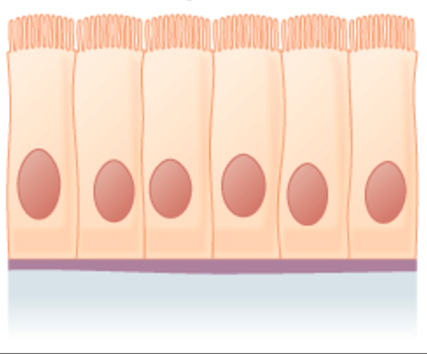
Epithelial tissue structure:
Desmosomes
Cells are connected via specialised contacts:
“Tight junctions” or “Desmosomes”
Prevent the passage of unwanted substances from the external environment, enables the epithelial surface to be one continuous sheet
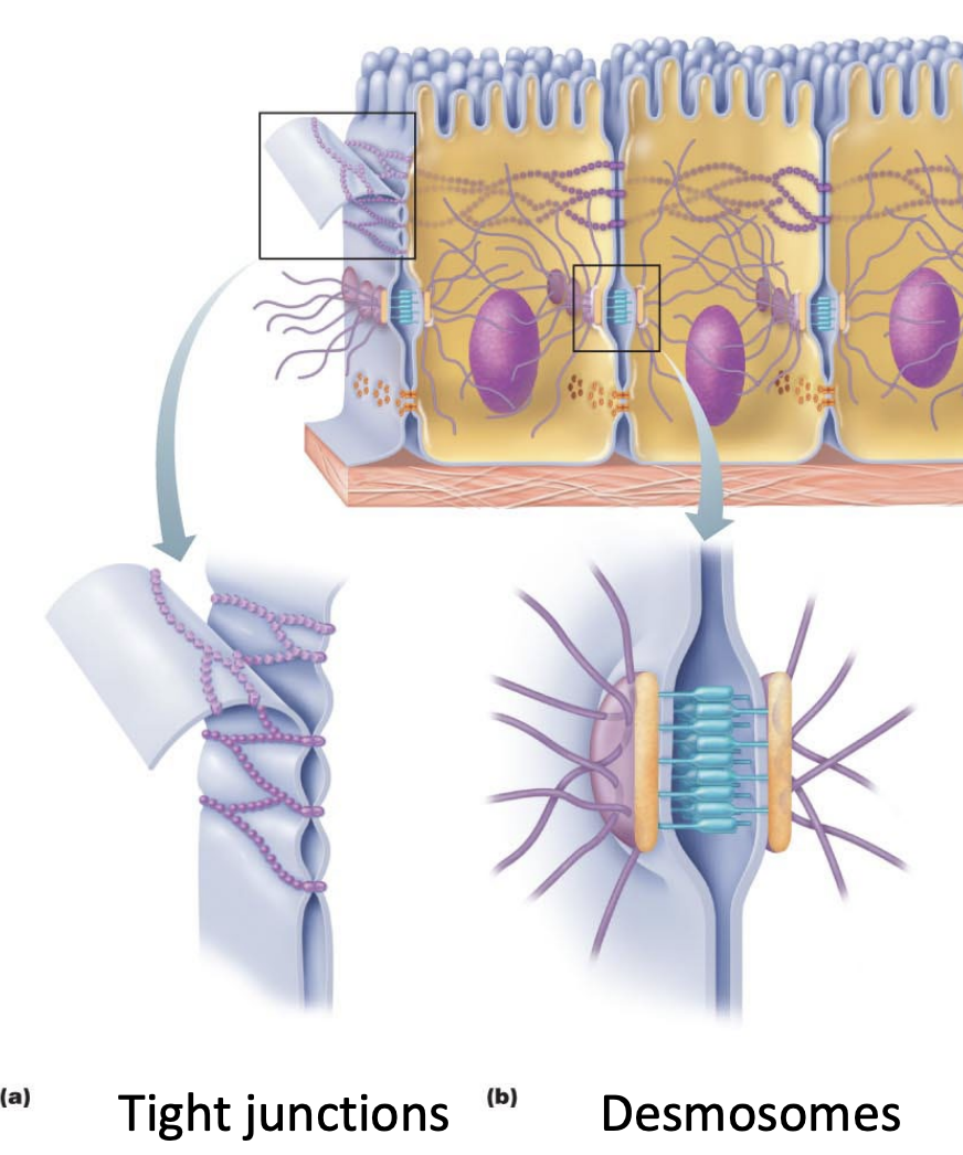
Epithelial tissue structure:
Basement Membrane
Basement membrane is connective tissue.
Anchors the basolateral surface of epithelial cells to the underlying tissue.
Epithelial tissue structure:
Blood circulation and Nerve innervations
The epithelial surface is avascular (does not have blood vessels); however it is innervated (has nerves throughout)
The cells most deep receive the most nutrients from adjacent blood vessels. However as they duplicate and are pushed further up they cannot receive the same nutrients, and start to die off
Nerves and tactile receptors are embedded within the epithelial tissue
Thus all epithelial cells have sensory perception
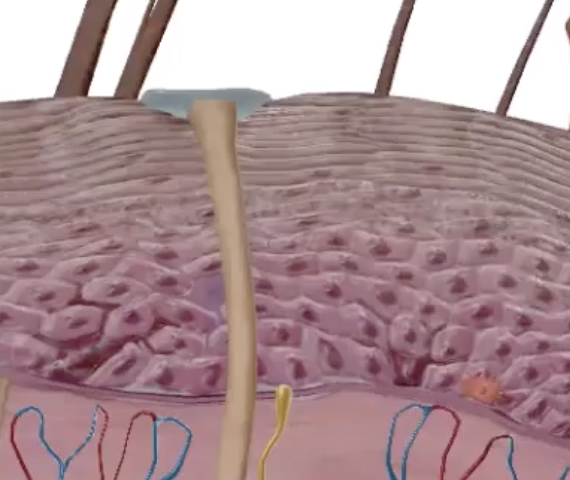
Are epithelial cells regenerative?
Yes, highly regenerative
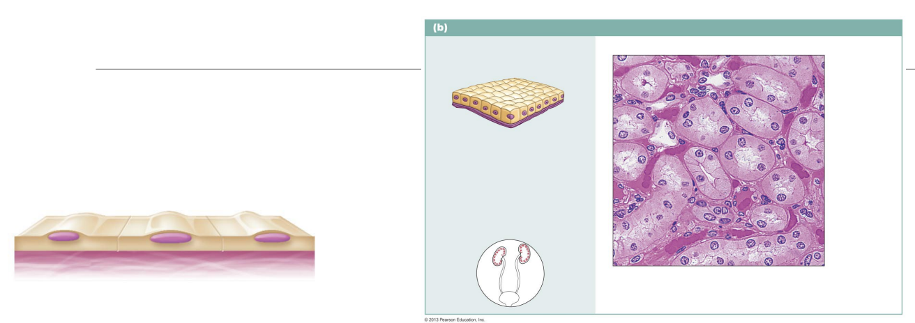
Simple Epithelia
Epithelial cells 1 layer thick
e.g alveolus, kidneys
Stratified Epithelia
Epithelial layers that are multiple layers thick
e.g bladder, tongue
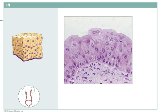
Epithelial Cell Shapes:
Squamous
Scale shaped.
Flatter and wider than it is thick.
Squamma - means ‘scale’

Epithelial Cell Shapes:
Cuboidal
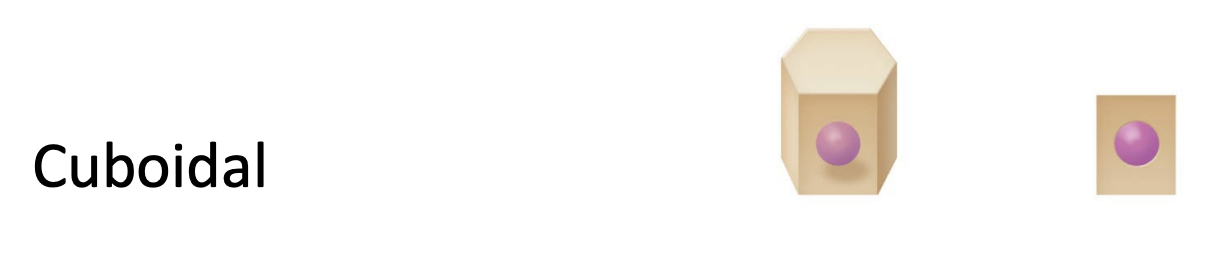
Epithelial Cell Shapes:
Columnar
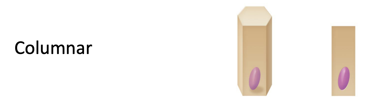
Taller than they are wide
Epithelial Cell Types:
Glandular epithelial cells
Secretes certain substances.
e.g mucus into the trachea
e.g acid to acid digestion in the stomach
even hormones/digestive enzymes
Epithelial Cell Functions:
Protection
Likely stratified epithelia
e.g skin
Epithelial Cell Functions:
Absorption
Likely simple epithelia
e.g small intestines
Epithelial Cell Functions:
Excretion
e.g kidneys and bladder
Epithelial Cell Functions:
Filtration
Likely simple epithelia
e.g kidneys
Epithelial Cell Functions:
Secretion
e.g mucus from cells lining trachea
Nervous Tissue
Excitable, non-contractile tissue. Can be electrically stimulated but cannot contract.
Sends & receives chemical and electrical signals
2 Types:
Neurones - Propagates signals through the nervous system
Neuroglia - Supports neuron function
Monitors the health of neurons, removing foreign bodies, ensures constant stream of nutrients available, can protect and insulate the axons.
Neuron structure:
Cell body
Contains all organelles, the cytoplasm
Neuron Structure:
Dendrites
Projections of the cell body, the receptive regions of impulses from other neurons or stimuli.
Neuron Structure:
Axon & Axon terminal
The longest projection of the cell body.
Electrical impulses travel down the axon, releasing as chemical signals (neurotransmitters) through the axon terminal to send signals to other neurons or muscle tissue.
Muscle Tissue
Excitable contractile tissue. Can be electrically stimulated (by local cells or nerves through neurotransmitters) and can contract.
Highly vascularised,
Cells are packed together.
Extensible - can stretch.
Elastic - recoil back to original shape
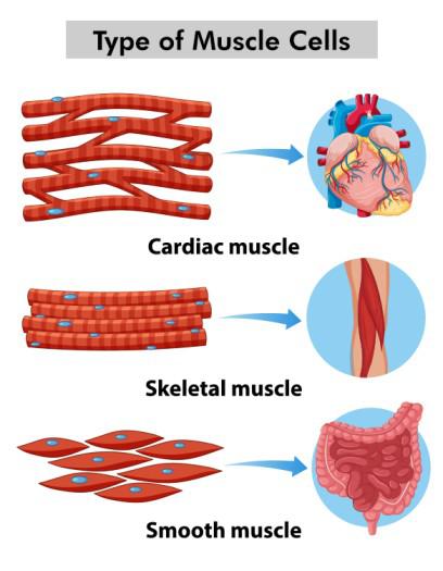
Muscle cell types:
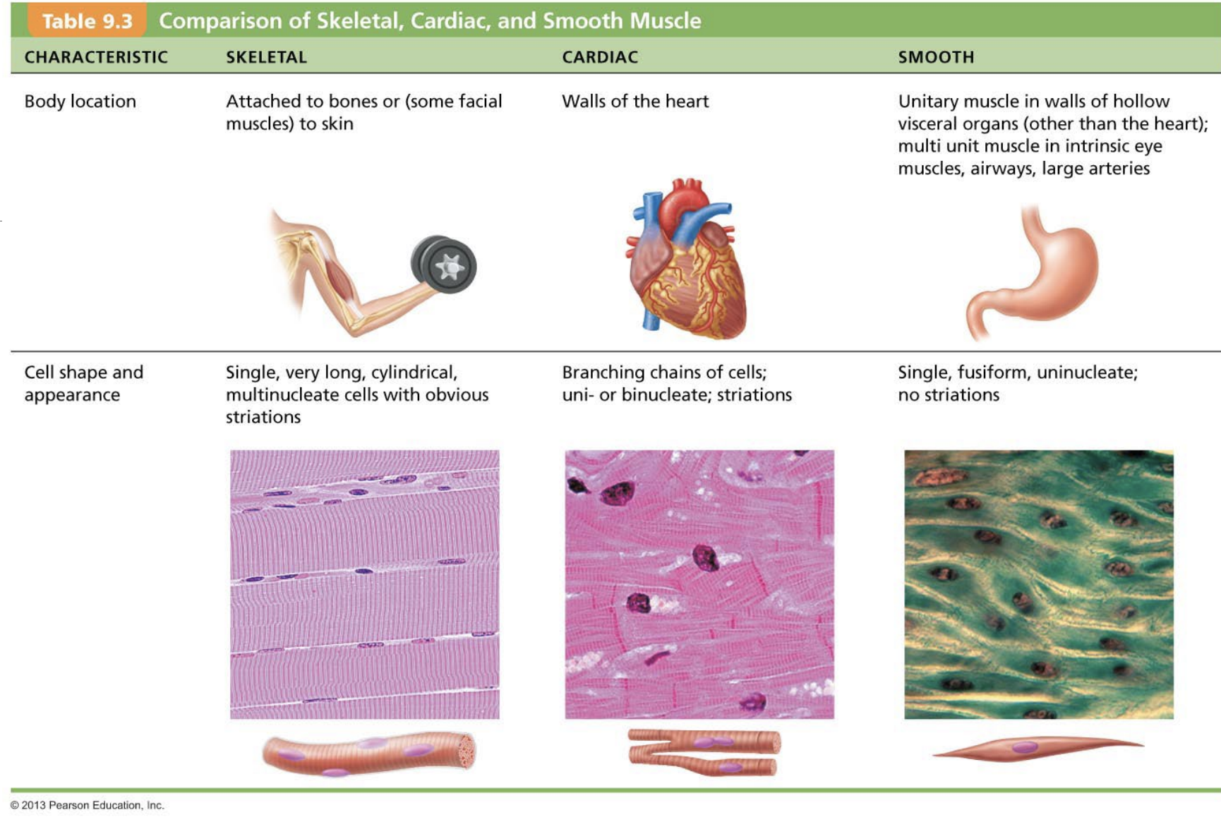
Skeletal Muscles
Skeletal muscle tissue
Majority is attached to the skeleton
All skeletal muscle tissue can be voluntarily moved
Multinucleated (formed through the fusion of myoblasts (progenitor cells))
Striated cells
Can span the entire length of a muscle
Muscle cell types:
Smooth Muscle
Smooth muscle tissue
Muscle of internal organs
Involuntary control
NOT striated
Can move around to accommodate the contractions of blood vessels
Single nucleus
Muscle cell types:
Cardiac muscle cells
Cardiac muscle tissue
Heart muscles that pump blood
Involuntary control
Striated
Single nucleus, connect to adjacent cardiomyocytes via intercalated discs (created to pull adjacent cells during contraction)
Ions can move between cells to ensure contraction, ensures a synchronous contraction of the heard (heartbeat)
Muscle Tissue:
Myofilaments
Actin and myosin that contract to shorten the muscle and release energy for movement.
Comparing muscle cell types
Connective tissue
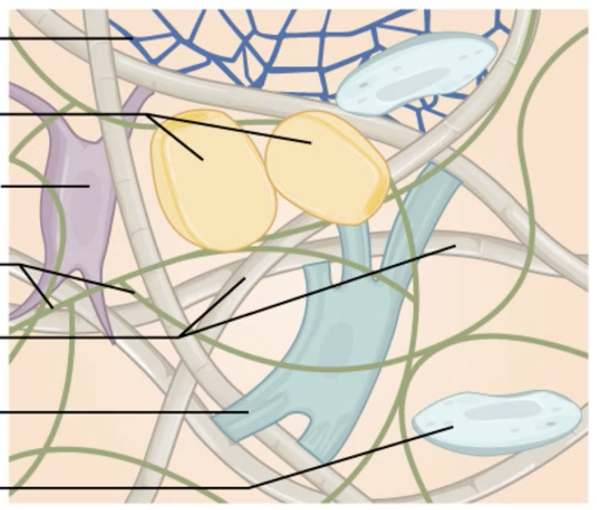
Cells dispersed within a extracellular matrix
Functions to connect, protect and support other tissues
Connective Tissue:
Extracellular Matrix
Ground Substance
Structure:
Interstitial fluid between cells
Proteoglycans - gives the fluid viscosity
Function:
Structure, hydration and intercellular communication
Connective Tissue:
Extracellular Matrix
Fibres
Collagen fibres:
Tough fibres that provide resistance to tensile forces (stretching)
Reticular fibres:
Finer collagen fibres that form delicate networks, support blood vessels and provide flexibility.
Elastin fibres:
Provides connective tissue elasticity, can stretch and recoil to original shape
Connective Tissue:
Extracellular Matrix
Cells
Fibroblasts:
Produce fibres
Chondroblasts:
Produce cartilage
Osteoblasts:
Produce bone
Connective tissue types:
Connective tissue proper
Loose connective tissue:
Adipose tissue
Made of fat cells. Used as energy stores, insulation or shock absorption.
Areolar connective tissue
Links body parts but still provides mobility. largest source of water for the body. Made mostly of fibroblasts & machrophages
Dense regular connective tissue:
Made mostly of collagen fibres running parralel to one another.
Run in one direction because the forces exerted on them apply in one direction, thus optimised to oppose tensile forces (stretching of muscles)
Found in muscle tendons, ligaments and aponeurosis (tendons for flat muscles such as back muscles)
Dense irregular connective tissue:
Dense, so made up of much elastin and collagen fibres to provide flexibility for movement
Found in deep fascia, joint capsules and skin’s dermis layer
Connective tissue types:
Cartilage
Found at the end of bones, with varying levels of collagen (fibrocartilage) and elastin.
made with chondrocytes from the extracellular matrix.
Withstands tensile and compressive forces due to the water absorbing and making the cartilage springy like a sponge that can squeeze and release water. (cartilage is 80% water)
Cartilage itself has no blood vessels or nerves, rather roaming chondrocytes provide nutrients from nearby blood vessels
Supportive connective tissue
Connective tissue types:
Bone
Withstands tensile and compressive forces
Specifically withstands conpressive forces better due to greater rigidity
made with osteoblasts which lay down collagen alongside calcium and phosphate ions in the extracellular matrix which hardens to form bone
upon crystalisation of bone occuring on all sides of osteoblasts, they become osteocytes
Has lost of blood supply with blood vessels and nerves running throughout, can repair quickly
Connective tissue types:
Fluid connective tissue (blood & lymph)
connective tissue because is contains cells (red blood cells, white blood cells & platelets) surrounded by an extracellular matrix (plasma)
also has soluble fibres called fibrin, which can turn insoluble to form blood clots