Nervous System I: The Central Nervous System
1/98
There's no tags or description
Looks like no tags are added yet.
Name | Mastery | Learn | Test | Matching | Spaced | Call with Kai | Chat |
|---|
No analytics yet
Send a link to your students to track their progress
99 Terms
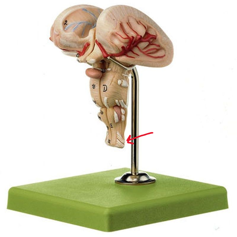
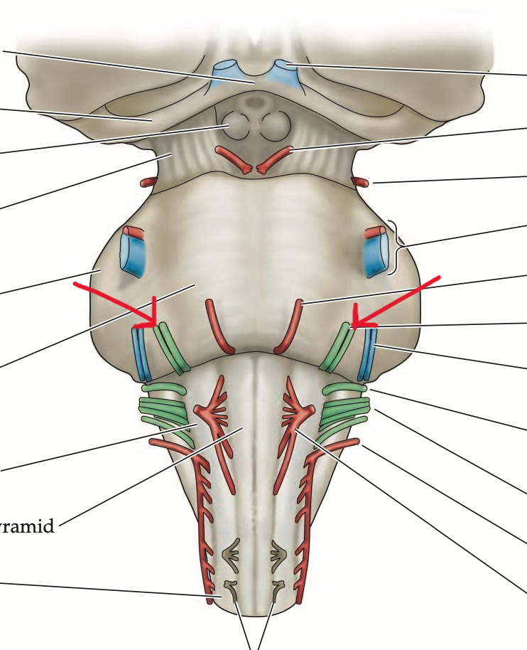
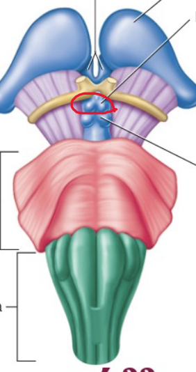
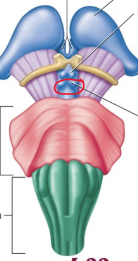
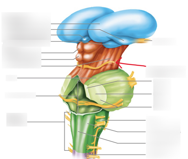
Medulla Oblongata
Structure. Very bottom of brain stem
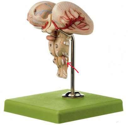
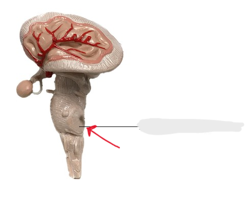
Olives
Structure
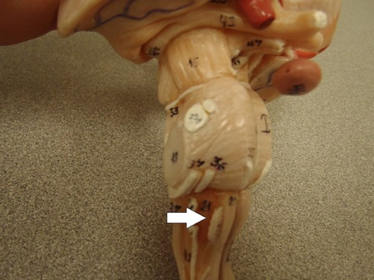
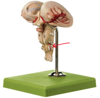
Pyramids
Structure. Front legs of the seahorse
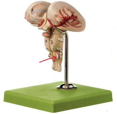
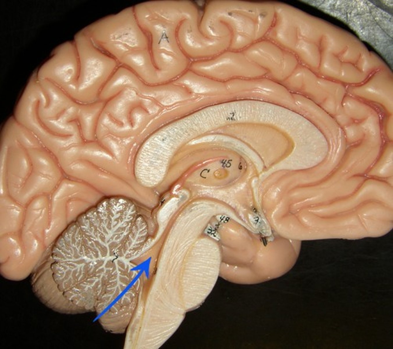
Inferior cerebellar peduncles
Structure. Back of the seahorse legs
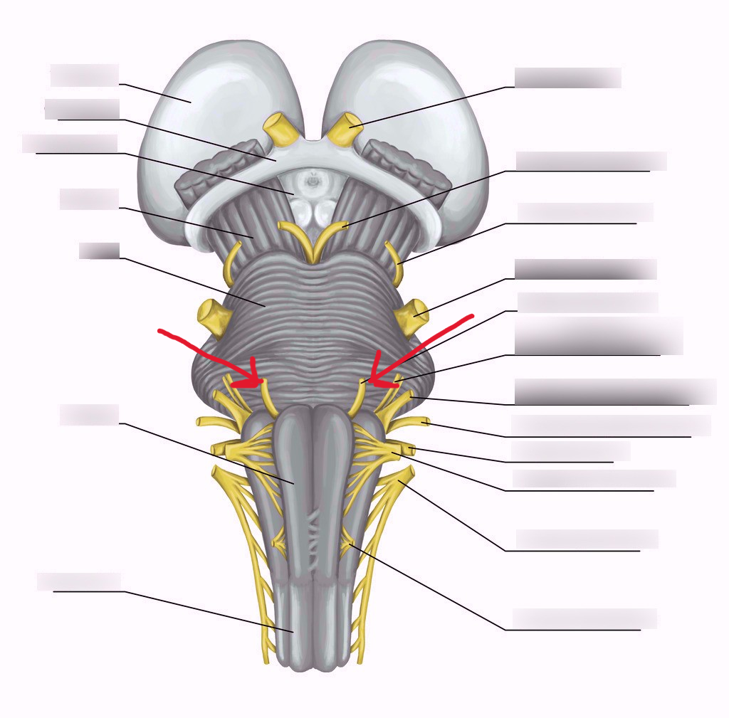
Glossopharyngeal nerve (CN IX)
Structure. To the left of the olives, little upper dot
Vagus nerve (CN X)
Structure. Just under the Glossopharyngeal nerve / Bottom dot
Hypoglossal nerve (CN XII)
Structure
spinal accessory nerve (CN XI)
Structure. V shape
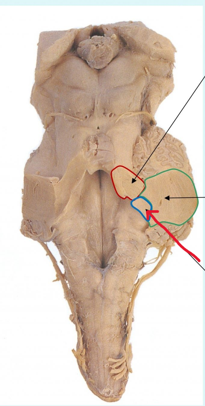
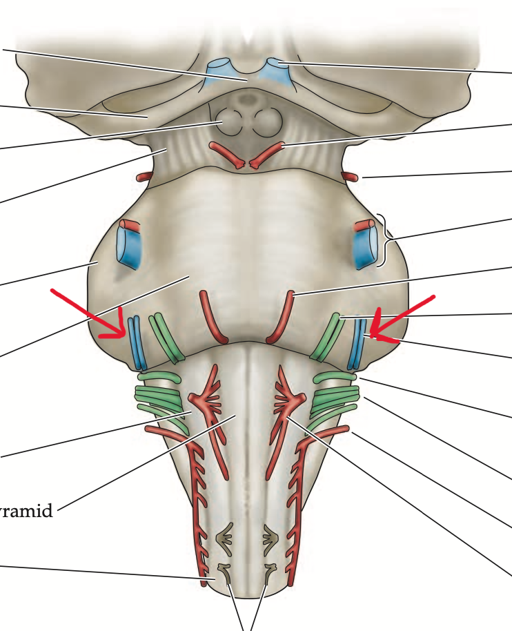
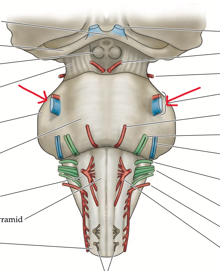
Pons
Structure
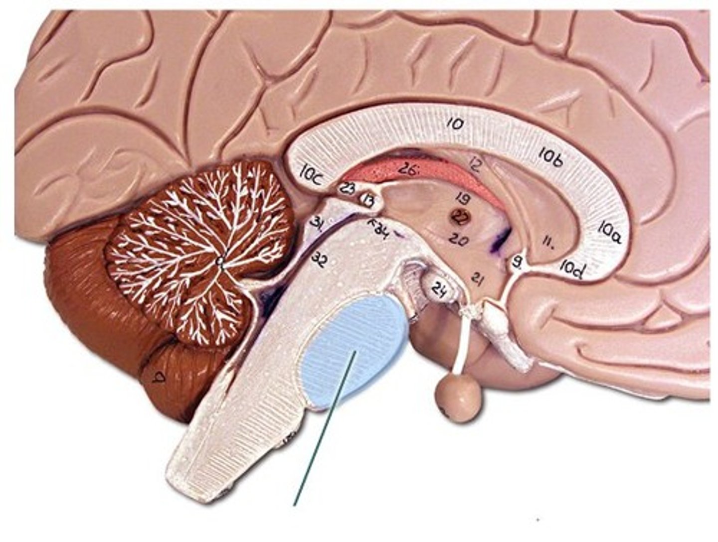
Middle cerebellar peduncles
Structure. Smooth area
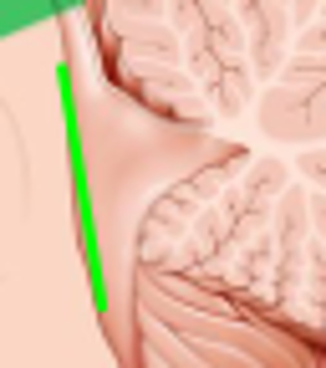
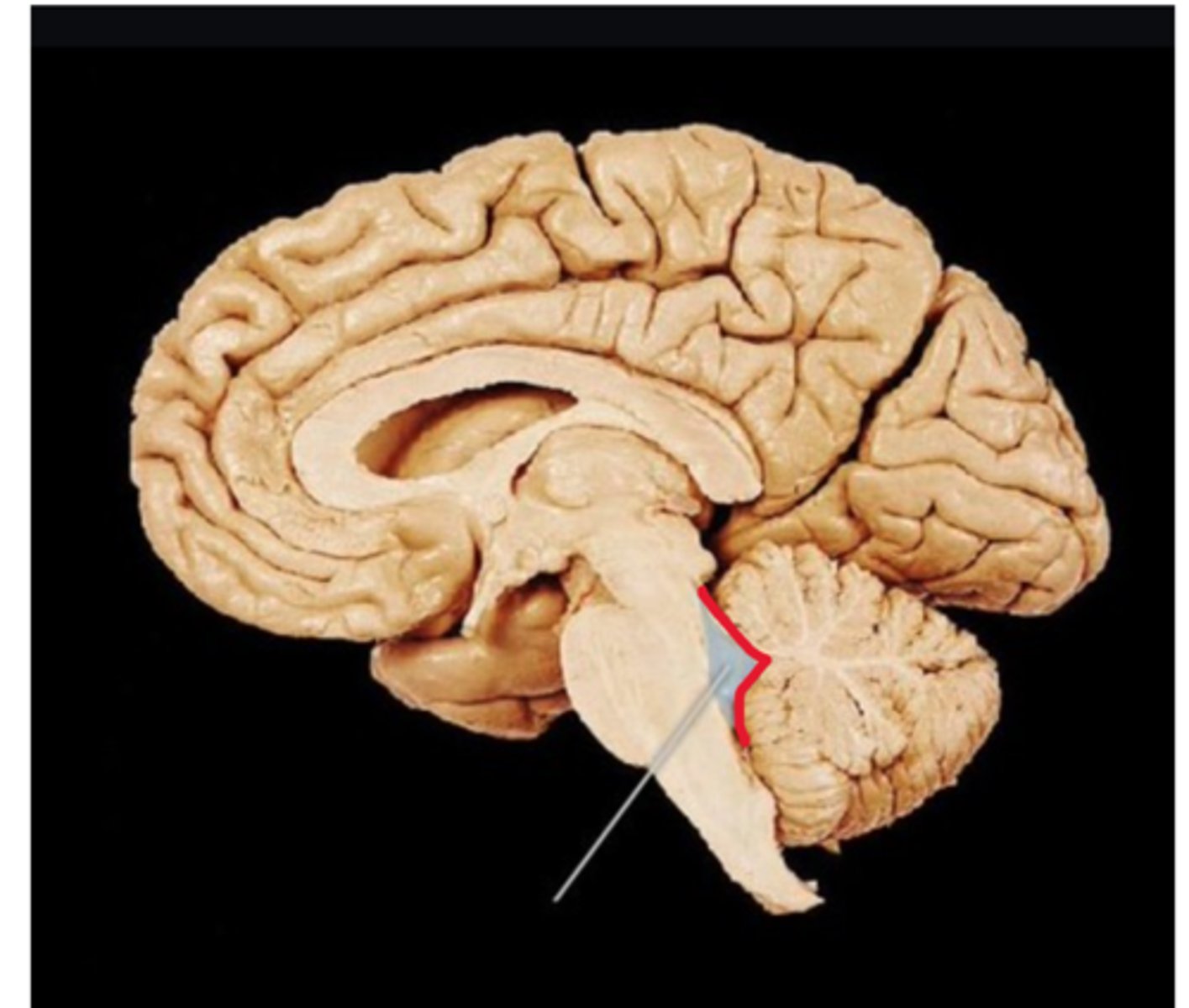
Superior cerebellar peduncles
structure. horseshoe shape
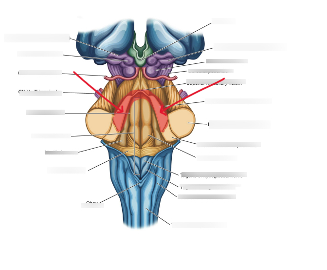
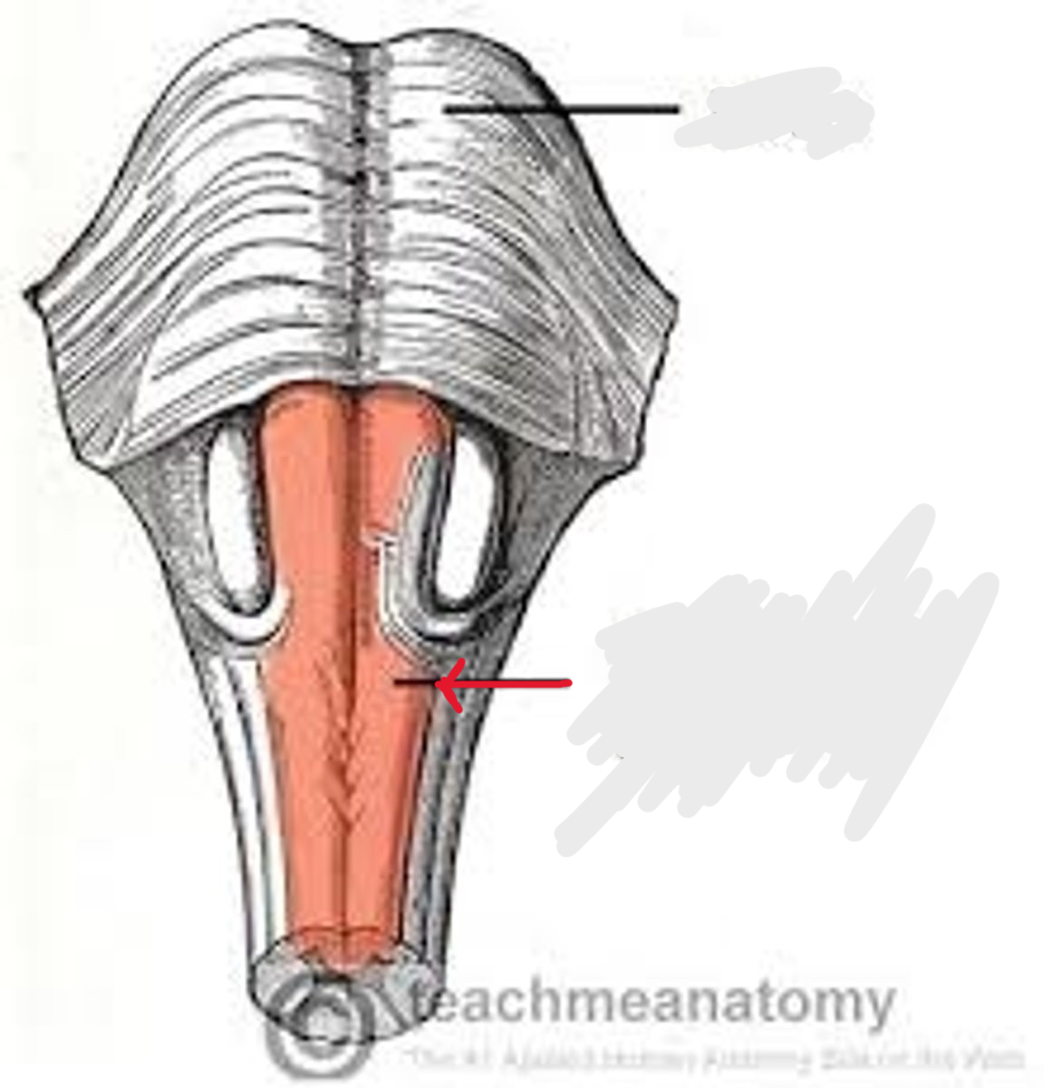
Fourth ventricle
Space. Triangle space next to pons/ cerebellum
Rhomboid fossa
Depression. Against pons /bottom of the triangle
Cerebellar part of the 4th ventricle
Structure. Sides of triangle against cerebellum
Abducens nerve (CN VI)
Structure
Facial nerve (CN VII)
Structure
Vestibulocochlear nerve (CN VIII)
structure
Trigeminal nerve (CN V)
Structure. snowman
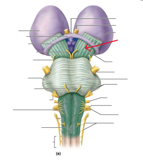
Corpora quadrigemina
collective structure. whole butterfly
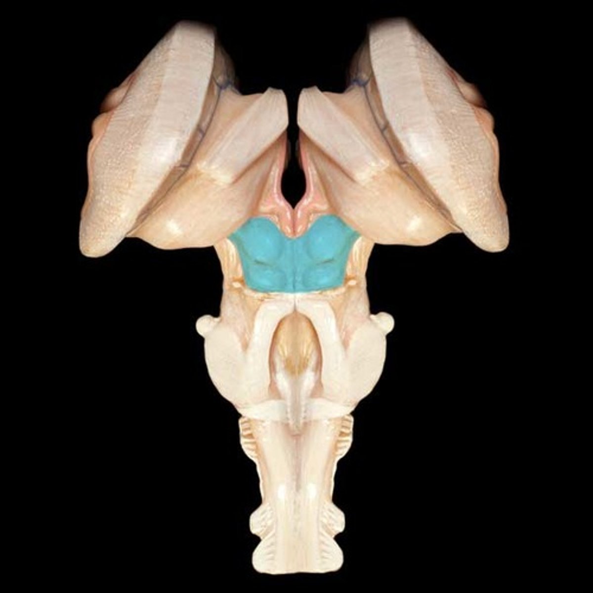
Superior colliculi
Feature. Top 2 wings of the butterfly
Inferior colliculi
Feature. Bottom 2 wings on the butterfly
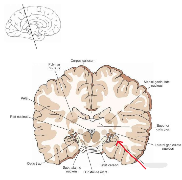
Crus cerebri
structure
Substantia nigra
structure. black line
Cerebral aqueduct
Space. Opposite side on the brain steak than the substantia nigra
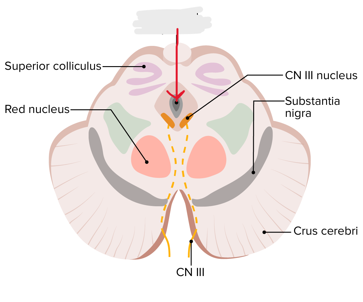
Oculomotor nerve (CN III)
structure

Trochlear nerve (CN IV)
Structure
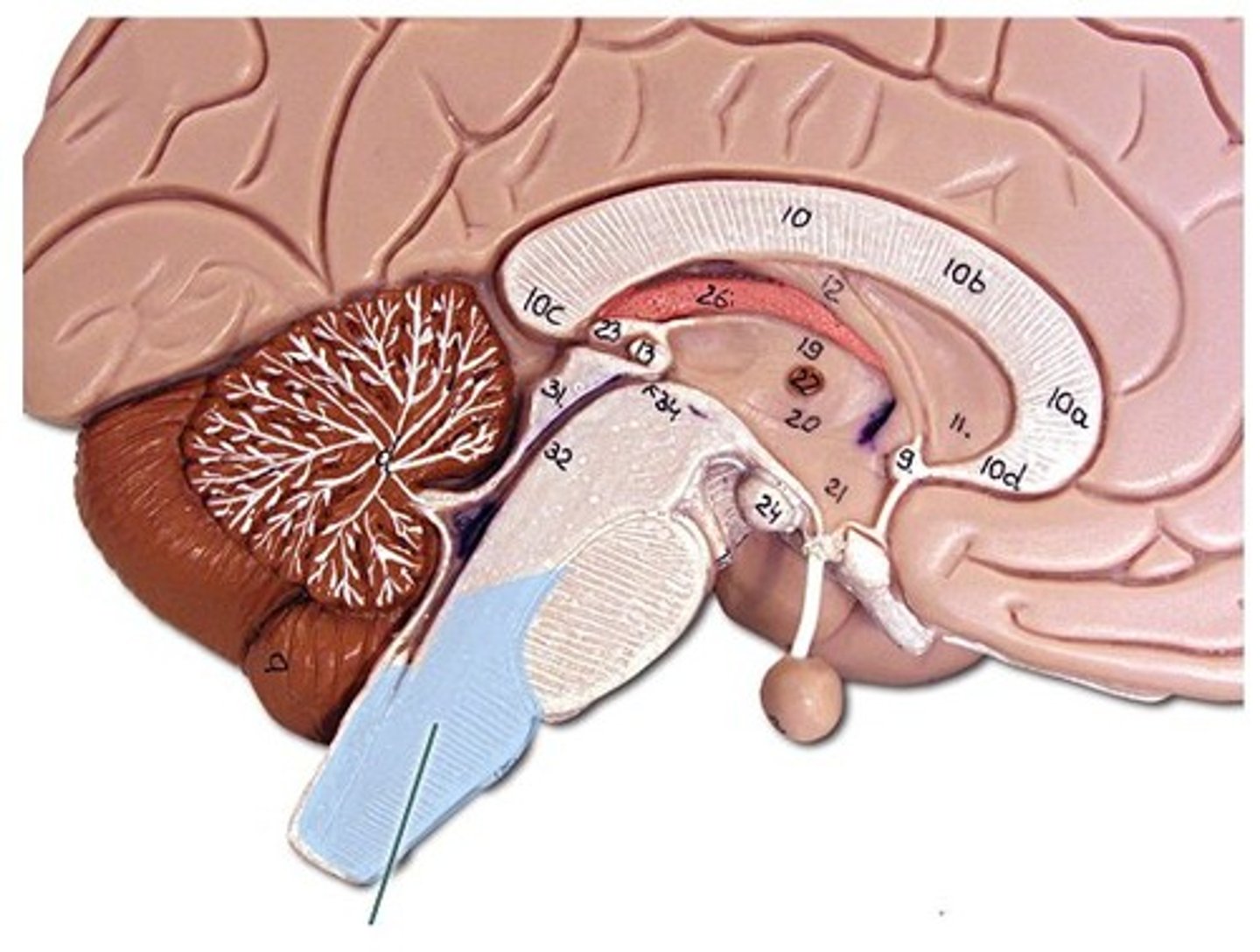
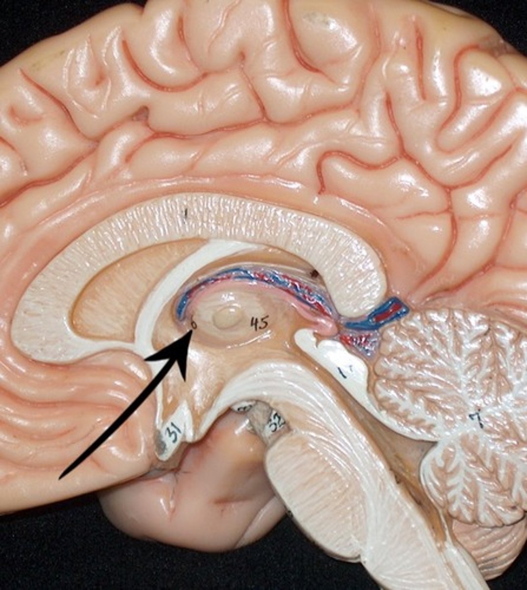
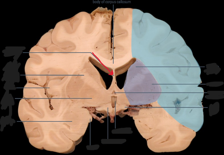
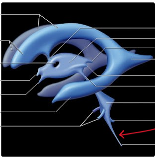
Thalamus
Structure. Eye on the seahorse
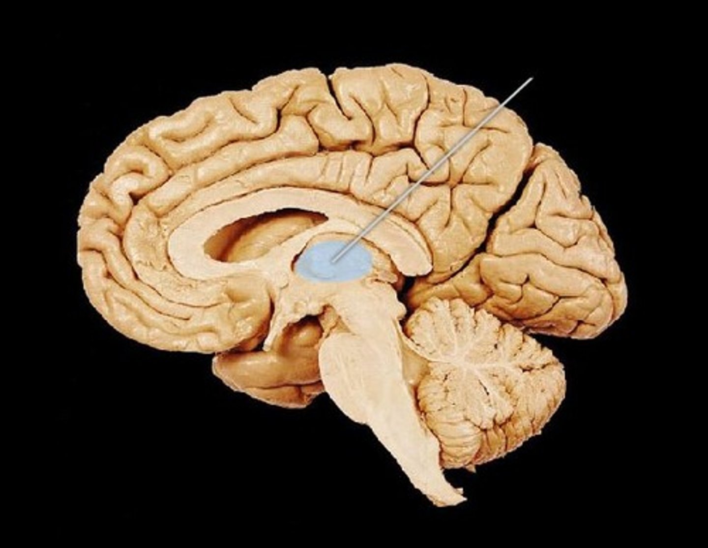
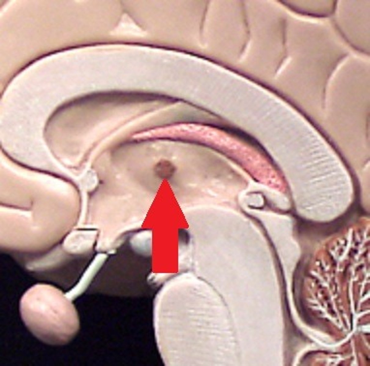
Interthalamic adhesion
feature. Pupil on eye, middle of the eye
Epithalamus
structure
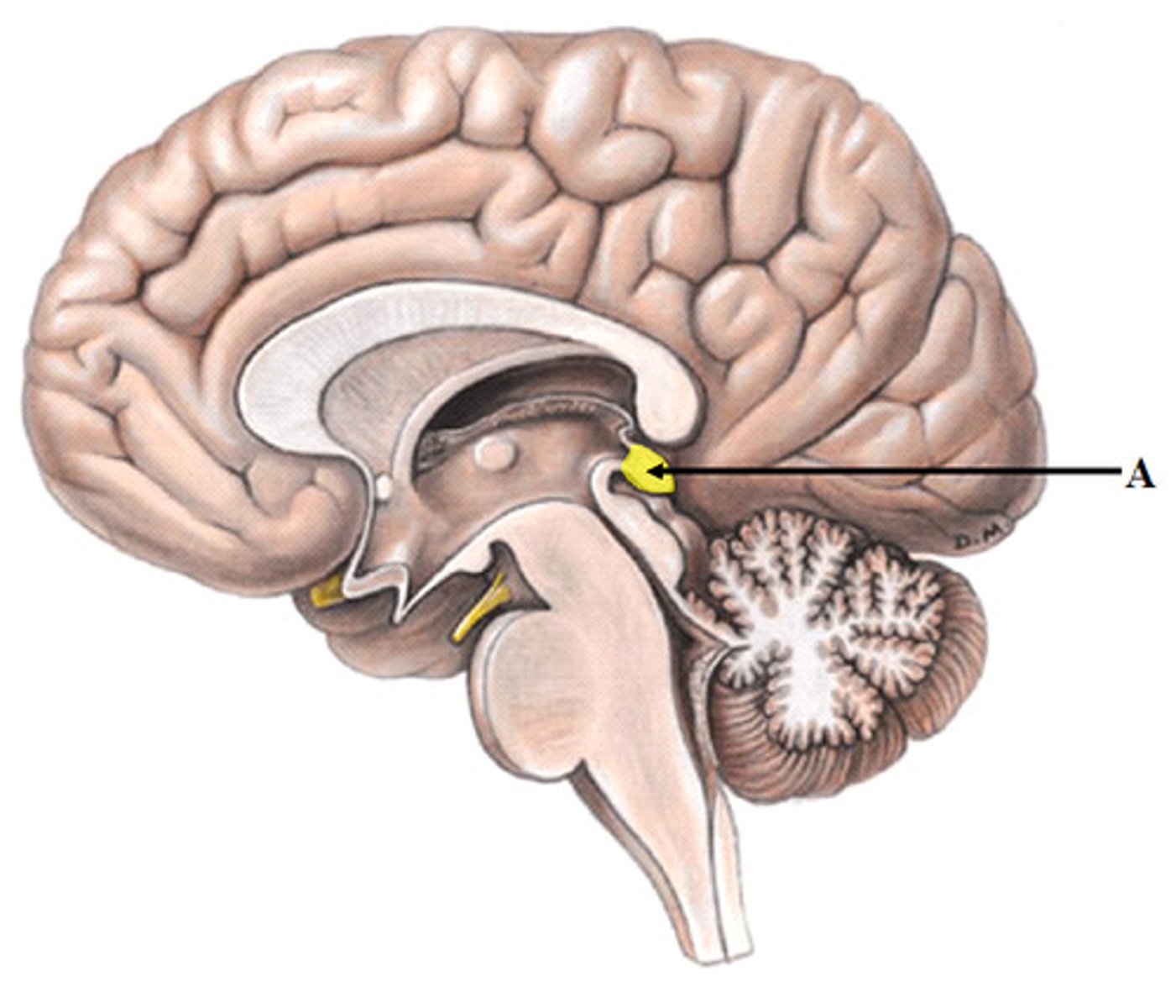
Pineal gland
structure
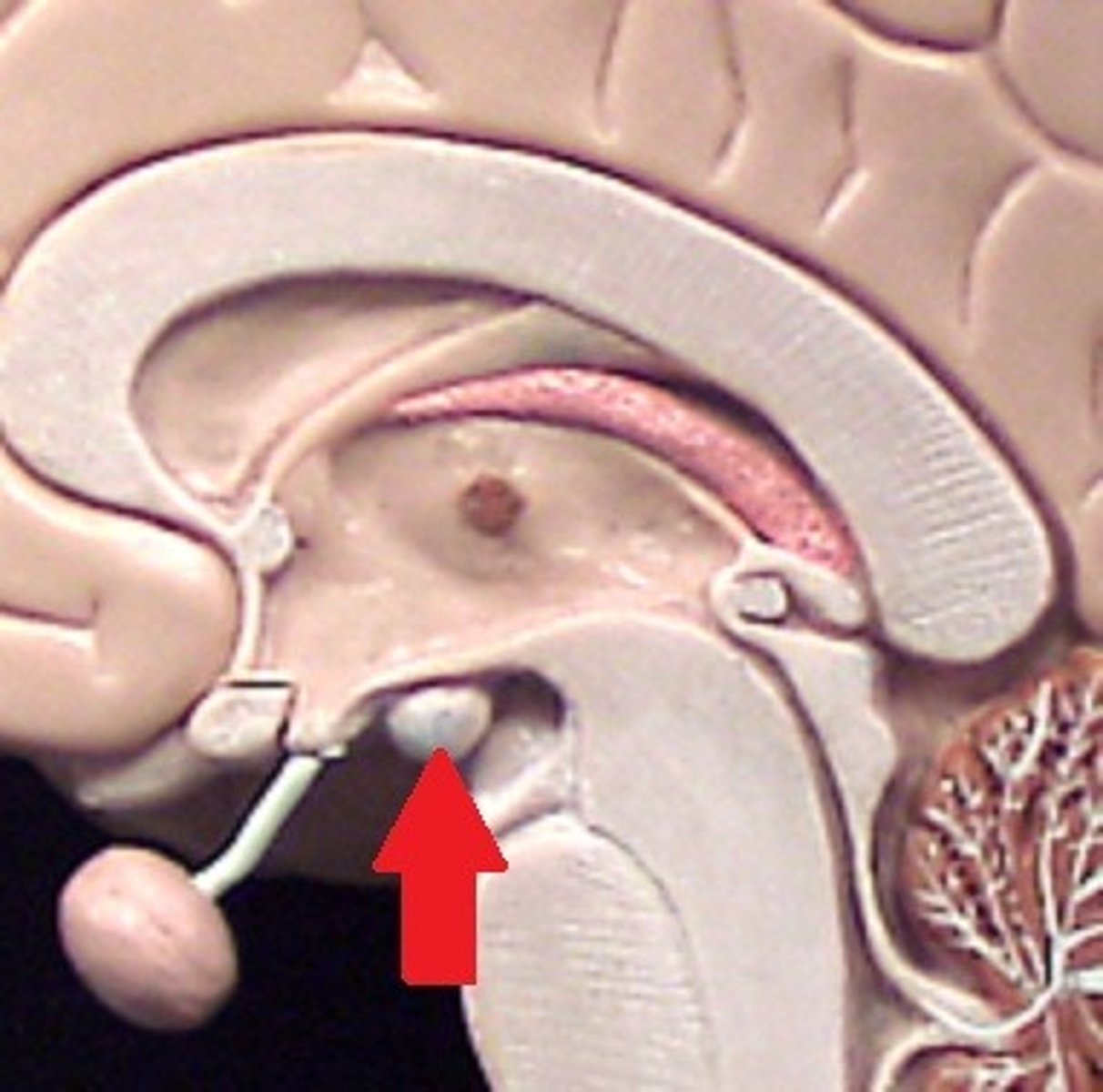
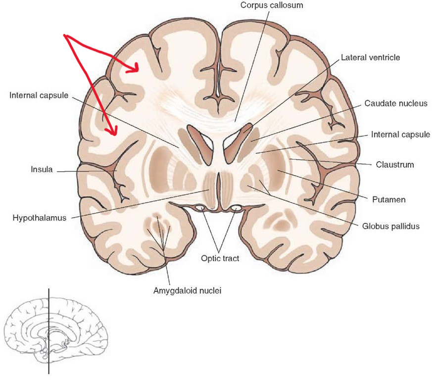
Hypothalamus
Structure. Nose of seahorse
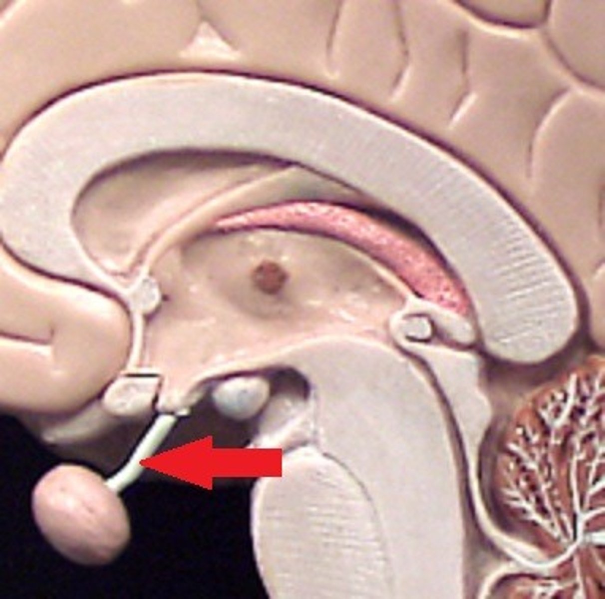
Infundibulum of the brain
Structure. Cherry STEM right before red part (cherry)
Mammillary bodies of the brain
Structure. Little bump near/ under nose
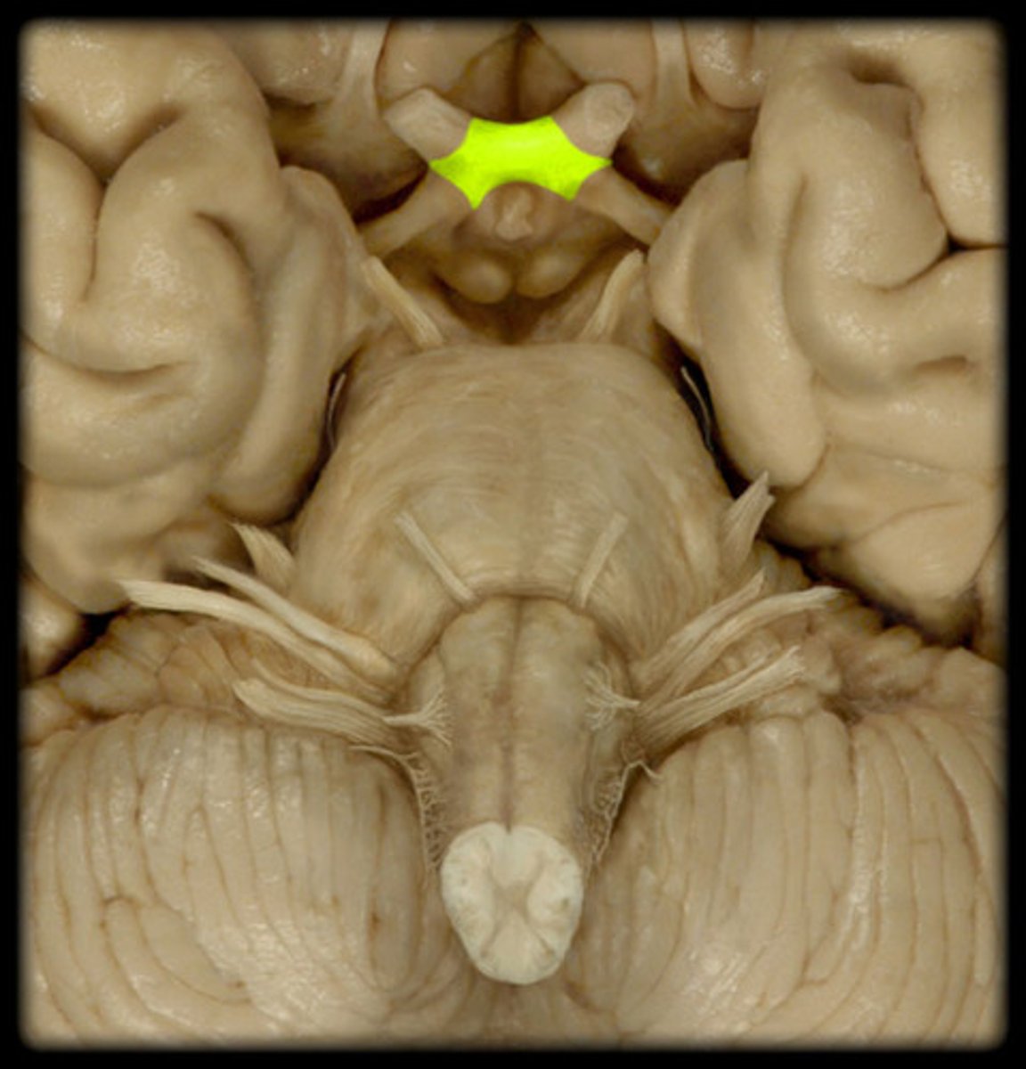
Optic chiasm
Structure. Shown on the brain/ X-shape
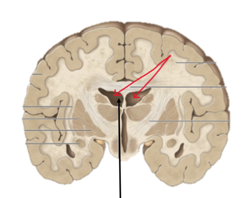
Third ventricle
Space
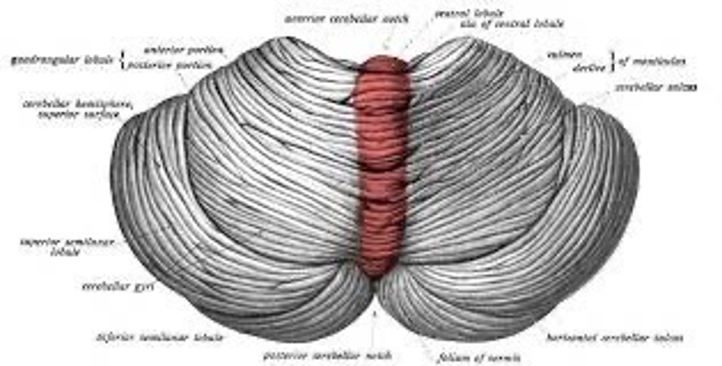
Cerebellar hemispheres
structure. 2 Balls
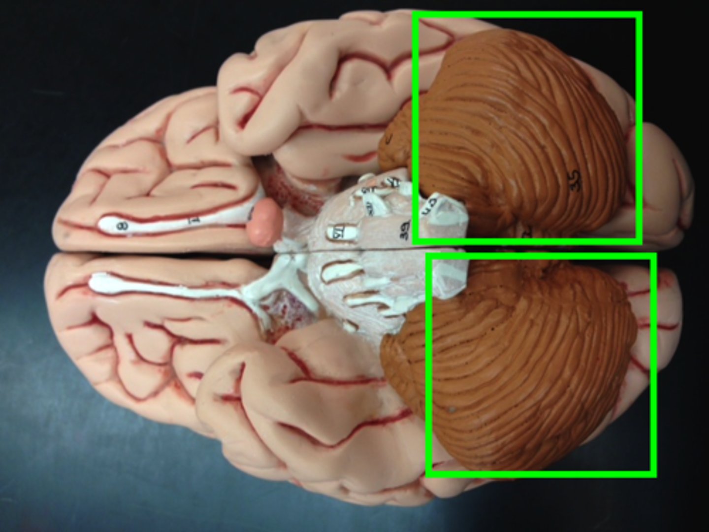
Folia of the cerebellum
Ridge. Ridges on top of balls
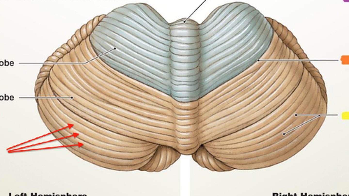
Fissures of the cerebellum
Depression. Space in balls

Vermis of the cerebellum
Division. Line separating balls
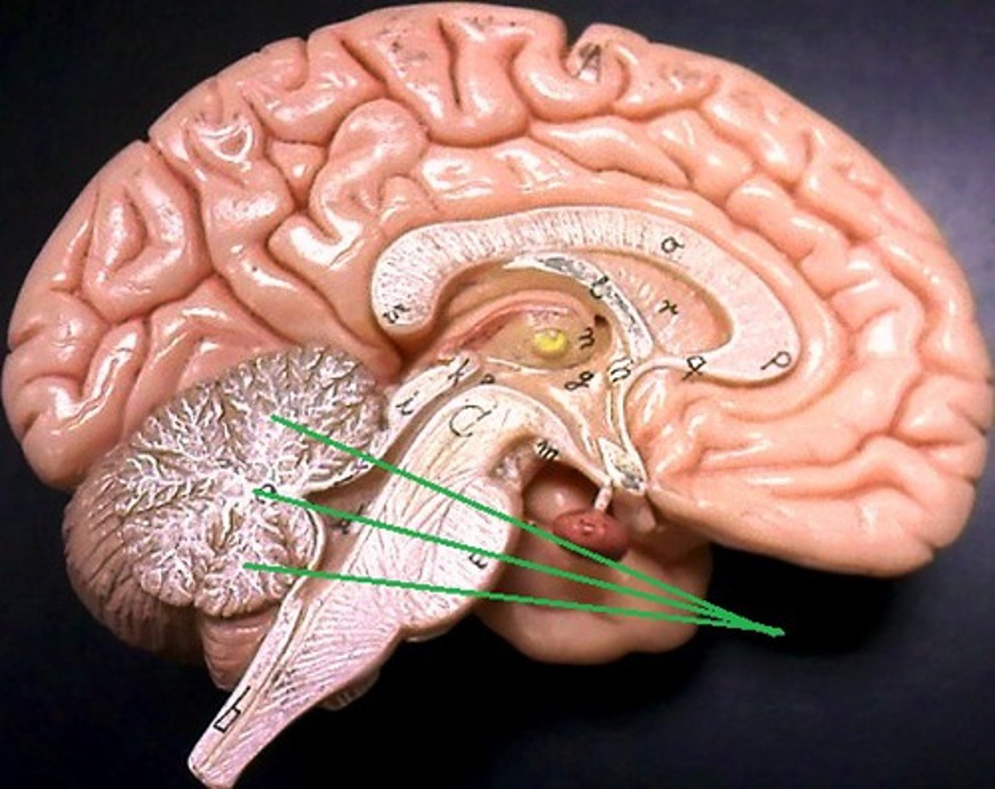
Cerebellar cortex
Structure. Tan around white
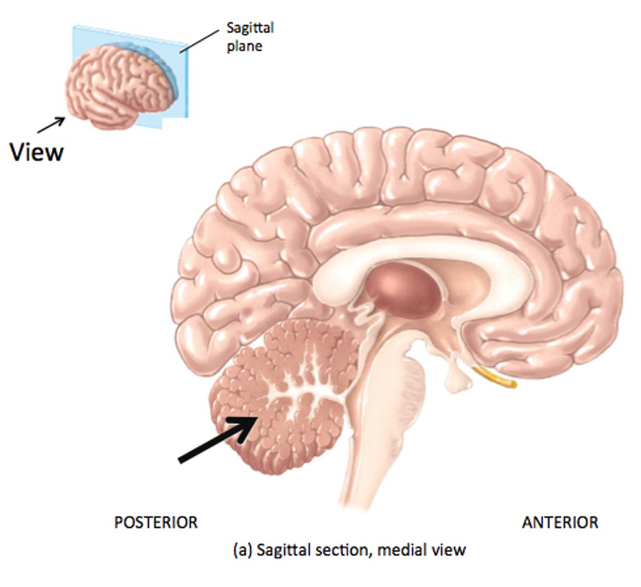
Arbor vitae
Feature. white snowflake
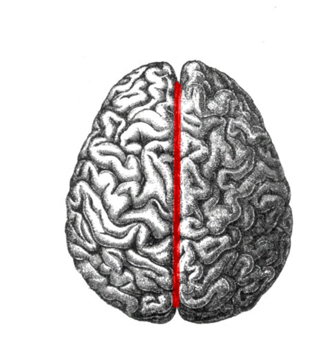
Cerebral hemispheres
Structure. Halves of brain/ Will put hand over one half

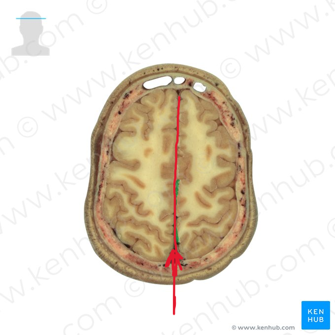
Longitudinal fissure
space
Cerebral cortex
Gray matter. Any gray on the outside of the brain steak
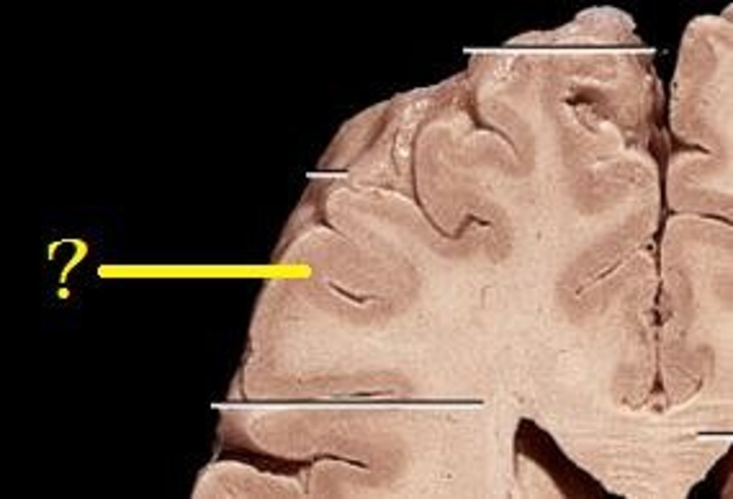
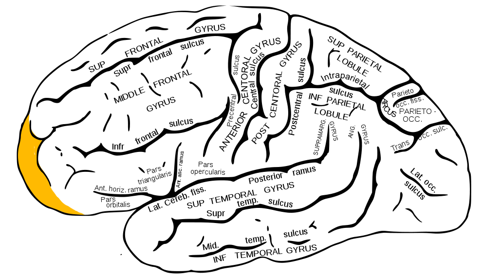
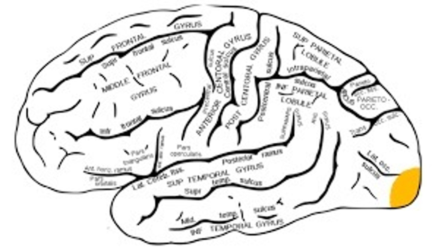
Cerebral gyri
ridges
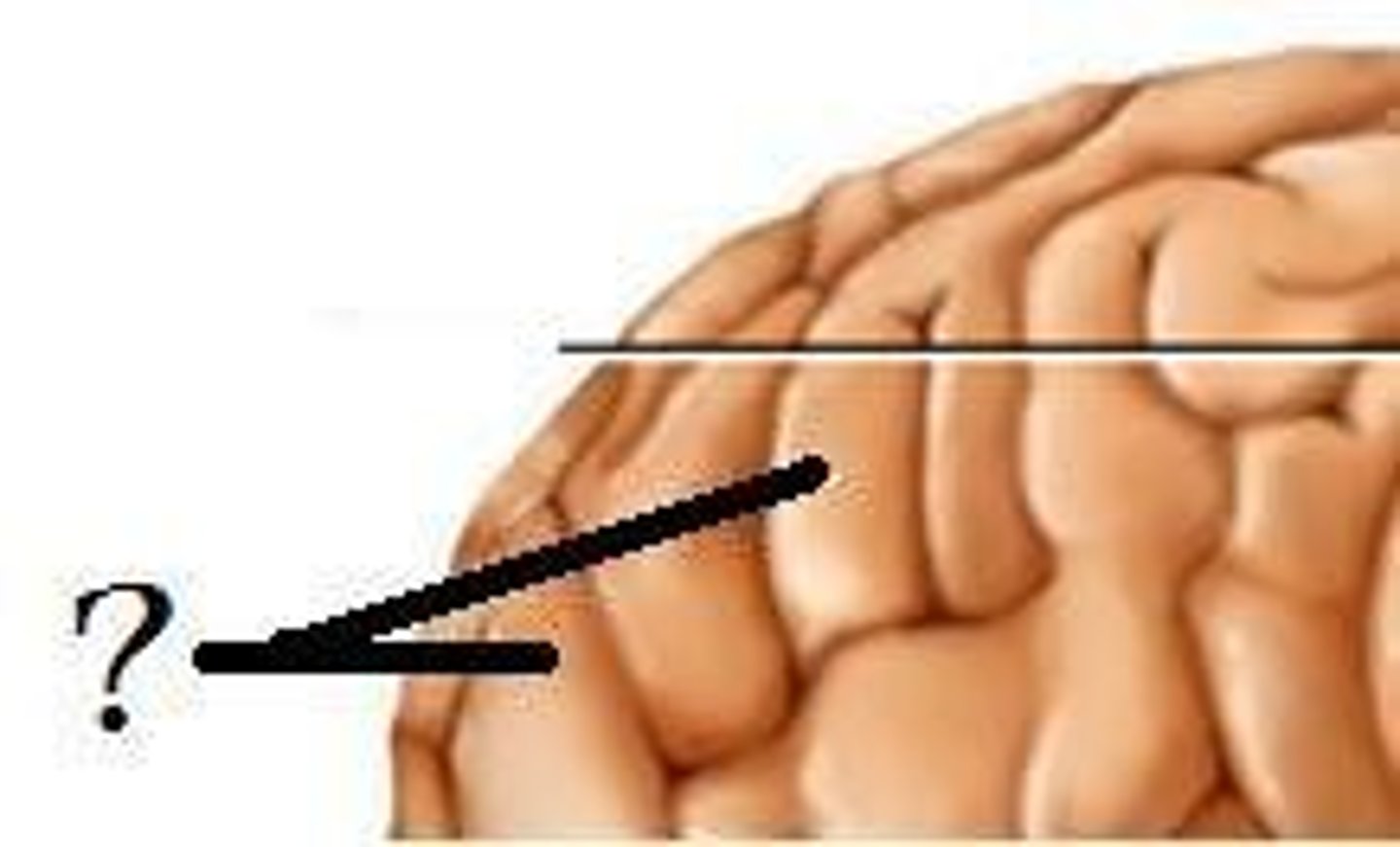
Cerebral sulci
depression. in between squiggles
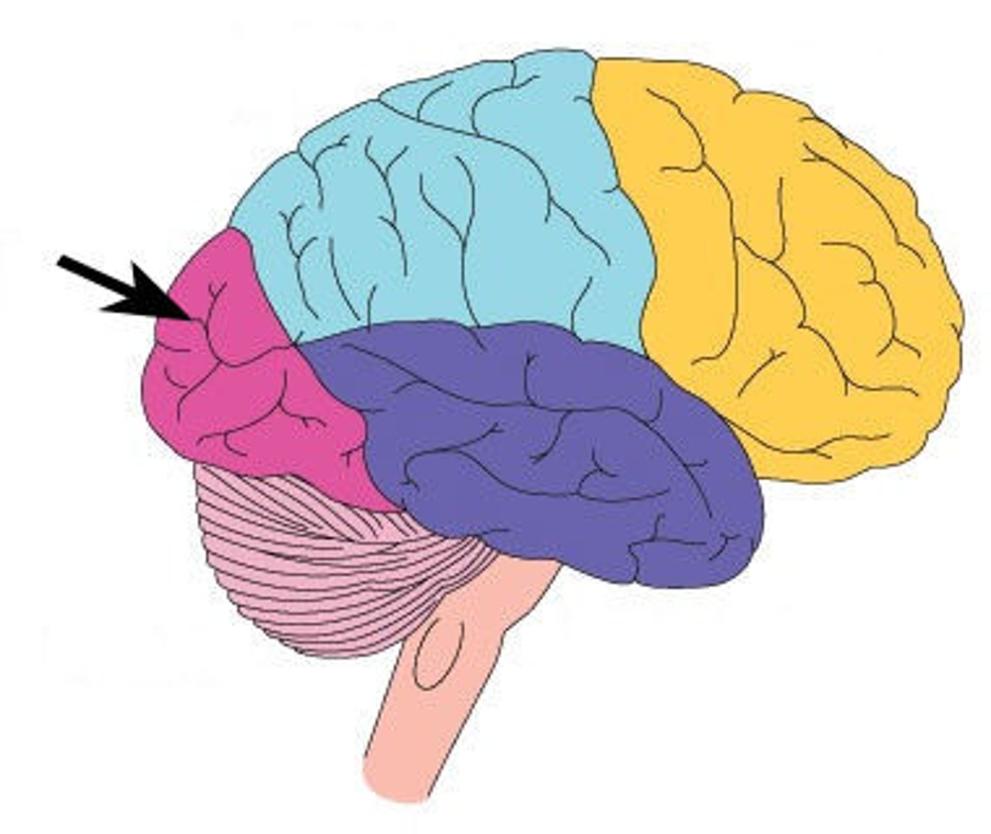
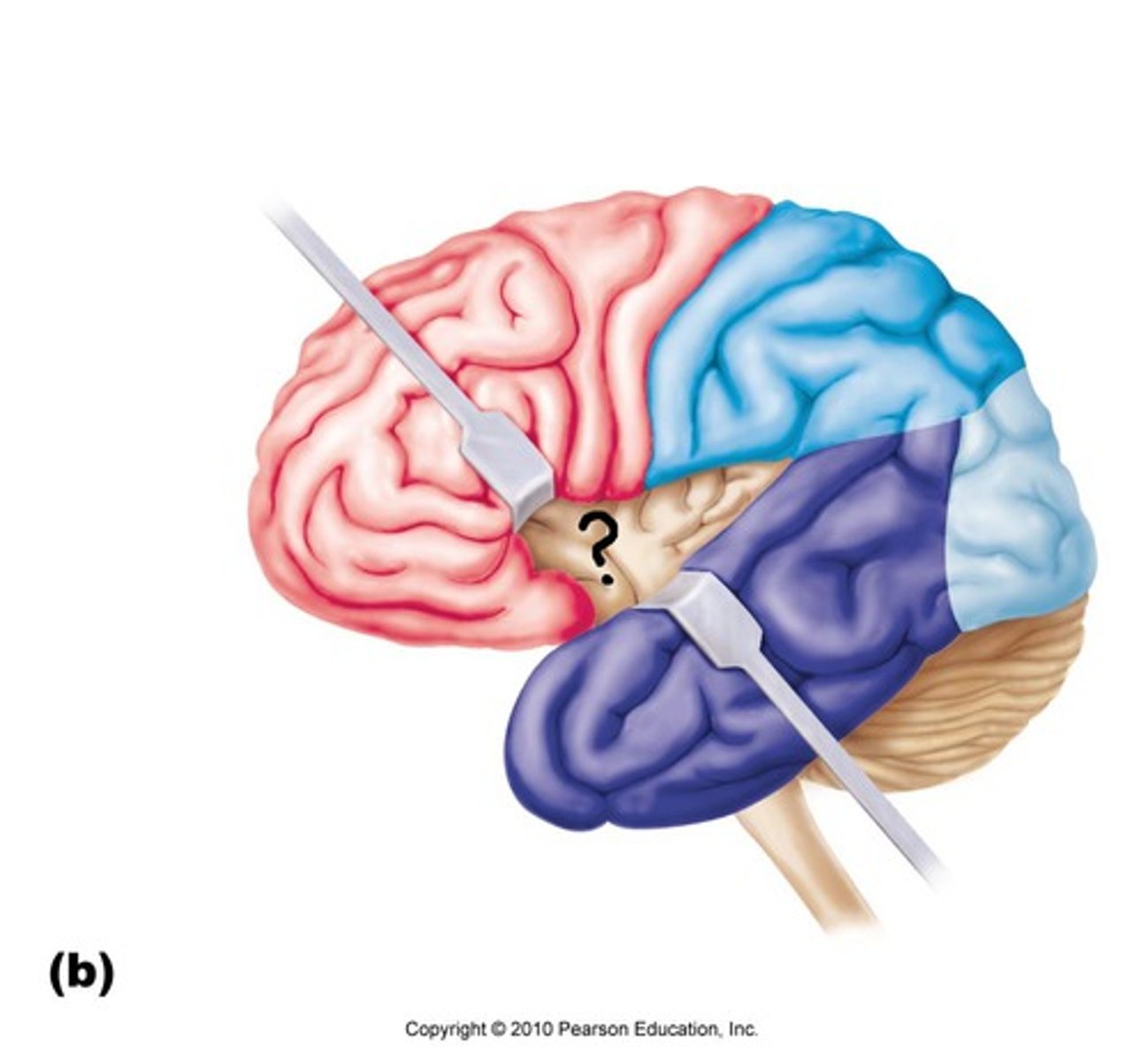
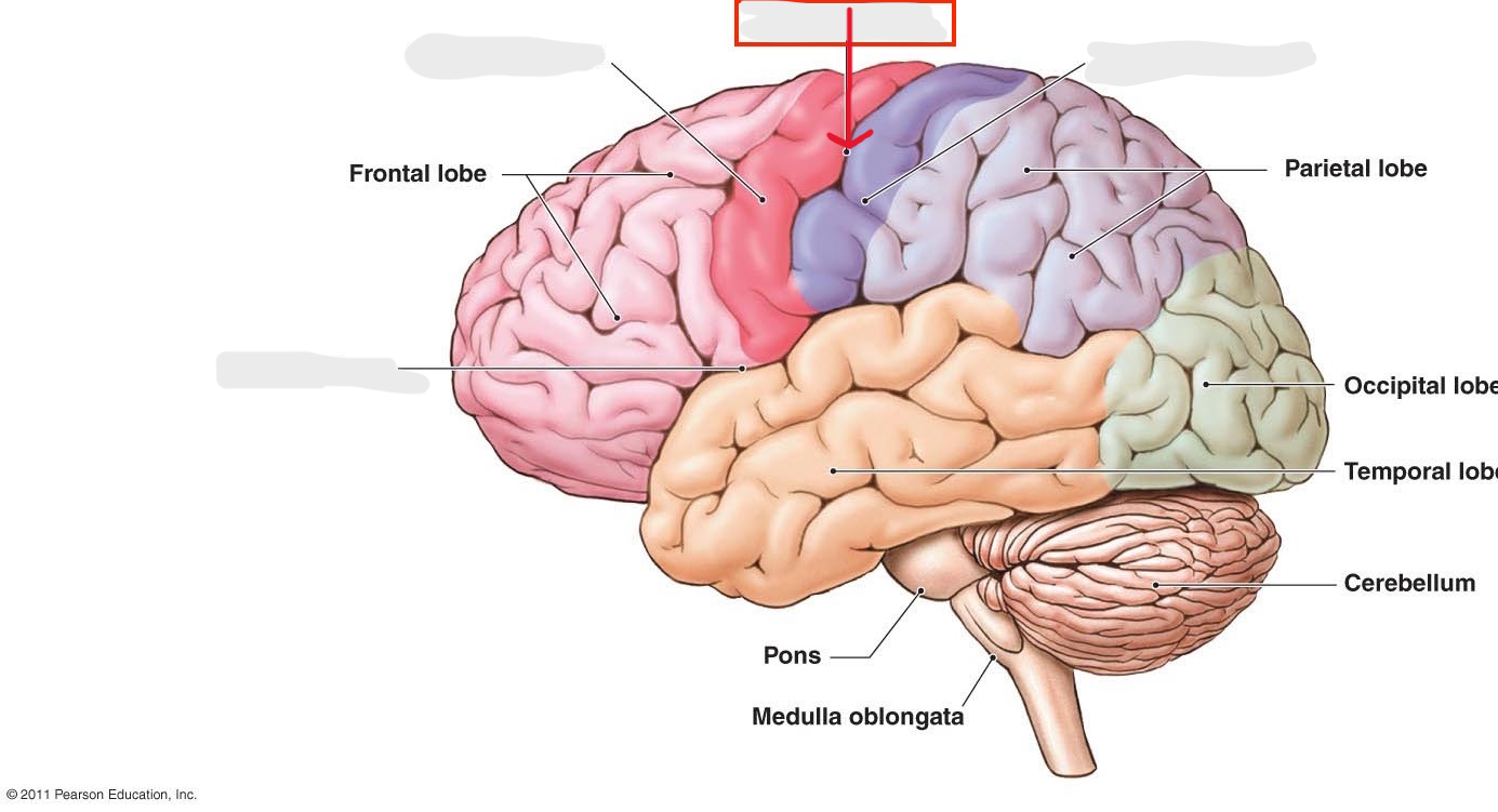
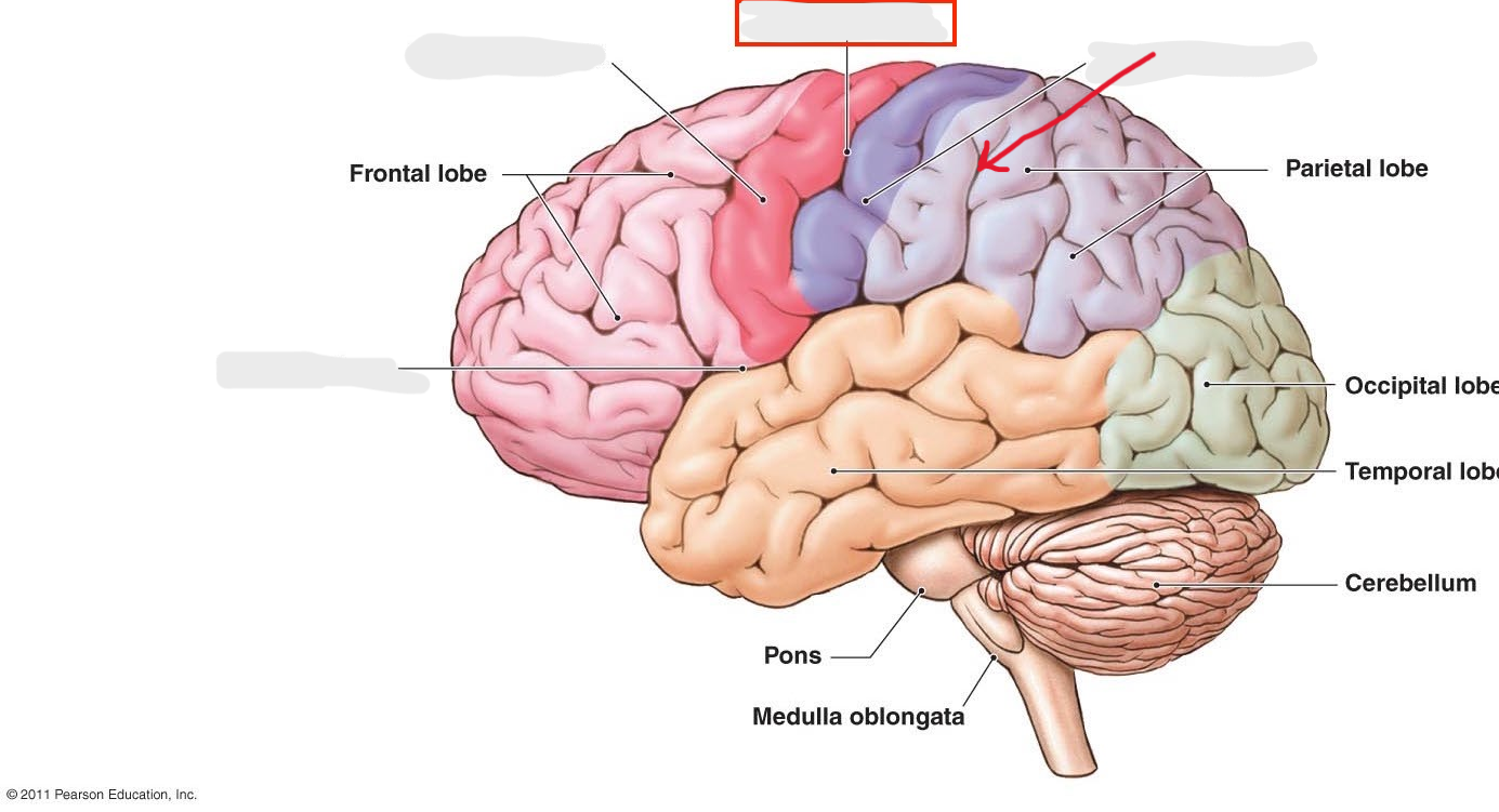
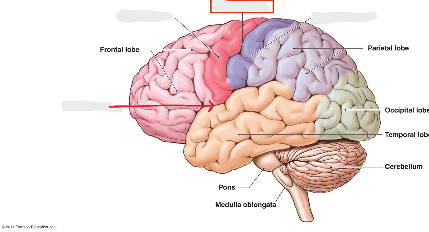
Frontal lobes
region
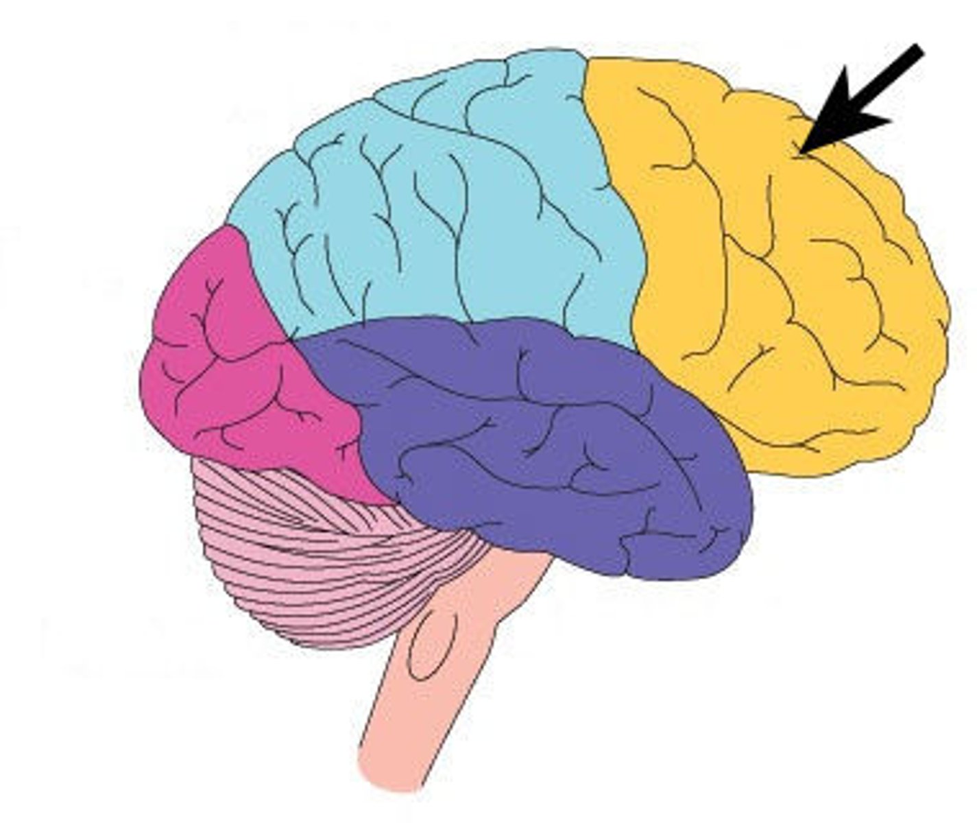
Frontal poles
feature. Will tap with 2 fingers on frontal lobe
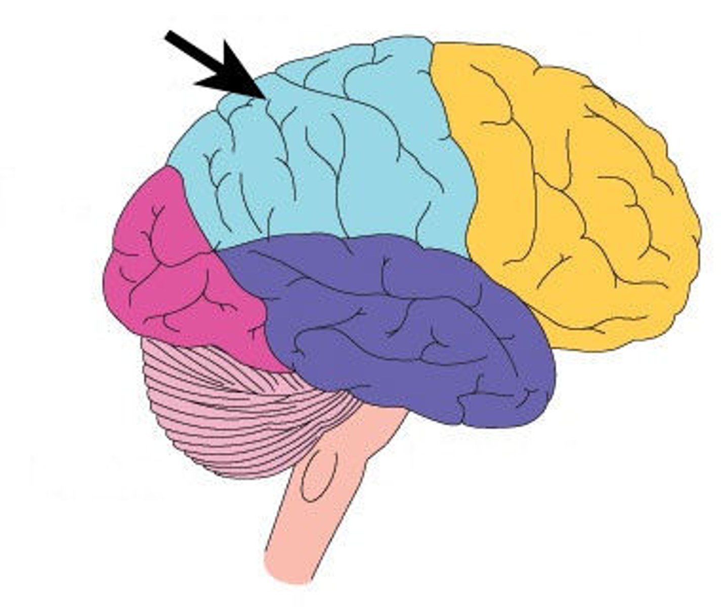
Parietal lobes
region
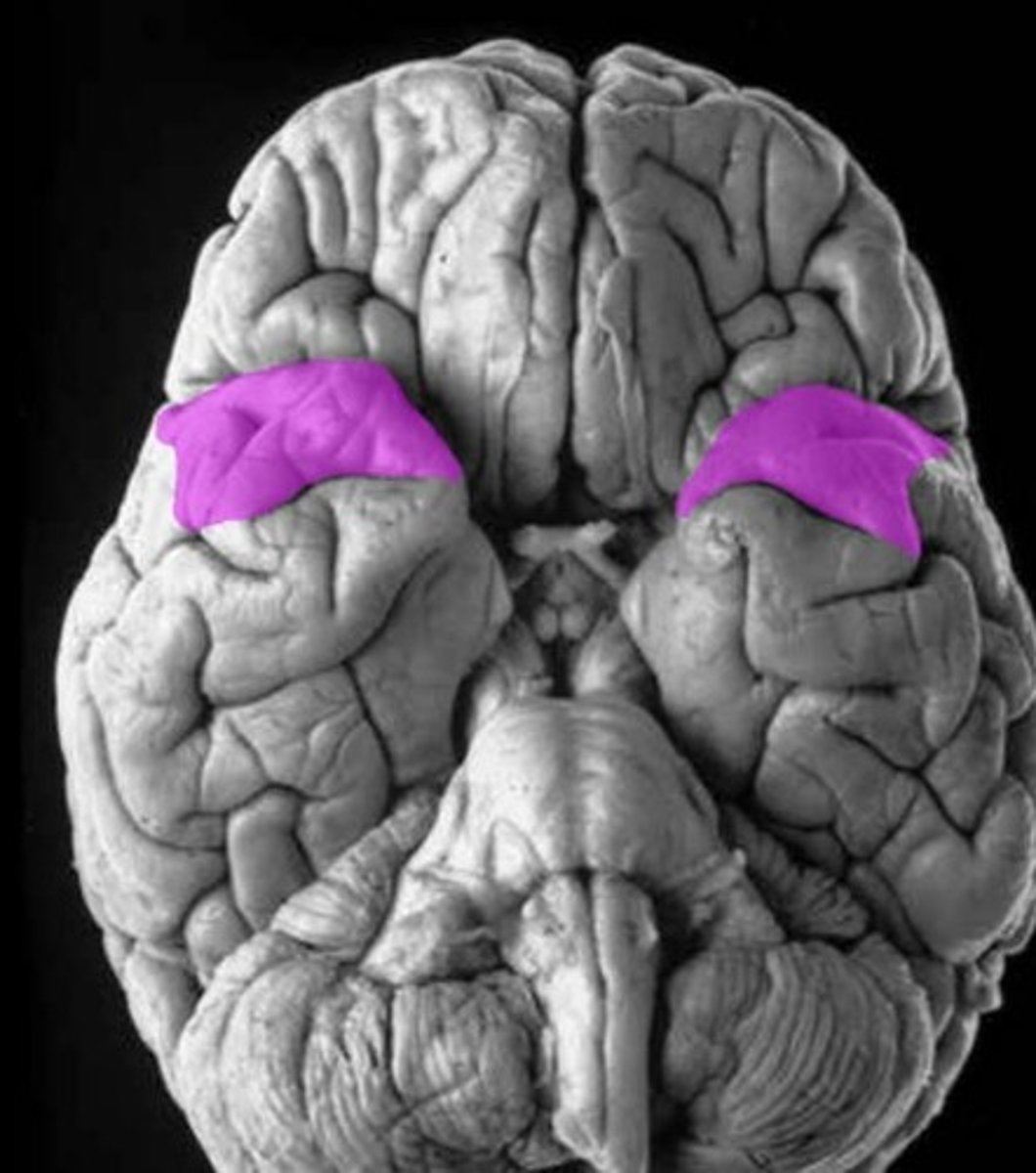
Temporal lobes
region
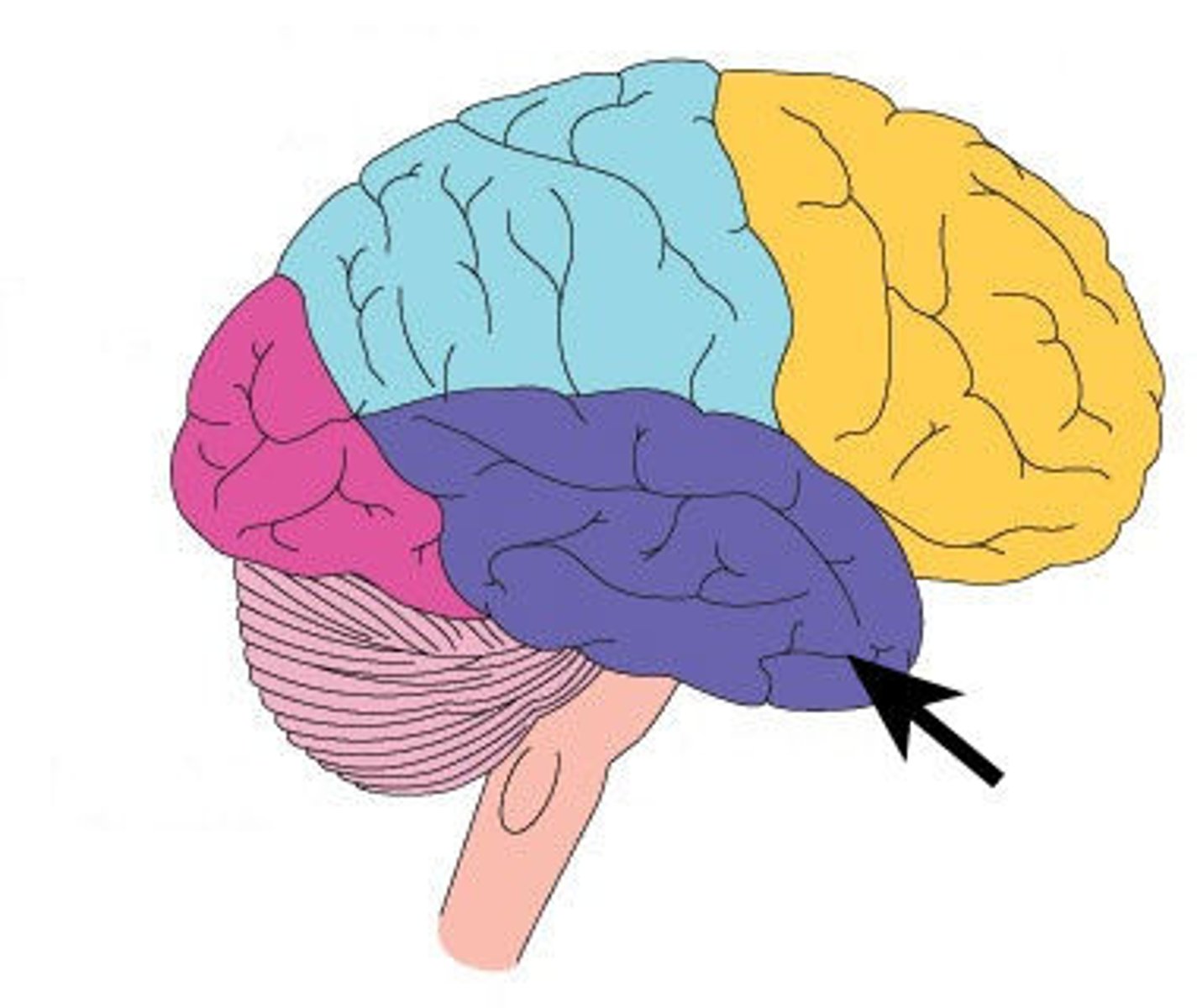
Temporal poles
feature. Will tap with 2 fingers on the bottom side
Occipital lobes
region
Occipital poles
feature. Will tap with 2 fingers
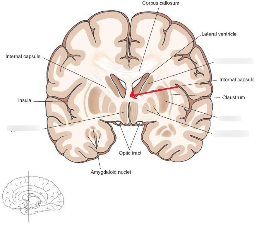
Insula
internal region
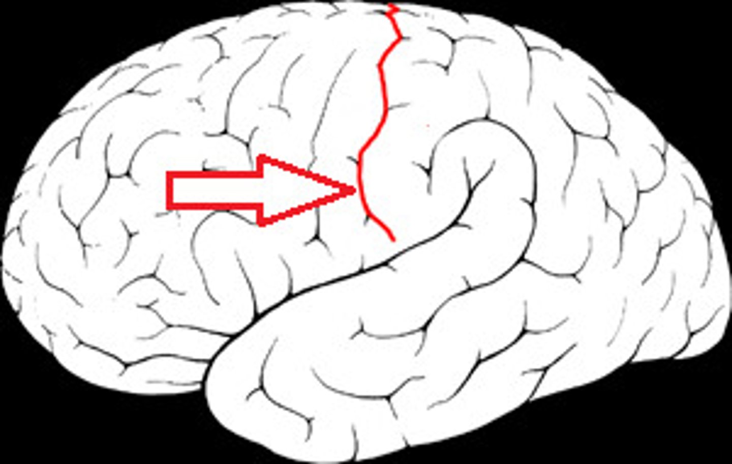
Central Sulcus
depressions
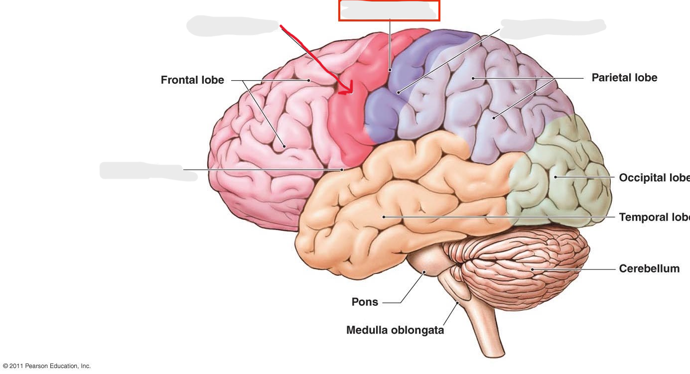
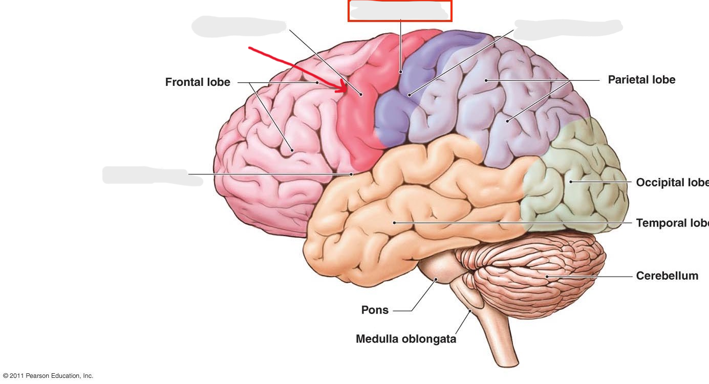
Precentral gyrus
ridge
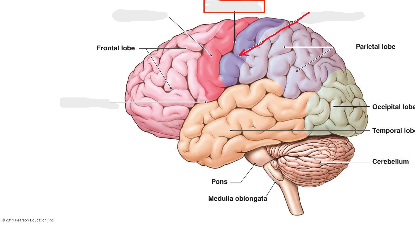
Postcentral gyrus
ridge
Precentral sulcus
depression
Postcentral sulcus
depression
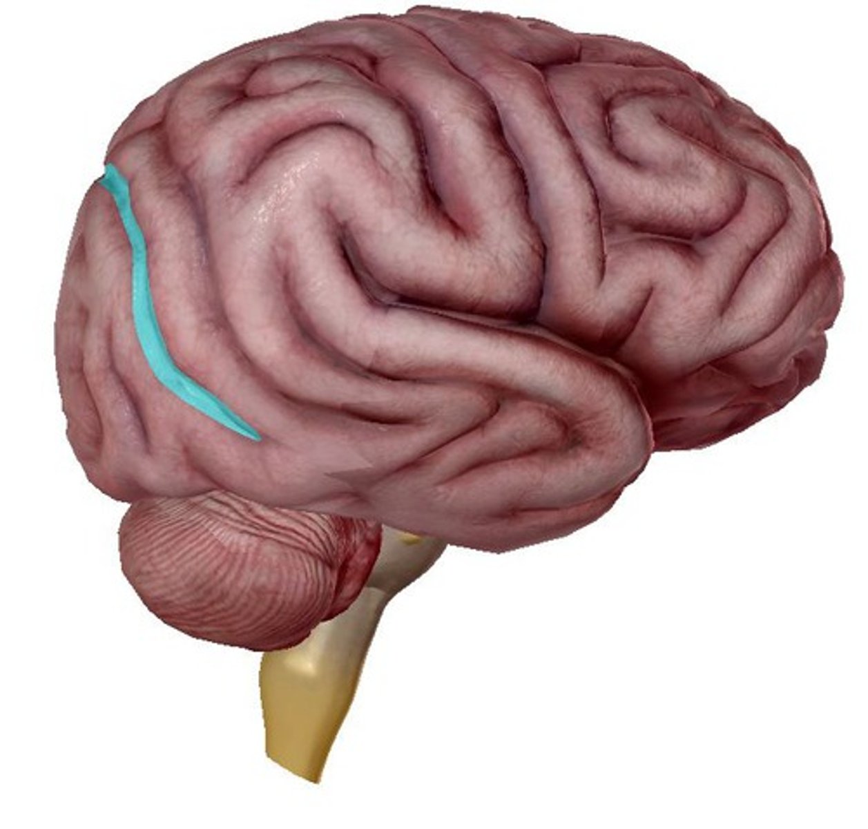
Lateral sulcus
depression
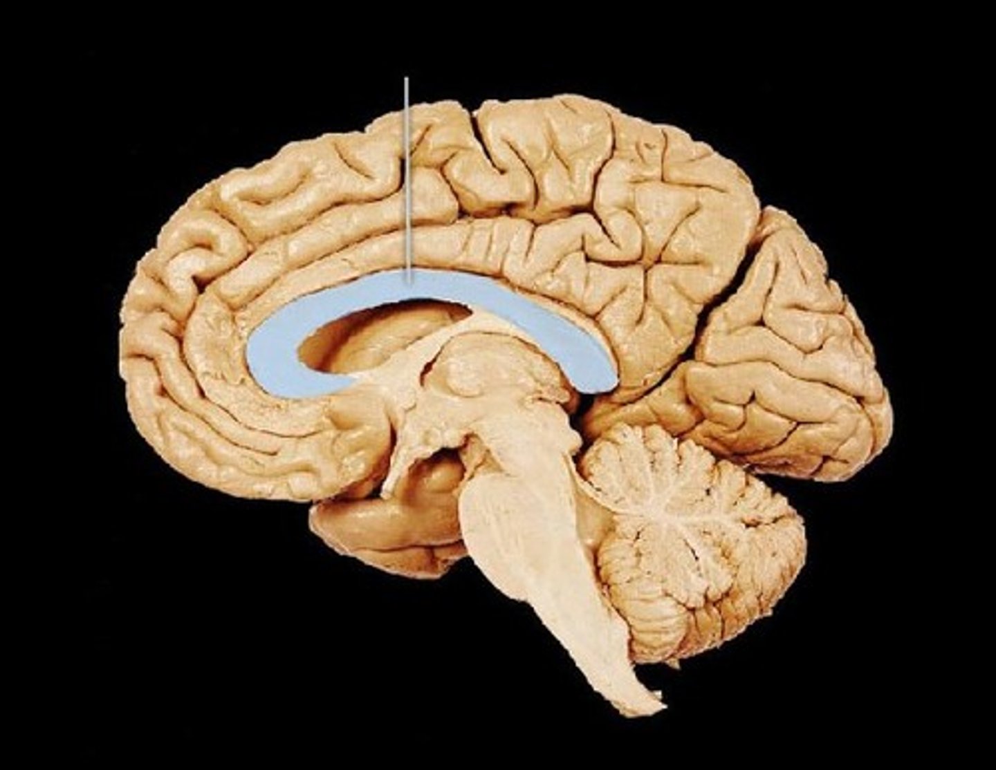
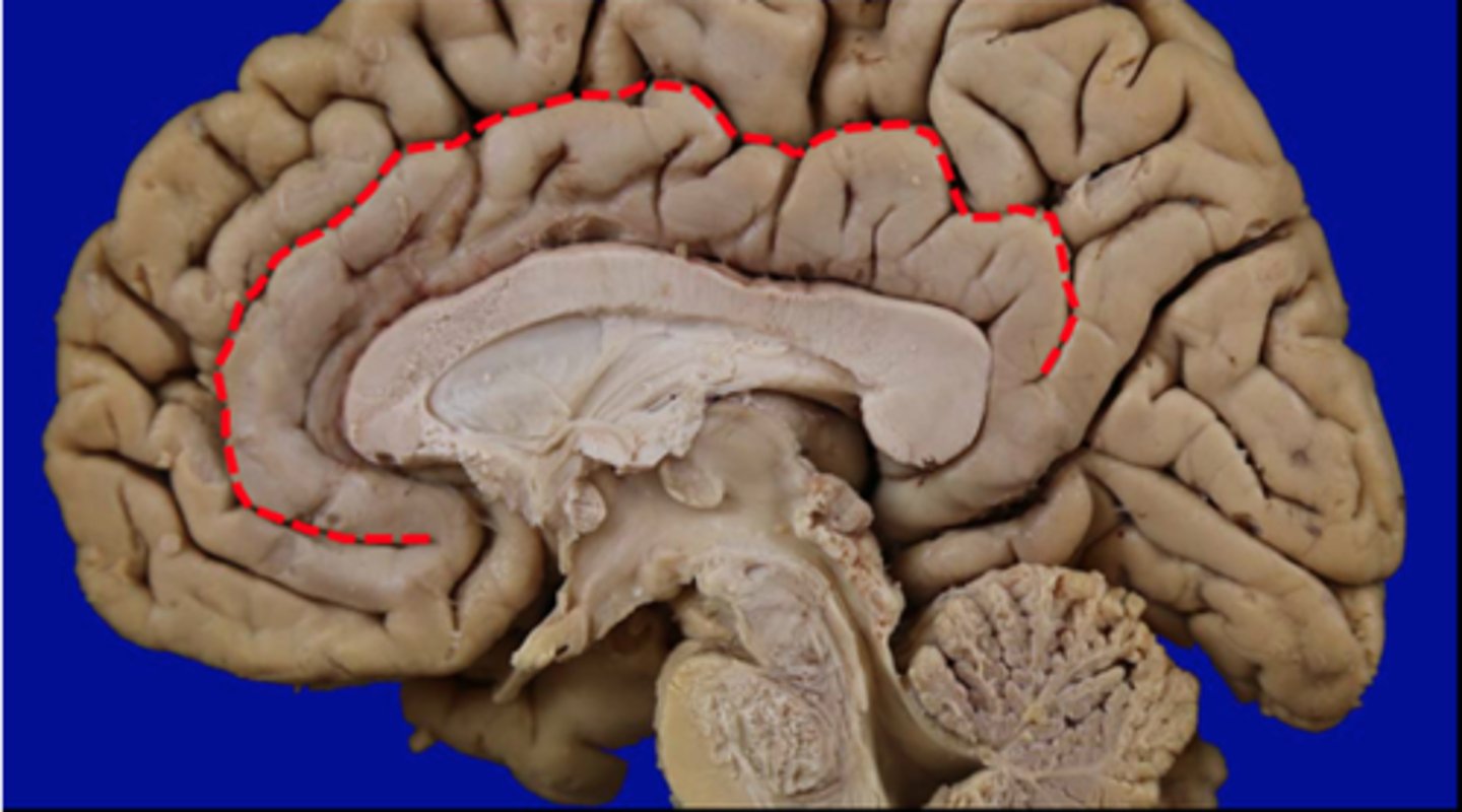
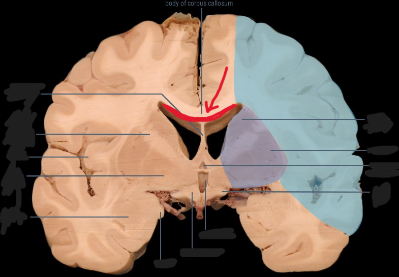
Corpus callosum
structure, first sombrero
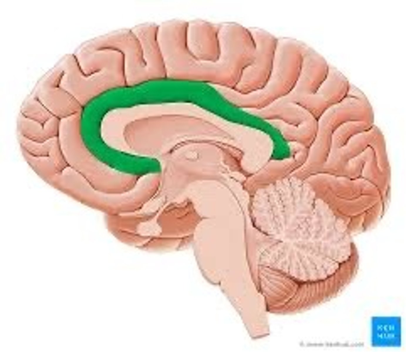
Cingulate gyrus
ridge, bigger sombrero
Cingulate sulcus
depression
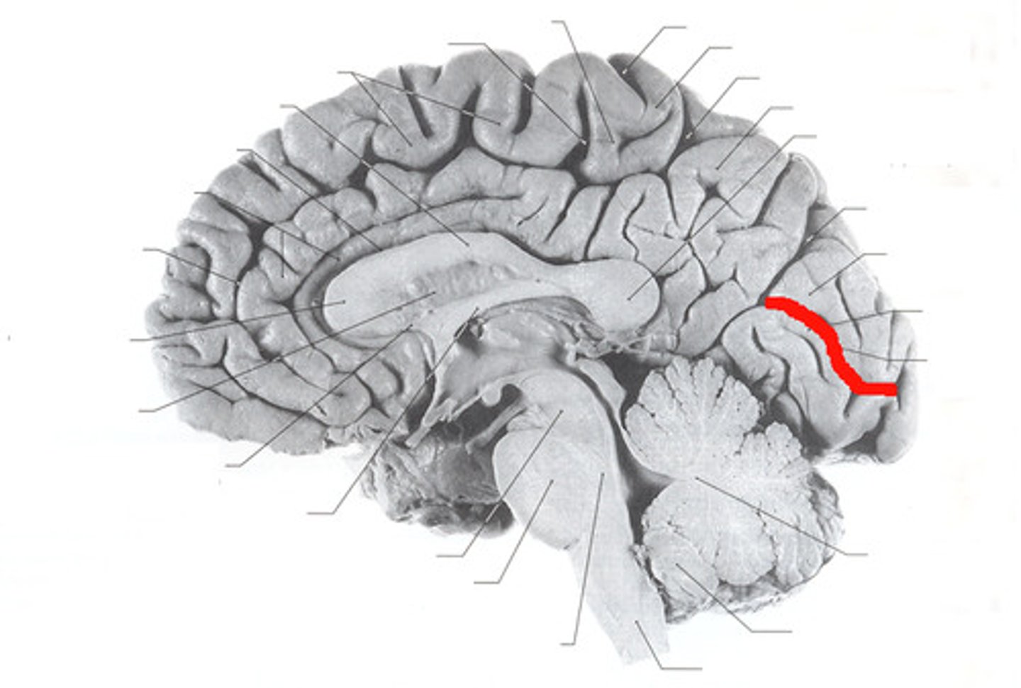
parieto-occipital sulcus
depression. between parietal/ occipital lobe
Calcarine sulcus
depression, straight out the back
Fornix
structure
Septum Pellucidum
structure
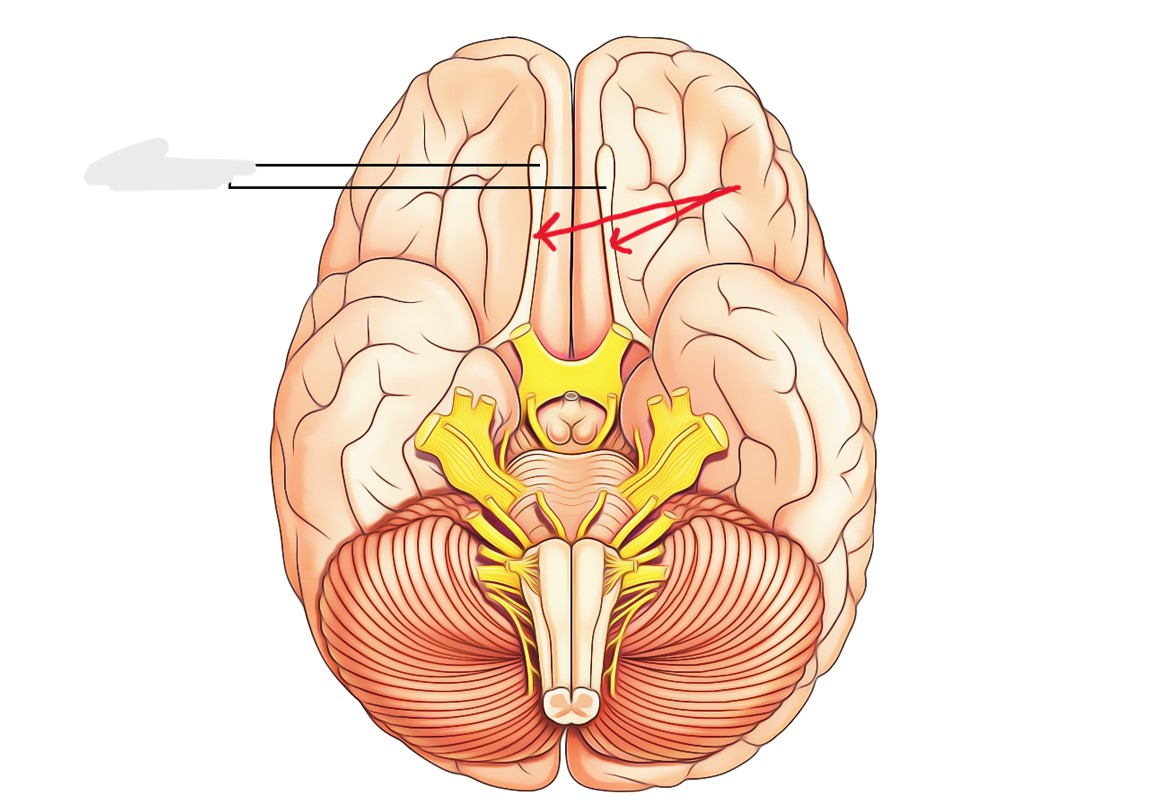
olfactory bulbs
feature
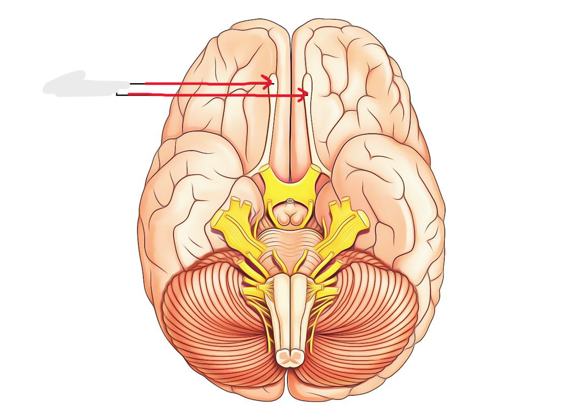
Olfactory tracts
structure
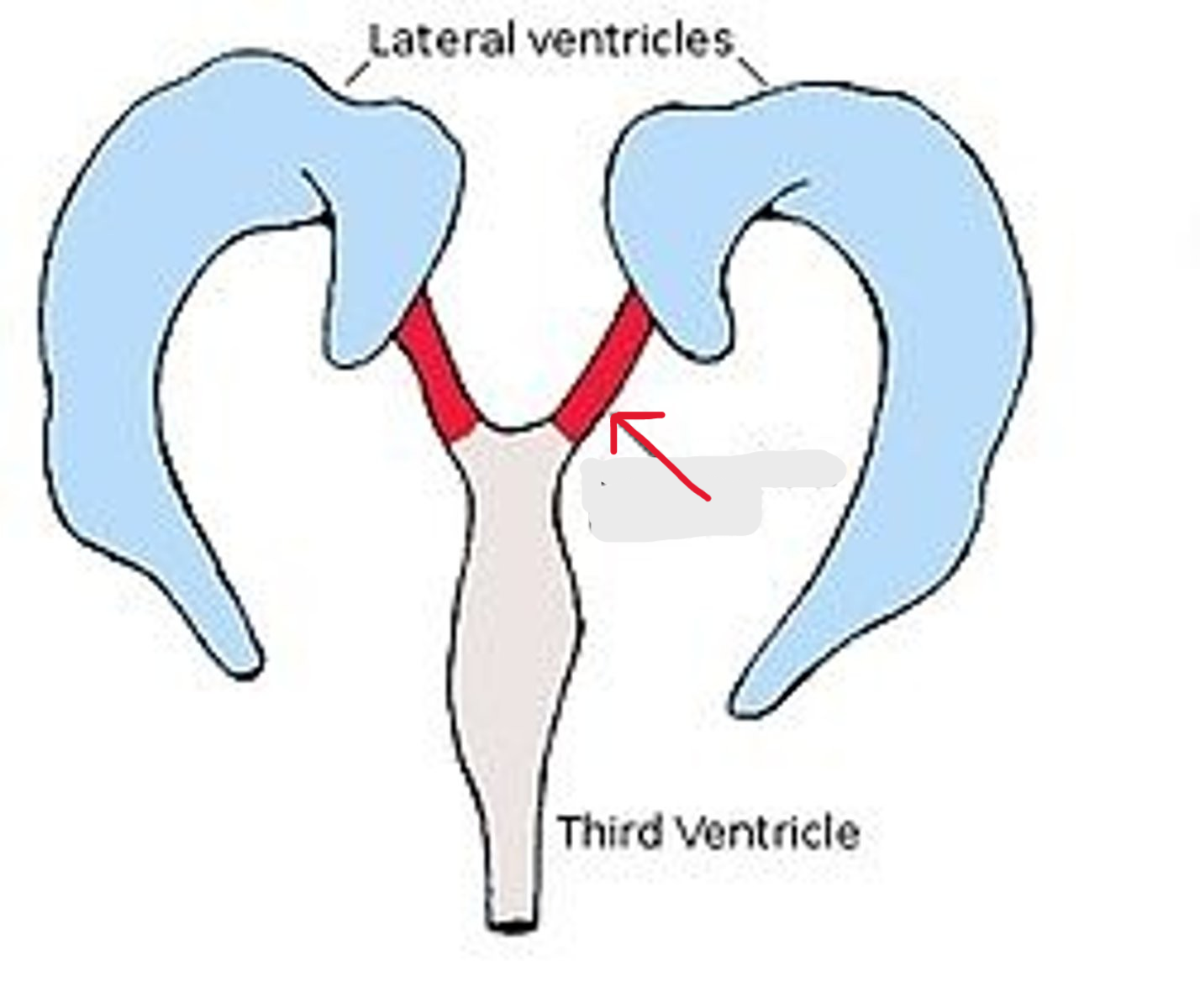
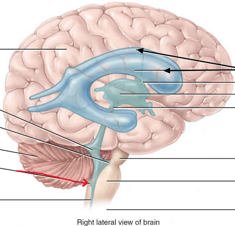
Lateral ventricles
spaces
Septal nuclei
union
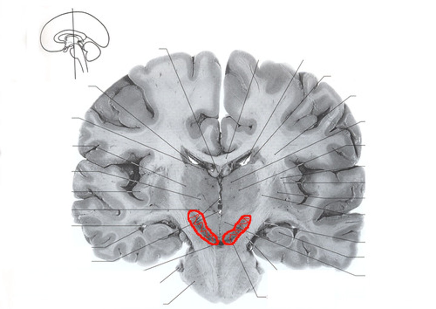
Hippocampus
structure. *cinnamon roll*
Corona radiata
white matter
Cranial dura mater
covering
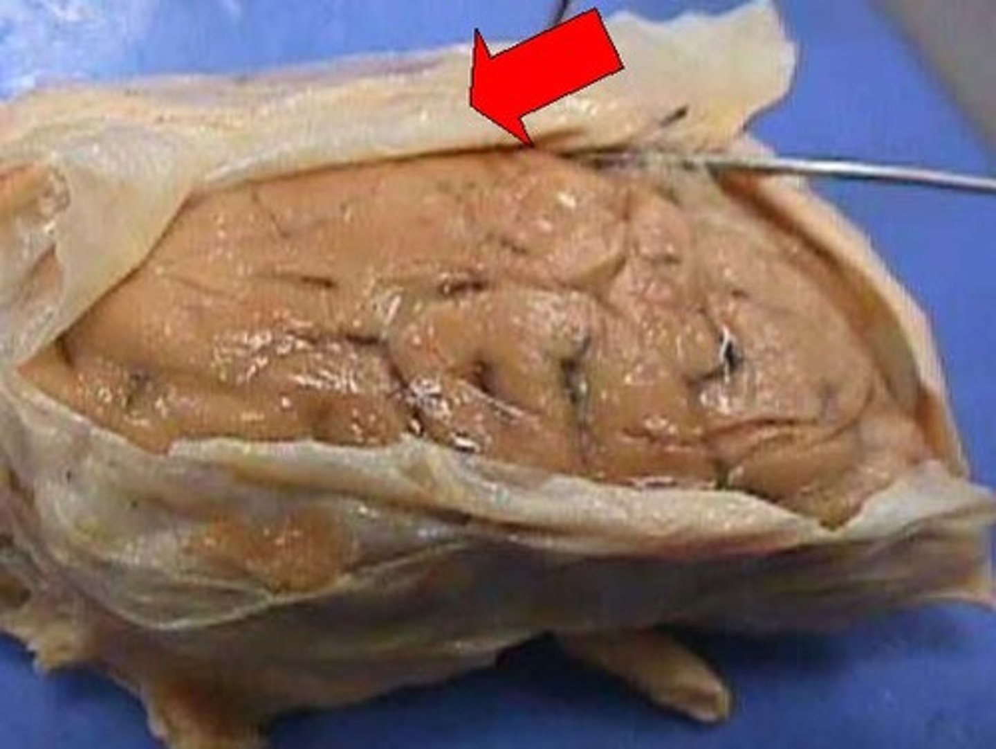
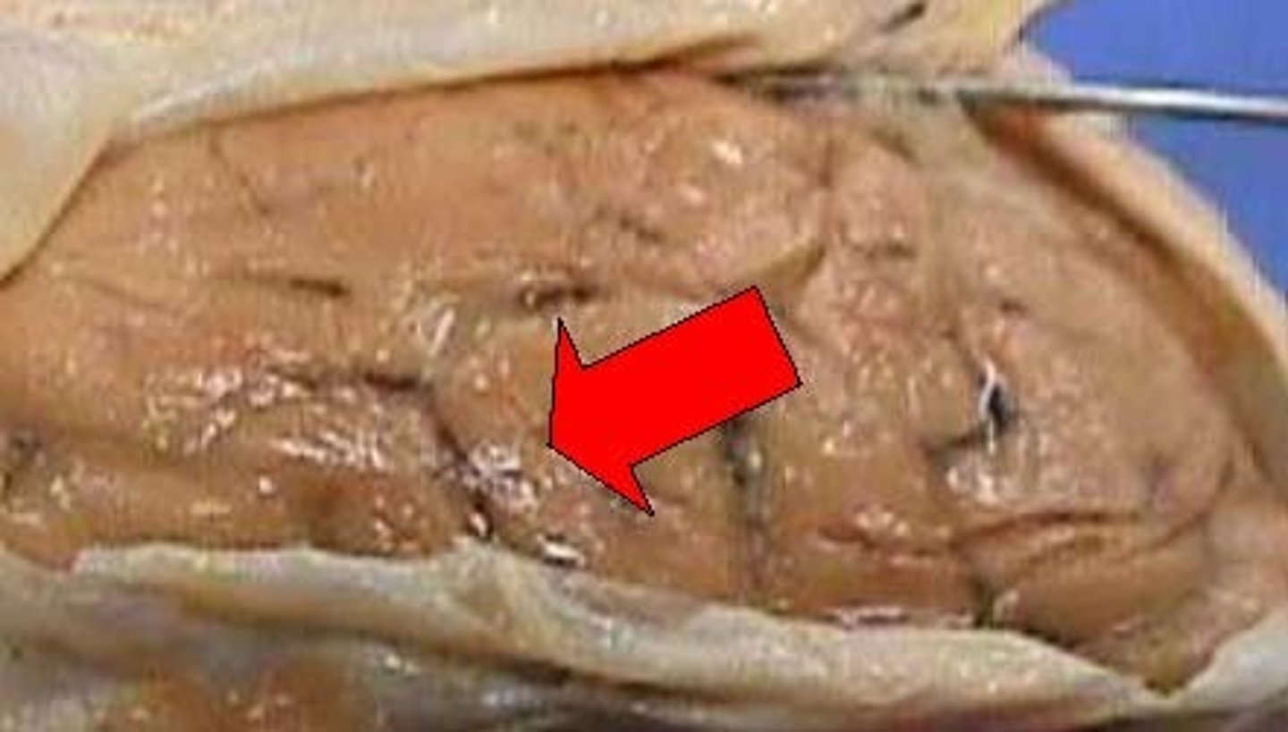
Cranial arachnoid mater
covering. webby like covering
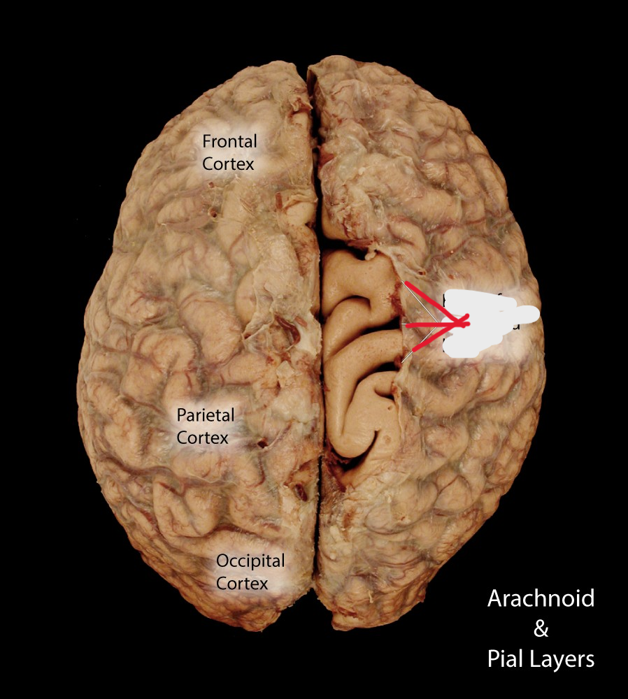
Cranial pia mater
Covering. tan portion of brain on bottom
Falx cerebri
Infolding. Thin sheet separates hemispheres
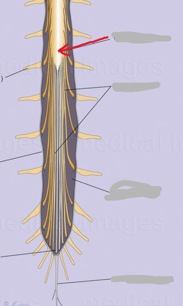
Conus Medullaris
Structure
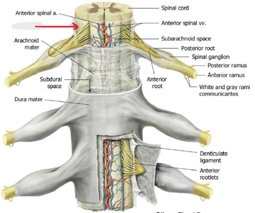
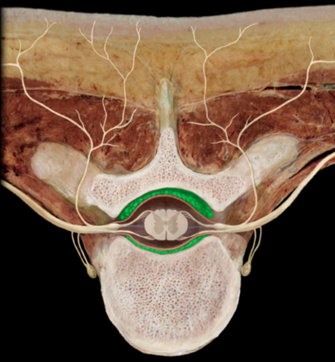
Ventral rootlets
structure
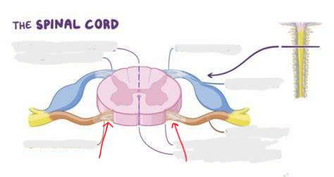
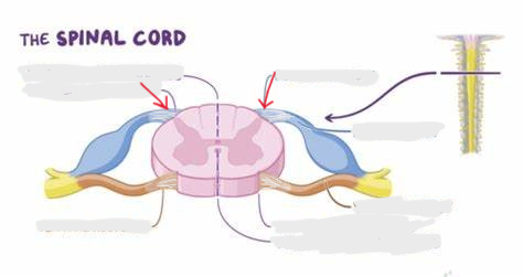
Dorsal rootlets
structure
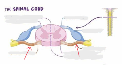
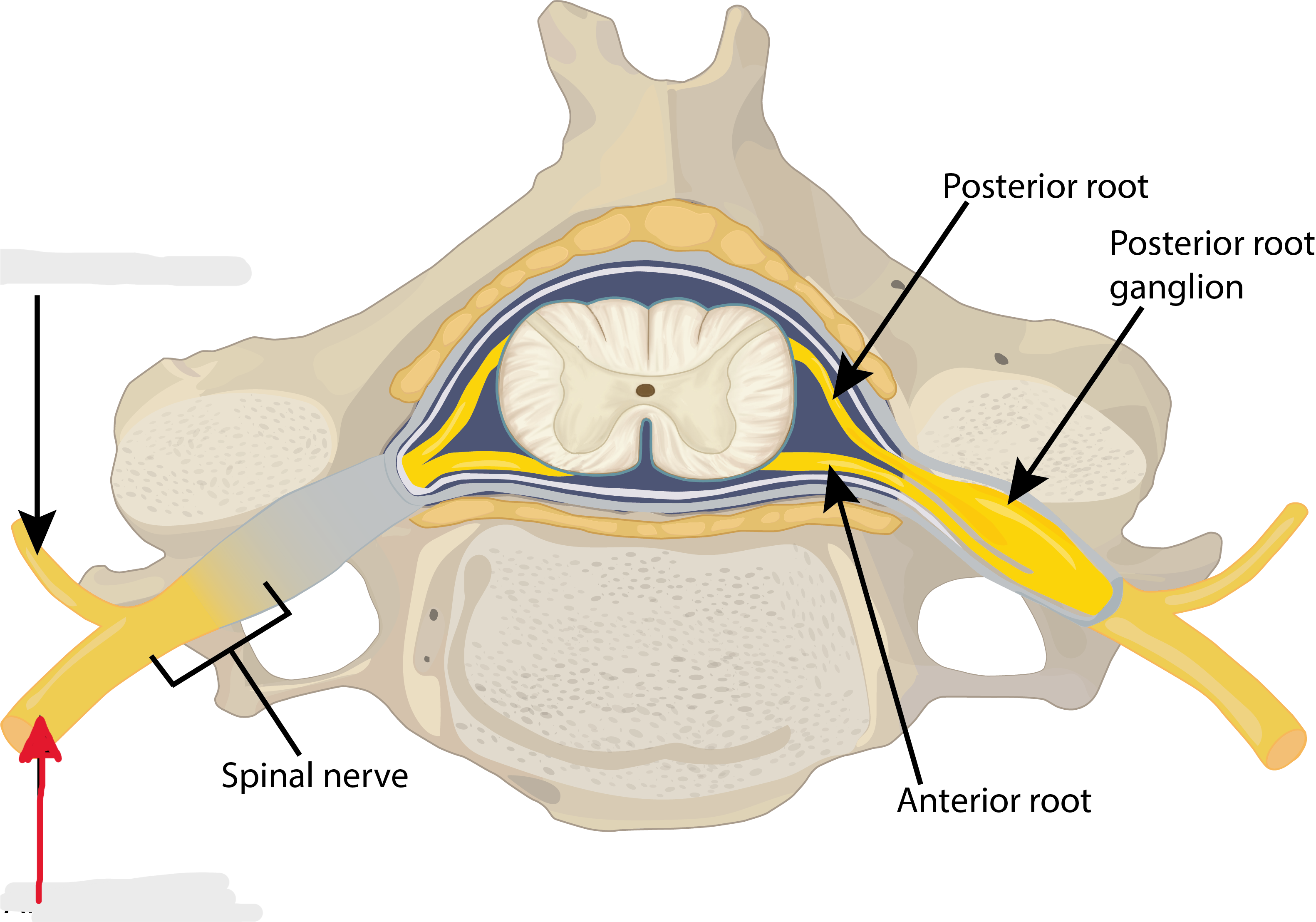
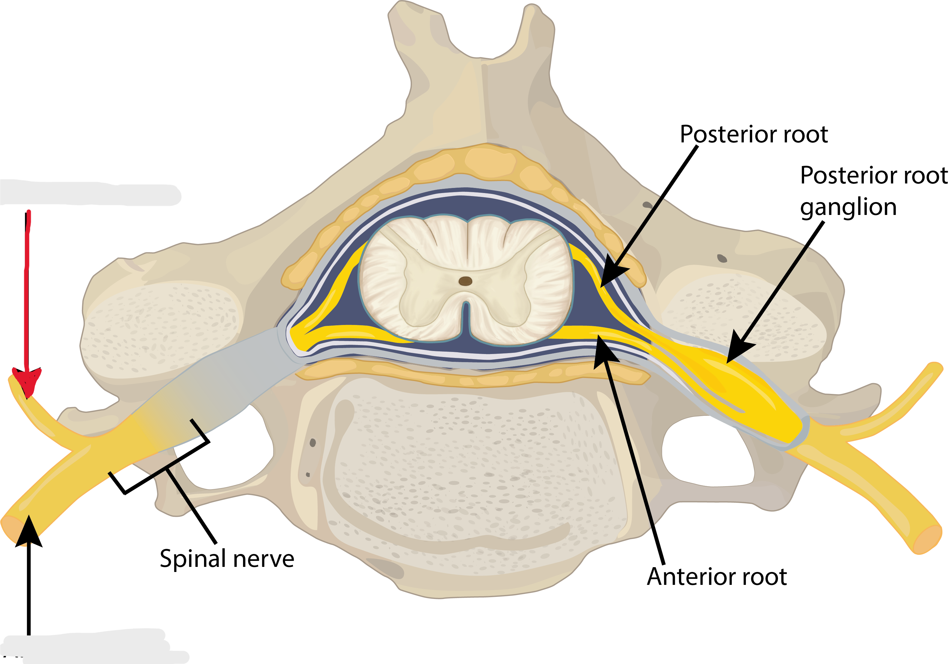
Ventral roots
structure
Dorsal roots
structure
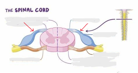
Spinal ganglion
structure
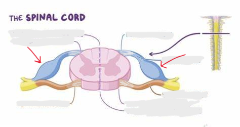
Spinal nerve
structure
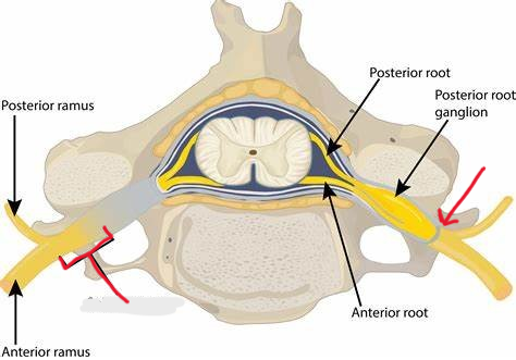
Ventral ramus
structure
Dorsal ramus
Structure. broken
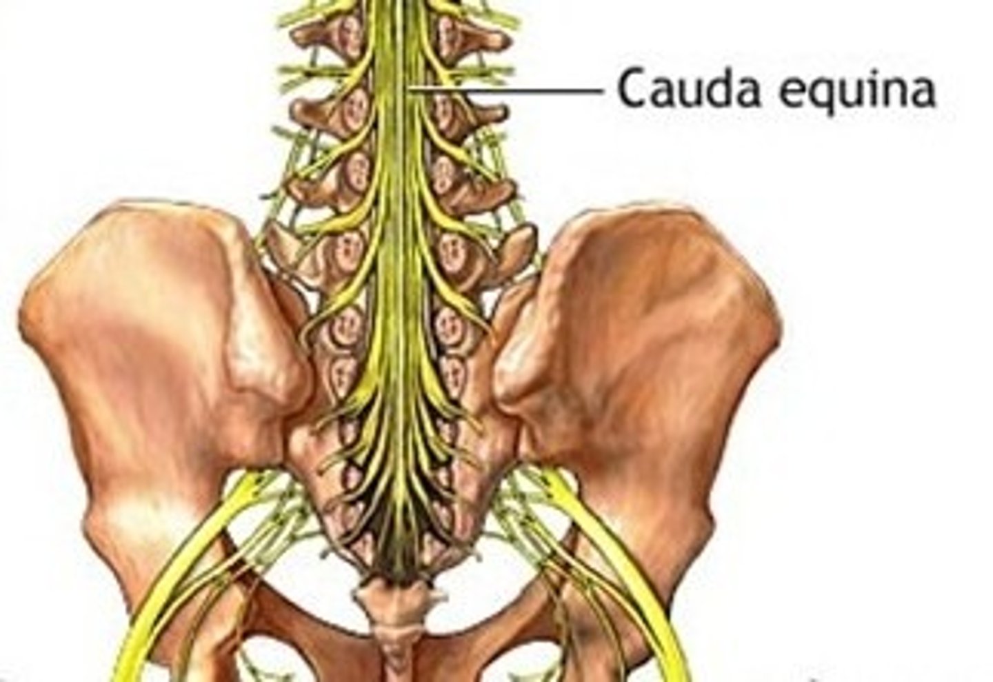
Cauda equina
Structure. horse tail
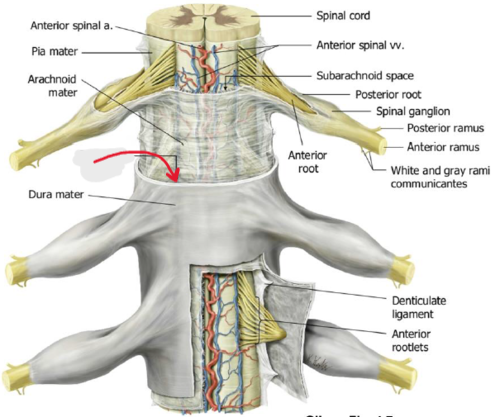
Spinal pia mater
Covering. will tap on spinal cord
Spinal epidural space
Space. will stick probe up under vertebrae/ above dura mater
Spinal subdural space
Space. under dura mater
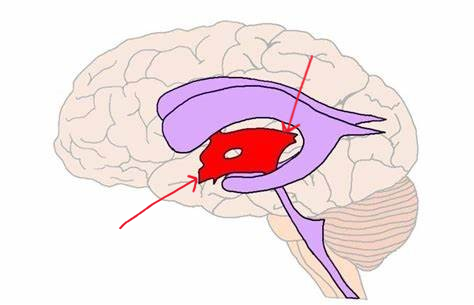
Choroid plexus
Structure. All pink on model
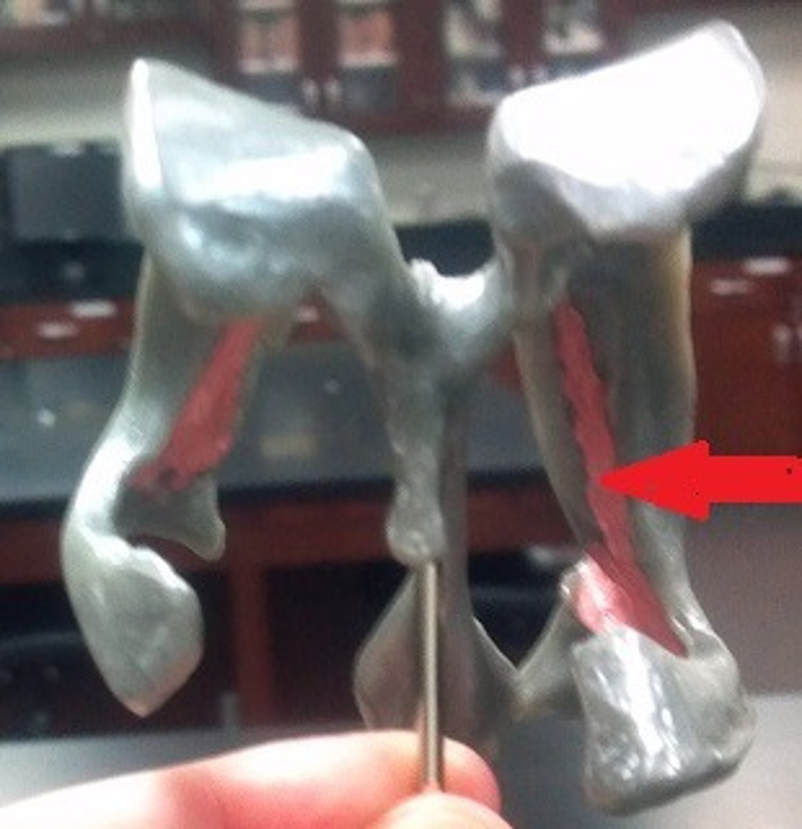
Lateral ventricles
Space. Ram horns on sides
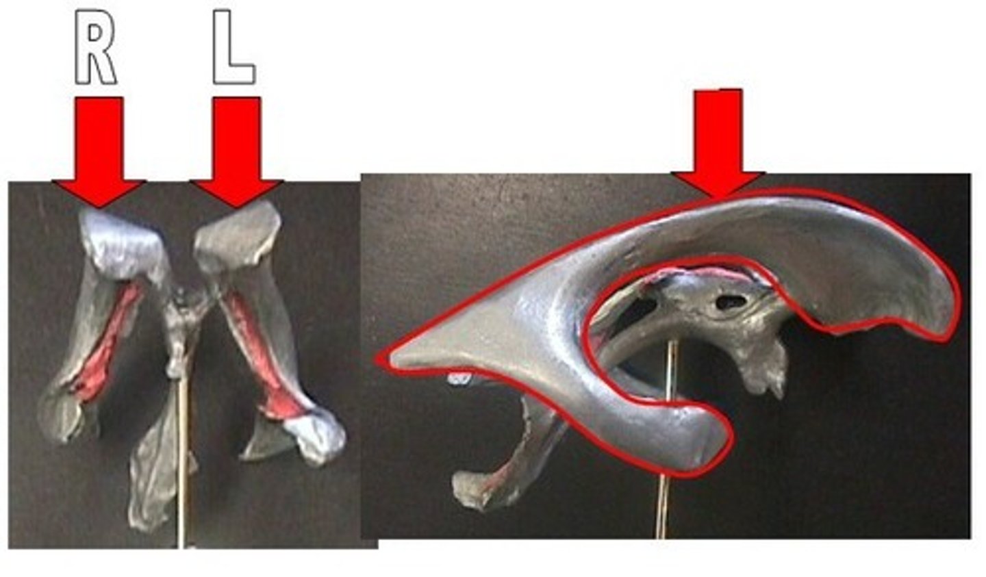
Interventricular foramina
Passageway. Connects top of ram horns to the center
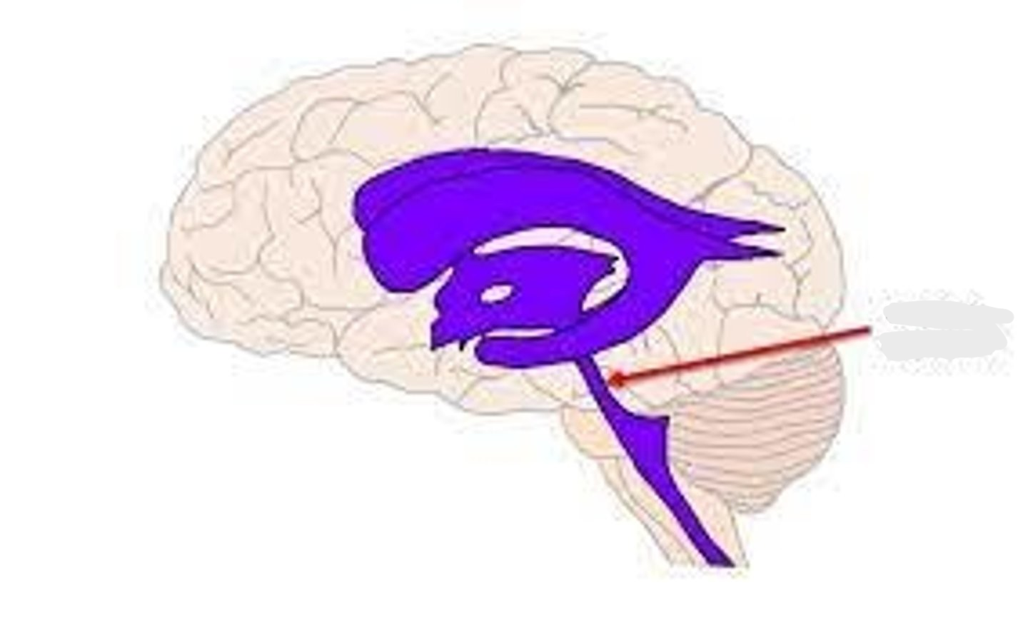
Cerebral aqueduct
Passageway. Looking at posterior side
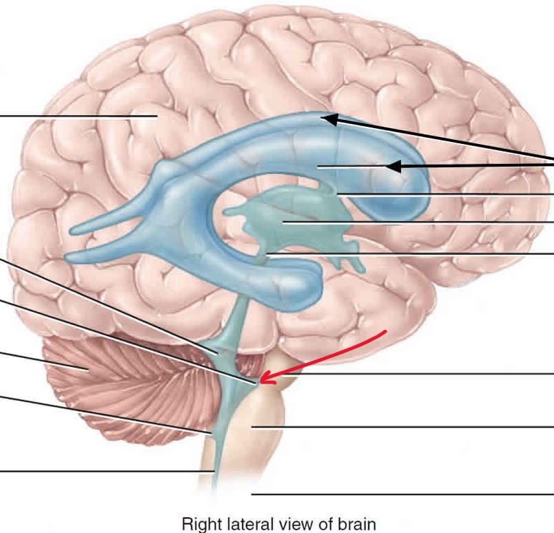
Fourth ventricle
space
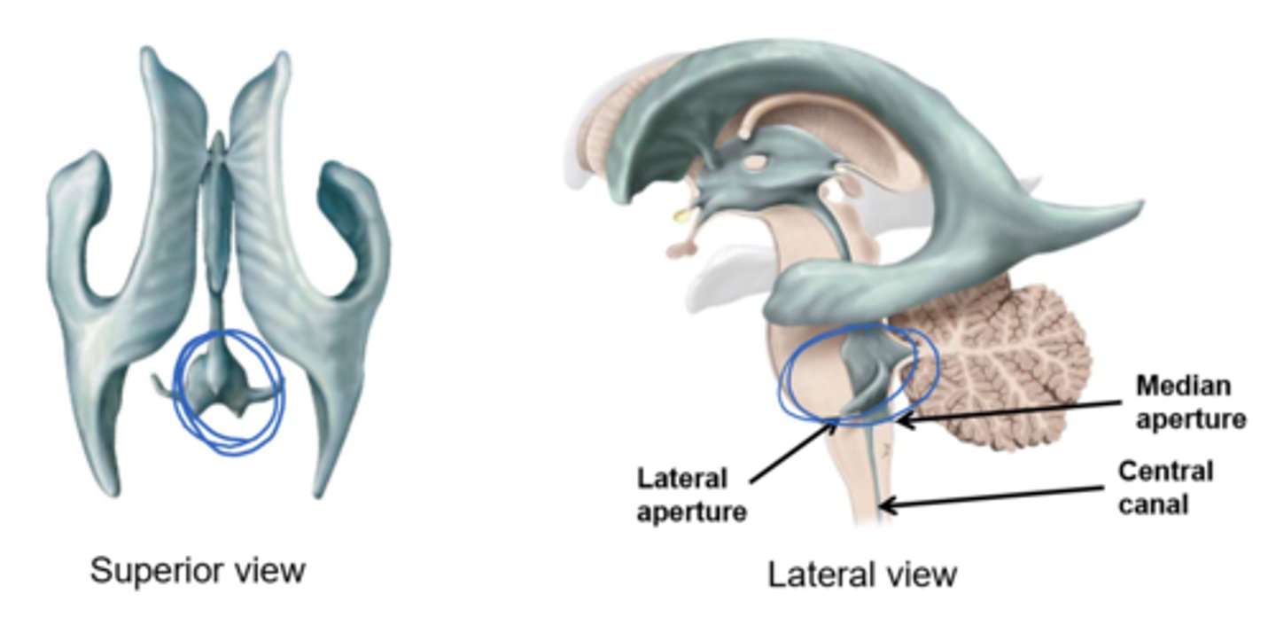
Central canal of spinal cord
Passageway. pointy end
Median aperture
passageway
Lateral aperture
passageway
third ventricle
space