22: Chronic Inflammation 1
 Call Kai
 Learn
Learn Practice Test
 Spaced Repetition
 Match
Match Flashcards
 Knowt Play
1/88

There's no tags or description
Looks like no tags are added yet.
Name | Mastery | Learn | Test | Matching | Spaced | Call with Kai | Chat |
|---|
No analytics yet
Send a link to your students to track their progress
89 Terms
duration of chronic inflammation
weeks-years
what are the main cells involved in chronic inflammation?
MACROPHAGES, lymphocytes, plasma cells, and antigen
what characterizes chronic inflammation?
FIBROSIS
what is the connective tissue used in fibrosis?
collagen
what causes granulation tissue formation?
desmoplasia, +/- fibroplasia, neovascularization
true or false: granulation tissue formation is a NORMAL PART OF HEALING
TRUE
causes of chronic inflammation
persistent infections, HS diseases, autoimmune diseases, prolonged exposure to toxin, isolation, defects in leuk formation
what is the SIGNATURE CELL of chronic inflammation?
MONOCYTES/MACROPHAGES
where do monocytes live?
bone marrow and spleen
what cell is this?
macrophage

when do macrophages form?
in response to foreign bodies or persistent ITC pathogens
describe epithelioid macrophages
larger, polygonal shape, can look like epithelial cells

what cell is this?
epithelioid macrophage
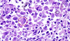
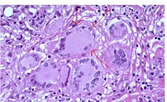
describe multinucleated giant cell
2+ macrophages form one large cell
what cells are these?
multinucleated giant cells
where are dendritic cells stored in larger amounts?
skin, mucosal surfaces of resp/GI tracts
unique feature of dendrites
have filipodia (arm)
pathway of dendritic cell
migrates to antigen -> phagocytize -> migrate to lymph organ -> presents to T/B cells
main function of dendritic cells
antigen presentation
what form of immunity do dendritic cells stimulate?
ADAPTIVE
what do lymphocytes play a key role in?
chronic inflammation
how long does it take lymphocytes to enter an area with UNRESOLVED inflammation?
24-48 hrs
what do lymphocytes surround?
granulomas and blood vessels
what do B lymphs differentiate into?
plasma cells
what do plasma cells do?
secrete Igs (antibodies)
different types of antibody
IgA/D/E/G/M
what do the Igs do?
bind/opsonize antigens, marking them for phagocytosis
where are plasma cells located?
bone marrow, lymph nodes, MMs, wound sites
true or false: plasma cells predominate in certain chronic conditions
TRUE
what do fibroblasts do?
produce collagen and EXCM proteins (important for healing)
what cytokine plays a MAJOR ROLE in fibroblast activity/collagen depositiion?
TGF-B
in what species are fibroblasts VERY RESPONSIVE to injury?
CATS
what cells are these?
fibroblasts

4 phases of wound repair
1. hemostasis 2. inflammation 3. proliferation 4. remodeling
what happens during first intention healing?
edges of NON-SEPTIC wound in close proximity to each other (suturing/bandaging ok)
outcome of first intention healing
little trace of wound over time, tensile strength almost the same
what happens during second intention healing?
cut edges too far apart, delays re-epithelization
what is the outcome of second intention healing?
ulcer or fibrin scar that is DIVITED and MUCH WEAKER tissue
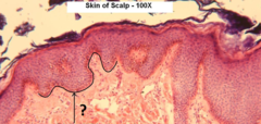
describe repair if the BM is DAMAGED
takes longer bc have to remake BM
what is being shown here?
basement membrane (BM)
HIF
hypoxia inducible factor
what does HIF do?
promotes angiogenesis and stimulates cell proliferation
what is angiogenesis?
creation of new blood vessels
new BVs are fragile and not as strong as the original vessels. what does this entail?
increased vascular permeability
most important/potent cell mediator in angiogenesis
VEGF-a
what does VEGF-a do?
stimulates endothelial cell proliferation
what is the counter mechanism that stops angiogenesis?
angiostatin
antigen is sequestered. what is formed?
granuloma or abscess
collagen is layed down. what is occuring?
fibrosis
what causes pus to form?
liquification of affected tissue
what cells/enzymes are used in abscess formation and WHAT DO THEY DO?
myeloperoxidase from neutrophils, liquifies/necroses tissue
what species LACK myeloperoxidase?
birds/reptiles (remember their abscesses are dry/crumbly bc of this)
inner surface of abscess
granulation tissue
outer wall of abscess
collagen
how long does it take to form an abscess?
weeks (so always chronic)
2 types of abscess formation
septic and sterile
true or false: color of exudate from abscess can sometimes tell us type of bacteria present
TRUE
what is a fistula?
track made to drain pus from abscess
phlegmon
no capsule, inflammation spreads along tissue plane
empyema
no capsule, pus accumulates in pre-existing space
granulomatous inflammation
diffuse sheets/spread out inflammation, tissues is overtaken by macrophages
what is this showing?
granulomatous inflammation

granuloma
mass of nodule cells (can be seen grossly)
major cell types in granulomas/matous inflammation
MACROPHAGES
mediator of granulomas
TH1
mediator of granulomatous inflammation
TH2
what is this showing?
granulomas (can see distinctly)

gross apperance of nodular (TH1) granulomas
gray/white, round/oval, firm/hard
what cell types are heavily seen in nodular granulomas?
macrophages, surrounded by fibroblasts/lymphs/plasma cells
caseating granuloma
cheese like appearance

non-caseating granuloma
lacks central necrosis, contains inflammatory cells

gross apperance of diffus (TH2) granulomas
gray/white, poorly demarcated from nearby tisse, firm
what species and organ is most commonly affected by diffuse (TH2) granulomas?
CATTLE and ILEUM
what is this showing?
diffuse (Th2) granuloma

what is this showing?
diffuse (TH2) granuloma histology

gross apperance of granulation tissue
granular appearance, firm
describe arrangment of granulation tissue
fibroblasts PARALLEL to wound, capillaries PERPENDICULAR to wound
what is this showing?
granulation tissue
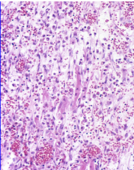
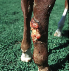
proud flesh
when granulation tissue becomes excessive/mass like
what is this showing?
proud flesh
scirrhous rxn
contraction of CT that tenses the surrounding tissue, can be restrictive/cause dysfunction!
what does scirrhous rxn looks like?
stress ball with the netting around it
gross apperance of scirrhous rxn
organ smaller + firm, looks like squeezed net stress ball
what is this showing?
scirrhous rxn

re-epithelialization
when epithelial cells at periphery proliferate
what is re-epithelialization VERY RAPID in?
1ST INTENTION HEALING
true or false: in re-epithelialization, cells must move ALONG A BASEMENT MEMBRANE
TRUE (will otherwise result in ulcer)
what is this showing?
ulceration on histology

what is this showing?
re-epithelialization on histology
