Lecture 10: Common Disorders of Equine Hind Limbs
 Call Kai
 Learn
Learn Practice Test
 Spaced Repetition
 Match
Match Flashcards
 Knowt Play
1/57

There's no tags or description
Looks like no tags are added yet.
Name | Mastery | Learn | Test | Matching | Spaced | Call with Kai | Chat |
|---|
No analytics yet
Send a link to your students to track their progress
58 Terms
- hock > below hock > stifle > above stifle
What is the prevalence of structures involved with causing hind limb lameness in the horse?
- kick injury - if wound, sequestration likely
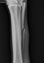
What is the common etiology of metatarsal 2 & 4 fractures (splint bones)?
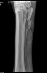
- radiographs
- observe wound; often not healed
How do you diagnose metatarsal 2 & 4 fractures (splint bones)?
- ostectomy (segmental vs. complete)
- only MT4 can be completely removed en bloc
- prognosis is excellent
How do you treat metatarsal 2 & 4 fractures (splint bones)? What the prognosis?
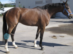
- drags toes
- fails to "track up" on lame leg
- lame leg adducts most
- rider feels difference in their connection
- reduced performance
What are gait characteristics for hock lameness?
- often none
- bone spavin (hard swelling)
- bog spavin (effusion, fluidy)
When a horse has hock lameness what could you find on joint palpation?
- flexion tests
- blocks (intra-articular)
- radiographs
- nuclear scintigraphy
- CT depending on size and location
How do you diagnose hock lameness in a horse
hock osteoarthritis
- distal intertarsal joint
- tarsometatarsal joint
What is the most common hind end lameness diagnosed in performance horses?
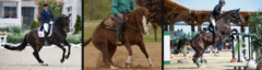
- athletic use
- genetic (juvenile spavin)
What is the etiology for hock osteoarthritis seen on this DLPMO?
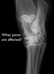
- + upper limb flexion
- BLOCKS to distal hock joints
- radiographs = do not correlate well with clinical lameness
How is hock osteoarthritis diagnosed?
- intra-articular corticosteroids
- shoeing --> squared toe
- facilitated ankylosis or arthrodesis
The prognosis for hock osteoarthritis, is good, how do you treat it?
- has to in LOW MOTION JOINTS, it will destroy high motion joints
Which joints do you have to put ethyl alcohol in if you are trying to facilitate ankylosis for OA treatment?
- bone spavin
What is this hard bony prominence on the hock?
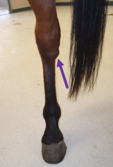
- OCD
- synovitis (poor conformation, hard work)
- OA (rare)
- blood = hemarthrosis
- infection = septic arthritis
What is the etiology for tibiotarsal effusion (bog spavin)?
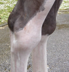
- effusion in tibiotarsal joint
- radiographs
- ultrasound
- diagnostic arthroscopy
How do you diagnose tibiotarsal effusion (bog spavin)?
- distal intermediate ridge of the tibia (DIRT)
- trochlear ridge (lateral > medial)
- medial malleolus
What are the common locations for hock OC? Rate from most to least common.
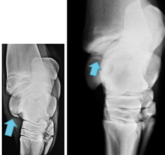
- drain joint and inject corticosteroids
- arthroscopy
How do you treat tibiotarsal effusion (bog spavin)?
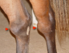
- good
- usually cosmetic problem if underlying issue is addressed
What is the prognosis for tibiotarsal effusion (bog spavin)?
- strain on plantar aspect of hock
- sickle hocked horse
- standardbreds overrepresented
What is the etiology for plantar ligament desmitis (Curb)?
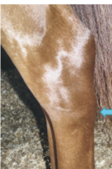
- characteristic swelling at the back of the distal hock joints in sickle hocked horses
- mild to moderate lameness
- blocks
- ultrasound
How do you diagnose plantar ligament desmitis (Curb)?
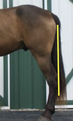
- usually just need rest
- good, but will always have swelling in that region
What is the treatment and prognosis for plantar ligament desmitis (Curb)?
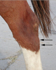
- caused by repeated trauma (stall kicker)
What is the etiology for calcaneal bursitis (capped hock)?
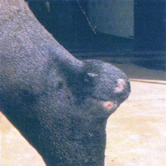
- characteristic swelling at the back of the calcaneus
- generally not lame
How do you diagnose calcaneal bursitis (capped hock)?
- drain and inject corticosteroids
- cosmetic fix: careful may cause iatrogenic infection
What is the treatment for calcaneal bursitis (capped hock)?
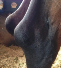
- excellent for function
- poor for comesis
What is the prognosis for calcaneal bursitis (capped hock)?
- septic calcaneal bursitis
What happens if calcaneal bursitis (capped hock) gets infected?
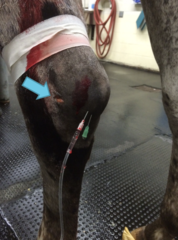
- wound over calcaneus
- infected calcaneal bursitis
(common area to be kicked)
What are the etiologies for septic calcaneal bursitis?
- VERY lame
- pressurize bursa
- contrast rads
- ultrasound
How do you diagnose septic calcaneal bursitis?
- flush bursa (tenoscopy best)
- regional limb perfusion
- may require opening bursa and resecting part of calcaneus
How do you treat septic calcaneal bursitis?
guarded
What is the prognosis for septic calcaneal bursitis?
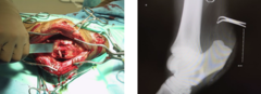
- wear and tear
- conformation (straight hocks)
What is tarsal sheath tenosynovitis (thoroughpin) caused by?
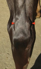
- characteristic swelling on the sides
- usually not lame
How do you diagnose tarsal sheath tenosynovitis (thoroughpin)?
- none is needed
- inject with corticosteroids?
- bandages are not really effective
How do you treat tarsal sheath tenosynovitis (thoroughpin)?
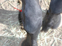
- excellent for athletic use
- guarded for cosmesis
What is the prognosis for tarsal sheath tenosynovitis (thoroughpin)?
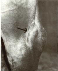
- special muscles that when the hock is extended the stifle is flexed
- involves peroneus tertius, SDFT, and gastrocnemius mm
What is the reciprocal apparatus of the equine hindlimb?
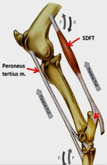
- peroneus tertius rupture --> due to tarsus being extended while the hock is being flexed
- affects the reciprocal apparatus
What could be wrong with this equine pelvic limb?
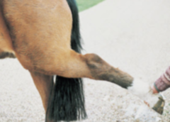
- trauma (foals usually)
- need full limb hind leg cast
What is the etiology of a ruptured peroneus tertius?
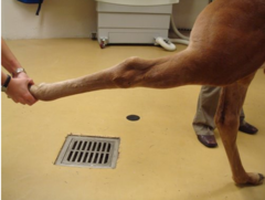
- ability to extend the hock with a flexed stifle
- lameness and swelling initially
- tarsus extends more than usual at trot
How do you diagnose a ruptured peroneus tertius?
- avulsion near the stifle - arthroscopy
- stall rest
How do you treat a ruptured peroneus tertius?
- good
- poor if involves stifle joint
What is the prognosis for a ruptured peroneus tertius?
- trauma
What are superficial digital flexor or gastrocnemius muscle/tendon ruptures usually caused by?
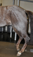
- dx by clinical signs - no flexion of digits, extension of tarsus (hock)
- dx via ultrasound
- treatment is long term splinting and rest
How are superficial digital flexor or gastrocnemius muscle/tendon ruptures diagnosed and treated?
- fair to poor
- good if it heals
What is the prognosis for a superficial digital flexor or gastrocnemius muscle/tendon rupture?
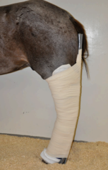
- fibrosis of the semitendinosus muscle caused by trauma
- ossification can occur in some cases
What is fibrotic myopathy?
- characteristic movement most evident at a walk - slap of leg in when walking
- hardening of semitendinosus mm. on palpation
How do you diagnose a fibrotic myopathy in horses?
- semitendinosus +/- semimembranosus tenotomy (more of a mechanical issue than a painful one)
- may not result in complete resolution of clinical signs
- minimal side effects
How do you treat a horse with fibrotic myopathy?
- fair for success of treatment
- horse will likely always have altered gait
What is the prognosis for fibrotic myopathy in horses?
- jump of lame leg in weight bearing phase - quicker, short step
- drags toe because leaves leg out behind
- increased lameness when limb is on outside of circle
What is the gait characteristics of stifle lameness in horses?
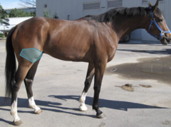
- joint effusion --> femoropatellar pouch easily palpable
- hind end muscle atrophy with chronic problem
What are the clinical signs of stifle lameness?
- flexion test: upper limb and caudal extension
- blocks
- radiographs
- ultrasound
- arthroscopy
- nuclear scintigraphy
How do you diagnose a stifle lameness?
- most common breeds: WB, TB, QH
- lesions: medial femoral cysts, trochlear ridge OCD
What are the common breeds and locations for stifle OC?
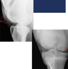
- effusion
- lameness when starts training
- radiographs
How do you diagnose a stifle OC lesion?
- surgery (wait until 1 year)
- debride
- cysts: fill with biologics and scaffolds, inject with corticosteroids, use a screw
The prognosis for a stifle OC lesions can vary and often lead to OA. What are options for treatment?
- underlying OC
- trauma
- athletic use
What are the etiologies for stifle OA?
- dx: stifle block
- treatment: intra-articular corticosteroids
- prognosis: fair
How do you diagnose and treat stifle OA?
- medial patellar ligament is caught over medial trochlear ridge when stifle extends 45*
- inability to release the stay apparatus so the patella does not disengages from the medial trochlea of the femur
- severe: stifle locked in extension
- mild: horse catches their stifle
What is upward fixation of the patella in horses?
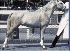
- weak quadriceps
- conformation: straight legs
What is the etiology of upward fixation of the patella?
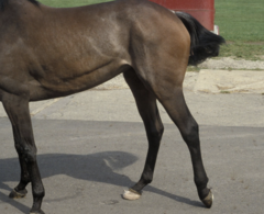
- immediate: back the horse up
- exercise strengthen quadriceps
- surgery: medial patellar ligament splitting or blistering, medial patellar ligament desmotomy = last resort
How do you treat upward fixation of the patella?