Cardiovascular and Muscle
 Call Kai
 Learn
Learn Practice Test
 Spaced Repetition
 Match
Match Flashcards
 Knowt Play
1/108

There's no tags or description
Looks like no tags are added yet.
Name | Mastery | Learn | Test | Matching | Spaced | Call with Kai | Chat |
|---|
No analytics yet
Send a link to your students to track their progress
109 Terms
3 Reasons why heart failure is less common in animals than humans
Lipid abnormalities are very rare in animals (and even high cholesterol → low risk of cardiac disease)
Idiopathic hypertension is rare in animals
2˚ hypertension is less significant the the 1˚ lesion (eg. CKD will kill before hypertension)
Structure and Function of the Heart
Left vs. right side
4 Chambers (+ functions)
4 Valves (+ functions)
L vs. R: Left side must pump blood to a more extensive capillary bed in the systemic circulation vs. the lungs for the right side of the heart
Systemic BP 3x pulmonary BP → L side of heart 3x bigger than R side to generate the higher BP
Chambers:
Right Atrium = Collects deoxygenated blood from systemic circulation
Right Ventricle = Pumps blood through lungs
Left Atrium = Collects oxygenated blood from lungs
Left Ventricle = Distributes oxygenated blood to systemic circulation
Valves: Cusps (flap of fibrous tissue) attached to papillary muscles by chordae tendinae which prevent backflow of blood
Right AV = Prevents backflow into RA
Pulmonic = Prevents backflow into RA
Left AV = Prevents backflow into LA
Aortic = Prevents backflow into LV

Describe THREE layers of the heart
Endocardium = Single layer of endothelium with small amounts of fibrous connective tissue for protection
Myocardium = Myocytes arranged in spiral bundles which form a highly efficient pump
Thickness depends on pressure required to pump blood
Most richly vascularised muscle in body (supplied by coronary arteries) = 1 capillary/myocyte
Exclusive reliance on oxidative metabolic
Epicardium = Outer layer of heart made of fibrous tissue attached to the visceral layer of the pericardium
Describe THREE features to observe during necropsy of the heart
Colour
Pale myocardium = Necrosis/fibrosis
Caution: PM muscle contraction (rigor mortis) → Squeezes blood out of myocardial vessels which produces pallor
Slice into myocardium: Pallor extending into lumen and papillary muscles = TRUE necrosis/fibrosis
Thickness of chamber walls and shape of heart
Normal: LV wall 3x thicker than RV wall due to pressure differences
Abnormal: LV wall = RV wall (1:1) (systemic and pulmonary BP are similar)
DDx: L → R shunt (eg. PDA, VSD, ASD) OR PS, pulmonary hypertension
Abnormal: LV > 3x RV wall (5:1)
DDx: SAS or HCM
Valves
Patency
Ensure valves are convex
Jet lesions

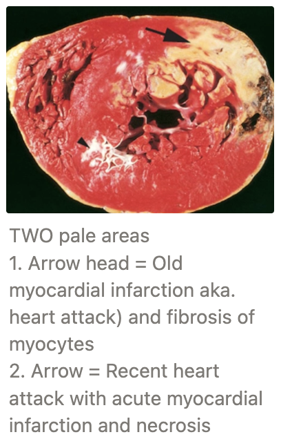
Response of heart to injury (6 steps)
Injury to myocardiocytes
Myocardium poorly heals via fibrosis ONLY (cannot regenerate)
Fibrosis reduces elasticity and contractility of the heart resulting in impaired BP necessary to pump blood forwards
3 - 5 fold functional reserve of heart by 6 compensatory mechanisms
Increase HR
Atrial dilation to increase venous return
Myocyte hypertrophy to increase contractility
Increased blood volume via RAAS
Increased peripheral resistance (vasoconstriction)
Redistribution of blood flow
CHF develops when compensatory mechanisms fail and the heart cannot propel sufficient blood
Cardiac decompensation = Final stage of heart disease where marked dilation and severe thinning of the heart walls prevents contractility to match (Starling's law no longer maintained) → Rapid progression of cardiac disease and death
Define congestive heart failure (CHF) and effects on forward vs. backward failure
Clinical syndrome caused by a variety of chronic conditions that reduce heart function
Develops as the heart cannot adequately propel blood forwards
Little effect on CO and organ perfusion
Heart disease does NOT cause CKD
Significant effects caused by backing-up of blood behind the heart which cannot pump blood out fast enough
What is this?
2 Causes (+ DDx)
Pathogenesis
2 Clinical signs


LCHF = Pulmonary oedema
Causes:
Dysfunctional LAV or AoV (endocardiosis) → Regurgitation/insufficiency
Left ventricular myocardial necrosis → Weakness (myocarditis or fibrosis)
Pathogenesis:
Left side of the heart cannot adequately propel blood into systemic circulation
Blood collects in pulmonary circulation → pulmonary congestion
Increased pulmonary HP causes fluid to leak from pulmonary capillaries → Pulmonary oedema
Pulmonary Oedema (foam on cut surface with swollen, wet and shiny lungs) = Acute LCHF
Increased permeability results in leakage of RBCs into alveoli
Alveolar macrophages enter the alveolar space to engulf this material
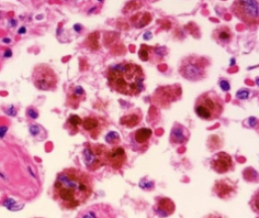
Haemosiderin is produced and stored in alveolar macrophages as a result of RBC breakdown
Heart Failure Cells = Alveolar macrophages containing granules of haemosiderin formed from RBC breakdown and indicates haemorrhage into the alveolar space
Tan lungs = Chronic LCHF
Clinical Signs:
Exercise intolerance
Nocturnal cough

What is this?
4 Causes (+ example DDx)
Pathogenesis
3 Outcomes
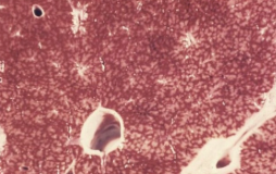
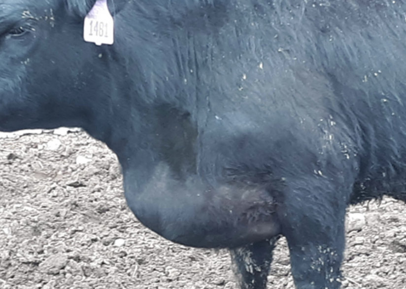
RCHF = Nutmeg liver and brisket oedema
Causes:
Cor pulmonale = RCHF that develops 2˚ to pulmonary hypertension → Increased afterload
Right side of the heart cannot generate sufficient force to propel blood through pulmonary circulation resulting in RCHF
DDx:
2˚ to LCHF (pulmonary hypertension due to congestion)
Equine heaves (high intrathoracic pressure)
Pulmonary fibrosis (high intrathoracic pressure)
Heart-filling defects (eg. pericardial effusion)
Dysfunction of the pulmonic or right AV valves (endocardiosis)
Right ventricular myocardial necrosis (myocarditis or fibrosis)
Pathogenesis:
Right side of heart cannot adequately propel blood into pulmonary circulation fast enough
Allows blood to collect in systemic circulation → systemic congestion
Systemic hypertension
Outcomes:
Oedema
Dog = Ascites
Cat = Hydrothorax
LA/horse = Brisket oedema and bottlejaw
Anasarca uncommon (do not live long enough for oedema to develop in all cavities AND SC)
Congestion of kidneys
Systemic venous congestion results in renal congestion
Reduced blood flow through the juxtaglomerular apparatus results in RAAS activation to increase GFR
RAAS promotes Na+ retention in the tubules which increases blood volume
Attempts to restore CO BUT exacerbates CHF instead due to volume overload → Oedema
Nutmeg liver
Congestion of systemic circulation
Dark red blood accumulates in the centrilobular veins of the liver which contrasts with the pale peripheral parts of the lobule (nutmeg liver)
As more blood accumulates, pressure in the central veins increases which causes adjacent hepatocytes to atrophy or die
Hepatocyte death results in cirrhosis (fibrosis of central vein)
Haemosiderin is also found within local macrophages as a result of RBC breakdown

What is this?
TWO types


Cardiac Hypertrophy (no hyperplasia as cardiac cells cannot regenerate)
Types:
Physiological hypertrophy (4) = Normal myocardial hypertrophy in response to increased exercise (eg. racehorse or greyhounds) which does NOT cause pathological changes
Pathological hypertrophy (4) = 1˚ OR 2˚
1˚ = Disorder of the myocardium (eg. idiopathic HCM)
2˚ = In response to non-myocardial disease process (eg. subaortic stenosis or hyperthyroidism)
Is there sufficient CK and AST leakage form cardiac muscle ONLY to detect disease on biochemistry?
NO! Many cardiocytes must die to detect → Death
proBNP
Definition
3 Causes of elevation
2 Disadvantages
2 Indications
Definition: Natriuretic peptide secreted by the heart to maintain fluid balance and BP regulation in response to:
Volume overload
Cardiac hypertrophy
Hypoxia
-ve:
Blood must be collected in a special pink-top tube containing protease inhibitor → Refrigeration → Sent to lab on SAME day
Cannot screen for myopathies without clinical signs of CHF (i.e. proBNP cannot distinguish between healthy patients and HCM cats with no CHF)
Indications:
Dogs or cats presenting with respiratory signs to distinguish between 1˚ respiratory disease (eg. chronic bronchitis) or heart failure
Analysing efficacy of treatment for heart failure
THREE types of congenital/genetic cardiac disorders
DDx for each
Most common DDx in dog vs. cat vs. cow vs. pig
Types:
Failure of foetal structures to close (4)
Patent Ductus Arteriosis (PDA)
Atrial Septal Defect (ASD)
Ventricular Septal Defect (VSD)
Tetralogy of Fallot (TOF)
Abnormal valve development (4)
Pulmonic stenosis (PS)
Subaortic stenosis (SAS)
Congenital valve malformations
Valvular haematocyst
Abnormalities in large blood vessels (3)
Persistent right aortic arch (PRAA)
Transposition of aorta and pulmonary artery
Ectopia cordis
Species Variation:
Dog = PDA, PS, SAS, PRAA
Cat = VSD, LAV malformation
Cow = ASD, VSD, transposition of aorta
Pig = SAS, VSD
Normal blood flow through a foetal heart and changes after birth
In utero: Blood shunted from R → L side of circulation allowing bypass of pulmonary circulation via
Ductus arteriosus = Connects the PA to the Ao
Foramen ovale = Opening between the LA and RA
Birth: Shunts close due to BP changes caused by lung inflation
Ligamentum arteriosum = Remnant of the ductus arteriosus which becomes non-patent at birth

What is this?
Signalment
Pathogenesis
3 PM features

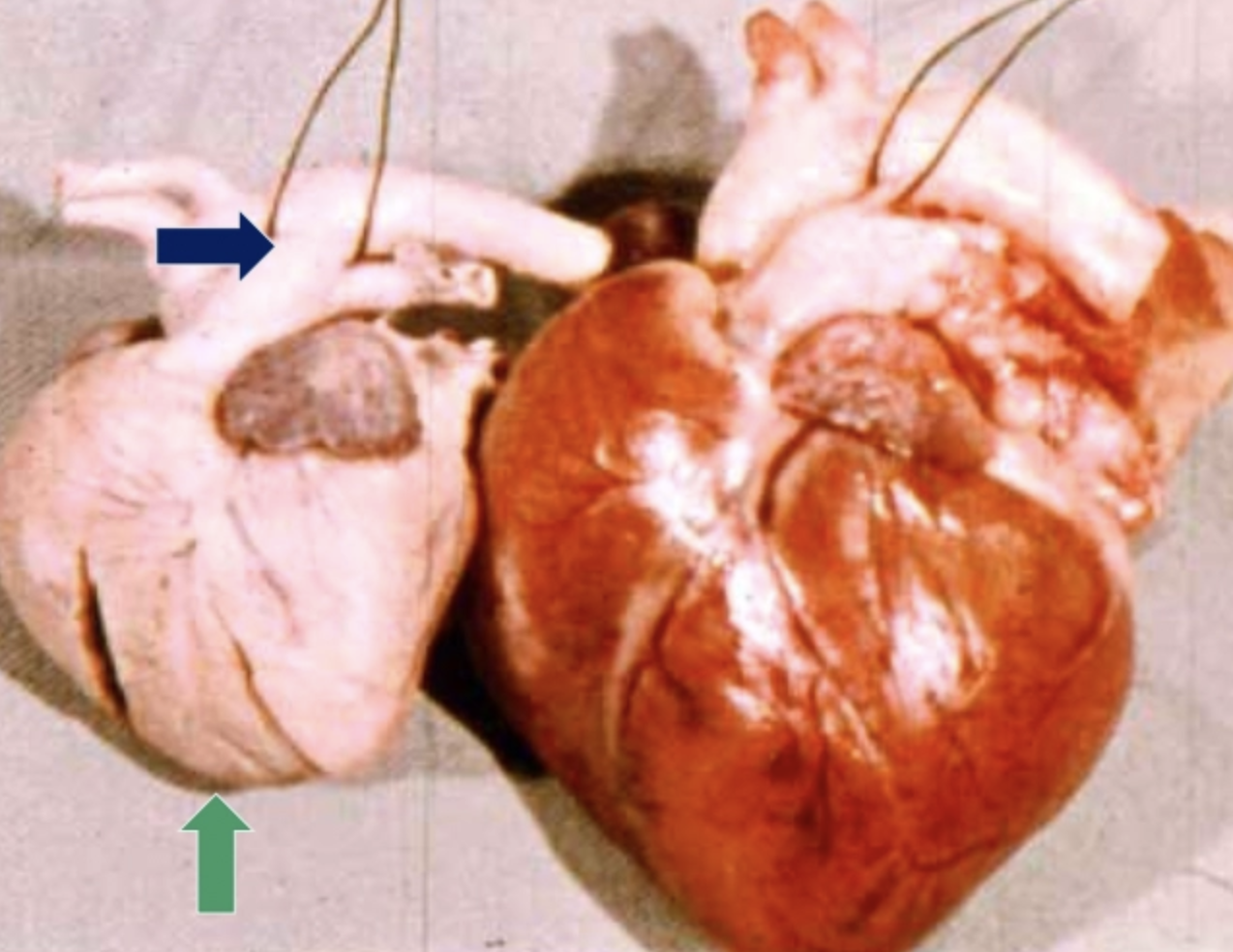
Patent Ductus Arteriosus (PDA)
Signalment: Dog (esp. female poodle)
Ductus arteriosus remains patent at birth (should become the non-patent ligamentum arteriosum)
Blood is allowed to travel from the higher pressure Ao → lower pressure PA
Increased blood in pulmonary circulation → Pulmonary hypertension and decreased systemic BP
BP right side of heart = BP left side of heart
Increased RV wall thickness to pump blood through increased afterload → Absence of 3:1 ratio
PM:
Dilated ductus arteriosus
Absence of 3:1 ratio of LV:RV thickness (1:1) resulting in loss of heart apex normally produced by enlarged LV wall
Fibrosis in later stages of disease

What is this?
Pathogenesis
PM

Atrial Septal Defect (ASD)
Pathogenesis:
Foramen ovale fails to close at birth/incomplete atrial septum development
Blood shunts from high-pressure LA to low-pressure RA resulting in pulmonary hypertension
BP between right and left side of heart are equal
Absence of 3:1 ratio (1:1 ratio)
PM: 1:1 ratio of LV:RV
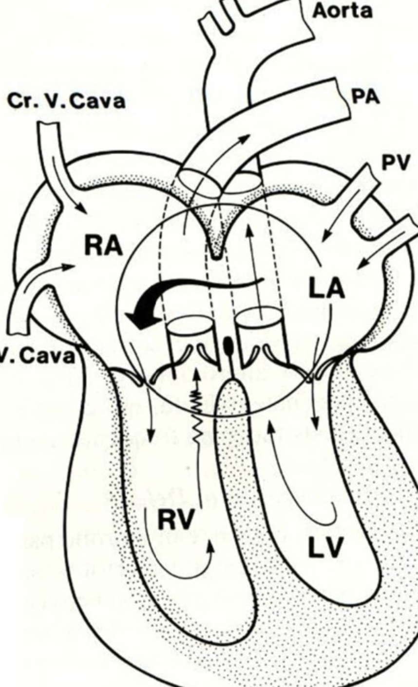
What is this?
Pathogenesis
Location
PM
Clinical signs


Ventricular Septal Defect (VSD)
Pathogenesis:
Incomplete development of ventricular septum
Blood shunts from high-pressure LV to low-pressure RV
Location: Defect immediately below AV valves
PM: 1:1 ratio of LV:RV
Clinical Signs: More severe shunting = More severe clinical signs

Tetralogy of Fallot
4 Cardiac abnormalities
Pathogenesis
3 Clinical signs
Prognosis
Abnormalities:
Ventricular septal defect
Pulmonic stenosis
Transposition of the aorta (shifted to the right and shares the LV and RV)
2˚ Right ventricular hypertrophy
Pathogenesis:
Pulmonic stenosis prevents blood flow through the pulmonary artery
This causes blood to flow from the the RV → VSD and aorta via aortic transposition (opposite from other congenital defects)
2˚ RV hypertrophy required to pump the blood into the high-pressure left ventricle through the VSD
Results in poorly oxygenated blood from RV being pumped into systemic circulation
Clinical Signs:
Cyanosis due to deoxygenated blood in systemic circulation (vs. other diseases with high O2 blood through body)
Dependent oedema
Nocturnal cough due to pulmonary oedema NO PAIN
Prognosis: Better to have ToF than VSD


What is this?
Pathogenesis
4 PM features

Pulmonic Stenosis (PS)
Pathogenesis:
Narrowing of the RV outflow channel → Increased RV pressure
2˚ RV hypertrophy required to generate enough blood pressure to squeeze blood through narrow valve
High pressure blood squirts from the stenotic pulmonic valve which causes dilation of the pulmonary artery and jet lesions
Dilation → slow blood hit by fast blood from squirting PS = turbulence and thrombosis formation
± Developing signs of RCHF with age depending on severity of PS as RV cannot pump blood forward fast enough
PM:
RV hypertrophy
Jet lesions in the pulmonary artery
Pulmonary artery dilation
± RCHF (systemic hypertension)

What is this?
Pathogenesis
Onset
5 PM features

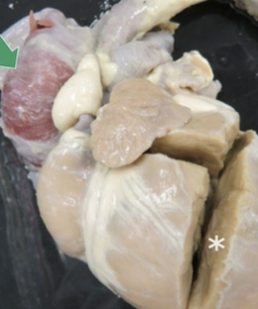
Subaortic Stenosis (SAS)
Pathogenesis:
Thick fibrous band below the aortic valve (defect in the endocardial cushions) narrows the outflow channel of LV → Increased LV pressure
2˚ LV hypertrophy required to generate enough blood pressure to pump blood to systemic circulation
High pressure blood squirts from the stenotic aortic valve which causes dilation of the aorta and jet lesions
Dilation → slow blood + fast blood from squirting SAS = turbulence and thrombosis formation
Thromboembolism travels through the coronary artery openings resulting in multifocal myocardial infarction
± Developing signs of LCHF due to failure of LV hypertrophy or sudden death due to myocardial infarct
Onset: May develop normally into adulthood before signs of CHF develop
PM:
LV hypertrophy (*)
Jet lesions in the aorta
Dilation of the aorta (green)
Multifocal myocardial infarction
± LCHF (pulmonary hypertension) NO kidney failure


Congenital Valve Malformations
Prevalence
Aetiology
Prevalence: Less common cause of reduced patency
Aetiology: Heritable syndromes of the left or right AV malformation reported in dogs and cats
What is this?
Definition
Signalment
Prognosis
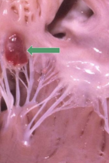
Valvular Haematocyst
Definition: Blood-filled cysts in calves
Signalment: Common in young ruminants
Prognosis: NOT serious and spontaneously regress after several months with no clinical signs
What is this?
Normal development
Pathogenesis
Clinical signs

Persistent Right Aortic Arch (PRAA)
Normal: In utero, foetus has L and R aortic arch → Regression of right aortic arch after birth
Normal: L = Ao → PA → Oesophagus = R
PRAA: L = PA → Oesophagus → Ao = R
Pathogenesis:
Persistent right aortic arch and regression of the left aortic arch in embryo
Oesophagus between the pulmonary artery and the aorta
Because the two vessels are on opposite sides of the oesophagus, contraction of the ligamentum arteriosum compresses the oesophagus
Chronic regurgitation leading to megaoesophagus proximal to constriction
Clinical Signs: No signs of heart disease (blood flow normal)
Chronic regurgitation beginning at weaning due to transition to solid foods which cannot pass through
Death due to aspiration pneumonia
Transposition of the AO and PA
3 Types
Prognosis
Types: Aorta or pulmonary artery are attached to the heart at an aberrant location
BOTH arteries in the same ventricle (eg. aorta and pulmonary artery originating from the RV)
One artery within BOTH ventricles (eg. Tetralogy of Fallot)
Arteries swap ventricles (eg. aorta originates from RV and pulmonary artery originates from LV)
Prognosis: Grave due to marked BP abnormalities → Death at birth or immediate post-natal period as incompatible with life
What is this?

Ectopia Cordis
Rare congenital disease of calves where the heart is located outside the thoracic cavity (entirely outside body or under skin) Calves may survive a few days if under skin
Cardiac Diseases
5 Pericardial DDx
4 Endocardial DDx
5 Myocardial DDx
2 Neoplastic DDx
Pericardial DDx:
Pericarditis (4)
VitE/Se deficiency (4)
Haemopericardium (3)
Enterotoxaemia (4)
Endocardial DDx:
Endocardial mineralisation (3)
Valvular endocardiosis (5)
Atrial thrombosis (3)
Endocarditis (4)
Myocardial DDx:
Nutritional deficiencies
Toxicity
Physical injury and shock
Hypertrophic cardiomyopathy (HCM)
Dilated cardiomyopathy (DCM)
Neoplastic DDx:
RA HSA in dogs (3)
Lymphoma in cattle (3)
Overview of the Pericardium
Normal anatomy (+ function)
2 Causes of reduced ventricular filling
Normal: Contains small amount of lubricating fluid to allow contraction and passive filling/relaxation of the heart
Causes of Reduced Ventricular Filling:
Pericarditis
Cardiac tamponade = Excessive fluid accumulation in the pericardial sac which compresses the heart and prevents it from fully relaxing/filling with blood
What is this?
Definition
Aetiology
DDx
Pathogenesis
3 Clinical signs

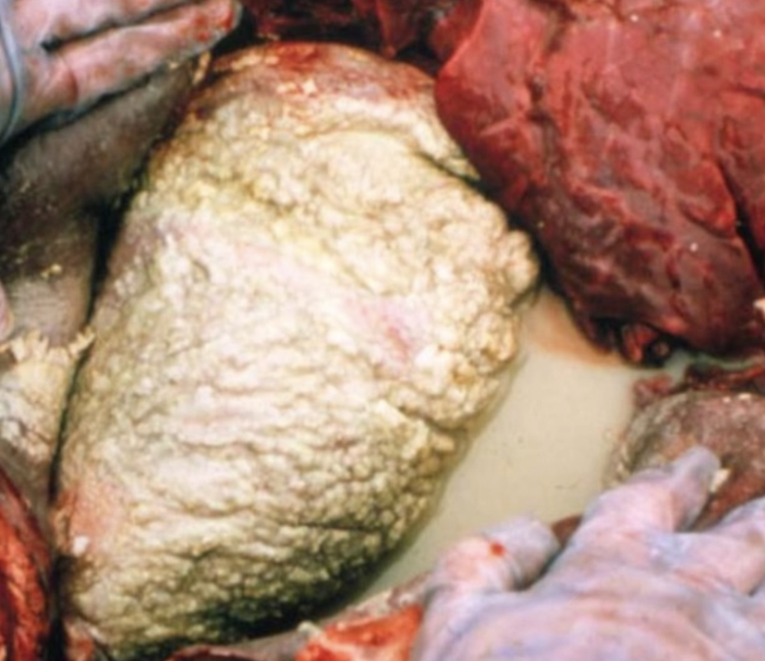

Purulent Pericarditis = Degenerate neutrophils in pericardial sac
Chronic = Organisation of adhesions
Aetiology: Pyogenic bacteria
DDx: Bovine Traumatic Reticulopericarditis (aka. Hardware Disease)
Cow ingests foreign body (eg. nail) which contains pyogenic bacteria on the surface
Peristaltic contraction pushes the foreign body from the reticulum → diaphragm → pericardium
Pyogenic bacteria colonise the pericardial sac
Cow often survives months before succumbing to RCHF or septicaemia
Clinical Signs: Present with RCHF due to defective heart filling
Dependent oedema (bottlejaw and brisket oedema)
Nutmeg liver
Anorexia and weight loss
What is this?
Definition
Aetiology
4 DDx
Pathogenesis

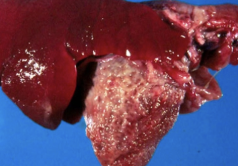
Fibrinous Pericarditis = Increased pericardial fluid containing flecks of fibrin
Aetiology: Haematogenous spread of non-pyogenic bacteria deposits in serous membranes → Fibrinous polyserositis (eg. pericarditis, arthritis, peritonitis, pleurisy)
DDx:
Pig = Haematophilus parasuis (Glasser's disease)
Horse = Streptococcus zooepidemicus
Cattle = Histophilus somni
Cat = FIP due to systemic vasculitis
Pathogenesis:
Bacteria in blood aggregate in serosal blood vessels of the pericardium and cause damage
Results in leakage of fibrin into the pericardial sac which forms adhesions between parietal and visceral pericardium
Mature fibrin contracts causing the heart to constrict which prevents adequate filling
Results in RCHF ± LCHF
Chronic fibrinous pericarditis = Organisation of adhesions
Bread-and-butter heart = Severe fibrinous pericarditis containing large aggregate of fibrin attached to the pericardial surface
What is this? (pig heart)
Aetiology
Pathogenesis
Outcome in other species

Mulberry Heart Disease
Aetiology: Vitamin E/Se deficiency
Pathogenesis: Oxidative damage to myocardial endothelial cells → Leakage of blood and fibrin → Red discolouration from pericardial fluid
Other Species: Myocardial necrosis and mild increased pericardial fluid with small amounts of fibrin (same effect as skeletal muscle)
Haemopericardium (3)
2 DDx in dogs
DDx in pigs/horses
Prognosis
Dog:
Ruptures RA haemangiosarcoma
Idiopathic haemopericardium (slower onset with signs of CHF)
Pig/Horse: Spontaneous rupture of proximal Ao
Connection of the proximal aorta to LV is weak
Strenuous exercise results in increased intracardiac pressure
Prognosis: Blood must be pumped under pressure into the pericardial sac → Rapid/spontaneous death
What is this? (lamb heart)
Signalment
Agent
Pathogenesis
PM features

Enterotoxaemia (4) aka. Pulpy kidney or overeating disease
Signalment: Young, rapidly growing lambs
Agent: Overgrowth of commensal = Clostridium perfringens Type D
Pathogenesis:
Increased CHO intake allows excess to spill into the proximal duodenum resulting in overgrowth of Cl. perfringens
Bacteria produce toxins which are absorbed into the bloodstream from the intestinal wall
Toxins travel around the body causing vascular damage
Damaged blood vessels leak fibrin throughout the body which is easiest to view in the pericardial sac as the smallest enclosed space of the body
PM:
Large fibrin clot in the pericardium = Only gross lesion highly suggestive of disease
Histology of brain = Definitive diagnosis
Renal autolysis = NOT specific for pulpy kidney as caused by high fever at death and stress-induced glucosuria
What is this?
TWO causes

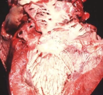
Endocardial Mineralisation (3) → No effect on heart function
Causes:
Cattle = Chronic debilitation (eg. Johne's Disease) → Endocardial fibrosis and mineralisation
Jet lesions = Focal mineralisations of the perivalvular endocardium caused by blood turbulence that results from insufficiency/regurgitation (squirts of blood hitting the endocardium)
What is this?
Signalment
Location
PM
Pathogenesis
Clinical signs


Valvular Endocardiosis (5) aka. Myxomatous mitral valve disease (MMVD)
Signalment: Almost all dogs >10yr have some degree of endocardiosis (most have no clinical signs)
#1 cause of CHF in dogs
Esp. CKCS
Location: LAV > RAV » Ao and PA
PM: Valves are smooth, shiny and nodular (vs. endocarditis)
± Atrial dilation
± Atrial jet lesions
Pathogenesis:
Mucoid or myxomatous degeneration of collagen fibres within valve cusps
→ Shortened cusps with areas of nodular thickening
Loss of valve patency and reduced function → Regurgitation
Backflow of blood from ventricle to atrium → Compensatory atrial dilation and endocardial jet lesions
LCHF (left AV valvular endocardiosis)
Clinical Signs:
Asymptomatic
Nocturnal cough and exercise intolerance with LCHF
Atrial Thrombosis (3)
Pathogenesis
DDx
Pathogenesis:
2˚ due to change of heart size, usually atria (eg. cardiomyopathy) OR any loss of patency of AV valves (eg. endocardiosis)
Results in slow and turbulent blood flow through the atria that is more likely to clot
Atrial thrombus impairs heart function and may result in rapid death if large
Pieces of smaller clots break off → Thromboembolism and infarction elsewhere in body

DDx: Feline HCM → Atrial dilation → Atrial thrombosis → Saddle embolism
What is this?
Signalment
PM
Histology
2 Risk factors
3 Outcomes

Valvular Endocarditis (4)
Signalment: Young production animals (more bacteria circulating the body)
PM: Large, yellow-grey, rough nodular vegetations (cauliflower appearance)
Histology: Layers of fibrin, trapped RBCs, bacteria and neutrophils
Risks:
More circulating bacteria from elsewhere in the body (eg. pneumonia or abscess)
Valvular turbulence (normally disrupts laminar flow allowing bacteria to strike valve cusps)
Outcomes:
ACUTE = Large vegetations prevent normal valve function and may block the valve orifice → Rapid death
Pieces of vegetations may break off forming multiple septic emboli
L Side (LAV/Ao)
Septic myocardial infarction (coronary arteries)
Septic renal infarction
R side (RAV/PA) = Pulmonary abscessation
CHRONIC = Survival of endocarditis → Fibrin contraction on valves and healing by fibrosis → Valvular regurgitation and CHF
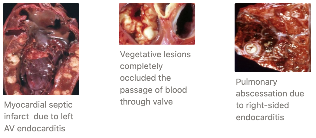
2 causes of myocardial infarct (stroke in animals)
Subaortic stenosis in dogs
Left-sided septic endocarditis
TWO myocardial infiltrations (2)
Adipocytes = Associated with obesity
Lipofuscin = Brown age pigment which accumulates in non-dividing cells
NEITHER associated with clinical signs
What is this?
2 Presentations
Clinical signs
PM
Histology
3 Causes (+ DDx)

Myocardial Necrosis (4)
Presentations:
Acute
Clinical Signs: Many myocytes affected → Acute necrosis and spontaneous death (rare in animals)
PM: White streaks of necrotic myocardium throughout
Subtle and difficult to detect due to similar appearance to PM rigor mortis and myocyte contraction
Histology: Swelling, hypereosinophilic and loss of cross-striations in myocytes
Chronic
Clinical Signs: Fewer myocytes affected → Survival of acute necrosis and signs of CHF due to heart repairing by fibrosis which prevents strength of forward pumping of blood
PM: Yellow, dry, chalky if marked mineralisation present which is more obvious than the reparative phase
Histology: Macrophage infiltration, fibrosis and mineralisation
Causes:
Nutritional Deficiency
VitE/Se = White muscle disease in lambs (RV) and calves (LV)
Potassium
Copper
Thiamine
Magnesium
Toxicity
Ionophores (eg. monensin toxicity in horses when fed cattle-feed)
Chemotherapy (eg. doxorubicin)
Physical Injury and Shock
CNS lesions and trauma (heart-brain syndrome)
Overexertion
Electrical defibrillation
Haemorrhagic shock
Cardiomyopathy (4)
Definition
2 Aetiologies
2 DDx
Definition: Degenerative disease of the myocardium → Reduced heart function and altered blood flow → Sudden death or CHF
Aetiology:
Idiopathic 1˚ disease with heritable component (no underlying disease process)
2˚ to another disease process (eg. feline HCM 2˚ to hyperthyroidism)
DDx:
Hypertrophic cardiomyopathy (HCM)
Dilated cardiomyopathy (DCM)
± Feline endocardial fibroelastosis
What is this? (cat heart)
2 Aetiologies
Signalment
4 Presentations
3 PM features
Pathogenesis


Feline Hypertrophic Cardiomyopathy (HCM)
Aetiologies:
Idiopathic in younger cats
2˚ to hyperthyroidism OR systemic hypertension with CKD in older cats
Signalment: Idiopathic = Male, large and ~4yr
Canine HCM = Uncommon, large dogs present with sudden death
Presentations:
Sudden death due to:
Reduced chamber volume → Little blood pumped out of ventricle chamber → Compensatory increased HR (>200bpm) → Fibrillation OR
Thrombus blocks AV ostia
Die during GA = GA drugs strain CVS resulting in reduced compensatory HR → Increased CHF and reduced CO
Develop LCHF
Aortic thrombus (saddle emboli 20%) = Compensatory atrial dilation and AV dysfunction → Slow blood with turbulence in atrium → Atrial thrombosis → Aortic trifurcation → Infarct of hindlimbs
PM:
LV concentric hypertrophy (increased ventricular wall thickness) and reduced chamber volume
Compensatory dilated LA
AV valve disrupted and squashed together by hypertrophied ventricular myocardium
Pathogenesis:
Degenerative myocardium results in pressure overload due to reduced contractility
LV concentric hypertrophy results with compensatory dilated LA
Marked thickening of LV wall results in left AV valve dysfunction
Regurgitation of blood into dilated LA (heart murmur)
Results in turbulent and slowed blood flow → Atrial thrombosis → infarct of the brain, kidney and aortic trifurcation
What is this?
Signalment (species, breed, sex, age)
Aetiology
Location
Pathogenesis
PM

Signalment: Large dogs (eg. Great dane), male, 5 - 7yr
Also breed-specific Dobermans and Boxers
Aetiology: Idiopathic #1
Cat = Taurine deficiency (grain-free diet) → Chylothorax (heart occludes thoracic duct)
Location: Variable (R/L/both)
Pathogenesis:
Ventricular chamber volume increases
Ventricular wall becomes stretched and thinner
Thin ventricular wall cannot generate sufficient blood pressure to pump blood
→ RCHF/LCHF
Dilated ventricles → Predisposition to thrombi (eg. saddle thrombi)
PM: Globoid heart with chamber dilation and thinning of the ventricular walls (may collapse on PM)
Feline Endocardial Fibroelastosis
Signalment: Burmese and Siamese cats ~4m
Aetiology: Idiopathic or associated with previous endocarditis
Pathogenesis: Diffuse endocardial thickening with little myocardial damage → Reduced chamber volume
Cardiomyopathies in pigs/cattle
Pig: Idiopathic HCM and DCM reported
Cattle: Occasional DCM
Myocarditis (2)
ONE: VIRAL INFECTION
Canine Parvovirus
Signalment: Puppy <2w (vs. enteric form @ ~12w)
Prevalence: Very rare as puppies are rarely born without maternal Ab against CPV due to widespread virus or vaccine of dam OR exposed to CPV immediately after birth
Pathogenesis: Parvovirus only infects rapidly replicating cells and replication of the cardiac myocytes cease at 2w of age
PM: Diffuse heart pallor and flaccidity
Histology: Necrosis and lymphocytic inflammation with large intranuclear basophilic viral inclusions
Prognosis: Survival results in CHF with age due to increasing demand of the heart
Encephalomyocarditis Virus
Signalment: Rare cause of myocarditis of piglets ≤ 4m
Source: Enterovirus carried by rats
Region: Northern North Island of NZ #1
TWO: BACTERIAL INFECTION
Aetiologies:
2˚ to bacteraemia
Lambs with staphylococcal omphalitis
ALL species with vegetative endocarditis
RARE 1˚ bacterial infection (healthy cardiac muscle is highly vascularised and difficult for bacteria to colonise)
Clostridium chauvoei (cardiac blackleg)
Mycobacterium tuberculosis
Clostridium piliformis (Tysser's disease)
Histophilus somni
THREE: PARASITIC INFECTION
Toxoplasma gondii
Signalment: Puppies and kittens
Histology: Multifocal necrosis with lymphoplasmacytic inflammation and protozoal tachyzoites
Neospora caninum
Signalment: Calves
Intermediate stages of tapeworm = Occasional eosinophilic myocarditis due to parasite degeneration
eg. Cysticercus ovis aka. sheep measles in the heart (no clinical significance)
Heart Base Tumours (2)
Aortic Body Chemodectoma = Neoplasm of the aortic body chemoreceptors
Develop in the wall of the proximal aorta → Occlude the Ao → LCHF
Signalment: Rare and only seen regularly in dogs
Behaviour: Malignant, infiltrative neoplasms which do not metastasise
Poor prognosis
Neoplasms of Ectopic Thyroid Tissue
Structure of the Vascular System
3 Components
3 Layers (+ functions)
Components: Same 3 layers with variation in relative thickness of each layer
Arteries
Veins
Lymphatics
Layers:
Tunica Intima = Innermost layer of the vascular system continuous with the endocardium of the heart
Consists of small amounts of fibrous tissue and lined by a single layer of endothelial cells which produce anti-coagulation factors
Tunica Media = Middle layer continuous with the myocardium
Consists of concentric bands of smooth muscle which produce factors stimulating repair of surrounding cells
Large artery = Smooth muscle contracts to propel blood
Small arteries and veins = Smooth muscle maintains vascular tone and BP
Tunica Adventitia = Outer tough and fibrous layer which is continuous with the epicardium
Prevents vessel rupture
4 Types of arterial diseases (+ DDx)
Aneurysms and Ruptures (3)
Thrombosis and Embolism (3)
Degeneration and Necrosis of Arteries
Arteriosclerosis (2)
Atherosclerosis (3)
Calcification (3)
Fibrinoid necrosis (3)
Arteritis (4)
Erysipelothrix of pigs
Feline Infectious Peritonitis (FIP) of cats
Malignant Catarrhal Fever (MCF) of cattle
Strongylus vulgaris of horses
Dirofilaria immitis of dogs
Autoimmune vasculitis
Aneurysms (3)
Definition
3 DDx
Clinical signs
Definition: Focal outpouching of artery caused by reduced wall strength
DDx:
Strongylus vulgaris in horses (RARE)
Copper deficiency in pigs
Essential for normal elastin development
Idiopathic aortic aneurysm of male turkeys
Clinical Signs: NOT defected until rapid, fatal arterial rupture (± compression of surrounding organs)
What is this? (pig and horse heart)
3 DDx
Location
Clinical signs
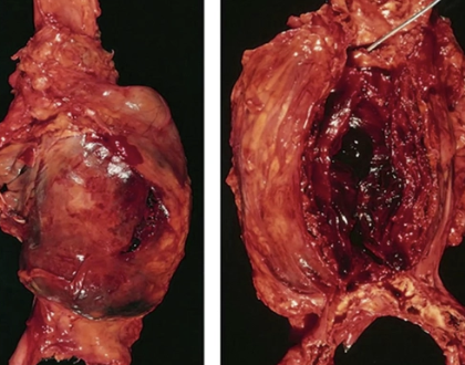

Arterial Rupture (Ao)
DDx:
#1 trauma
Spontaneous or exercise-induced proximal Ao rupture of pigs and horses
eg. Guttural pouch myosis in horses → Rupture of internal carotid artery → Rapid death
Arteritis caused by local inflammation or neoplasia
Location: Proximal Ao (within pericardial sac)
Clinical Signs: Rapid death due to haemopericardium → Cardiac tamponade
Most common cause of thrombosis in humans
Atherosclerotic plaques = Focal accumulation of fatty fibrous tissue within arterial elastic lamina → Loss of compliance
NOT clinically significant in animals (except parrots with high fat diet → infarction)
Seen in hypothyroid dogs but have no clinical signs associated (only important for diagnosis at PM)

Most common cause of thrombosis in animals and location
Atrial Thrombosis due to altered blood flow in atria caused by:
Alterations of heart shape (eg. cardiomyopathy)
Valvular damage → Regurgitant blood creating turbulence with the slow blood
Slowed blood flow due to CHF and compensatory atrial dilation
THREE outcomes of atrial thrombosis
Myocardial infarct (L)
Renal infarct (L)
Saddle thrombus (L)

What is this? (heart or parrot and Ao of cow)
2 Causes
Location
Clinical significance
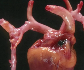

Arterial Calcification (3)
Causes:
Cow = Debilitation (eg. Johne’s disease)
Hypercalcaemia (eg. cholecalciferol toxicity, CKD)
Significance: No effect (Ao #1 = Non-collapsible pipe)
Fibrinoid Arterial Necrosis (3)
Definition
3 DDx
Definition: Necrosis of blood vessels resulting in oedema and haemorrhage within affected organs
DDx;
Pig and chicken vitE/Se deficiency
Pig:
Mulberry Heart Disease = Oedema and haemorrhage of the heart and pericardium
Hepatosis Dietetica = Haemorrhage of liver
Chicken: Oedema and haemorrhage in cerebellum and skeletal muscle
Oedema disease of pigs
Uraemia in dogs and cats = Fibrinoid necrosis of gastric and oral blood vessels
What is this?
Signalment
Pathogenesis
Clinical sign
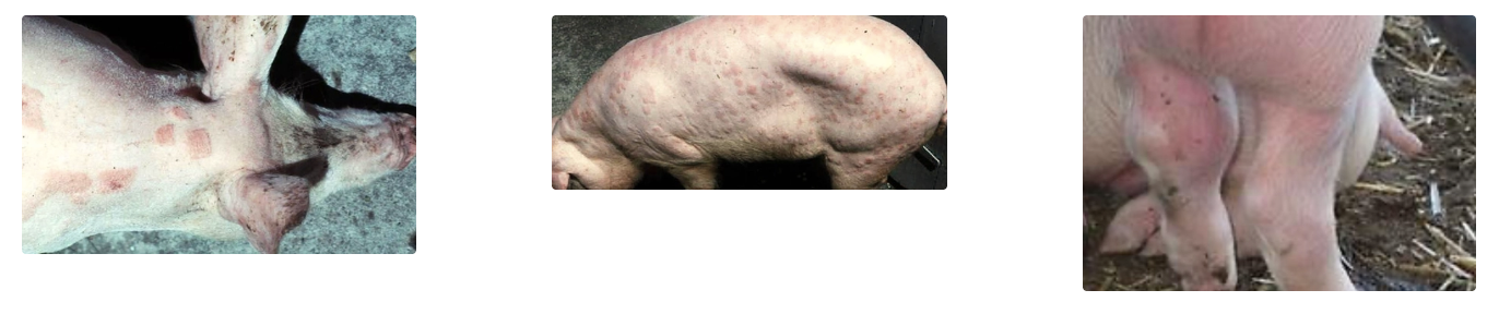
Erysipelothrix rhusiopathiae (4)
Signalment: Pigs and turkeys
Zoonotic
Pathogenesis:
Infection → Septicaemia
Bacteria damage endothelium of the superficial blood vessels
Leads to thrombosis and multifocal cutaneous infarcts
Septicaemia also predisposes the pig to endocarditis and arthritis as there are lots of bacteria circulating the blood
Clinical Sign: Well-demarcated areas of reddening (“diamond skin disease”)
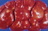
What is this? (cat peritoneal cavity, liver and brain)
Agent
Signalment
Pathogenesis
5 Organ systems affected
PM
2 Forms
6 Methods of diagnosis
Disadvantages

Feline Infectious Peritonitis (FIP)
Agent: Mutated feline enteric coronavirus (FCoV) = FIPV
FCoV is ubiquitous in cat intestinal tracts but few develop mutated virus
Signalment: Purebred cat <2yr
Pathogenesis:
Cat infected with feline enteric coronavirus which colonises the intestinal tract
Virus mutates to FIPV in few cats which enables it to leave the intestines and circulate the blood within macrophages
Loses affinity for enterocytes and hence no longer infective
Circulating FIPV form Ab-Ag complexes which become trapped in smaller blood vessel
Inflammatory cells destroy immune complexes AND direct damage of virus → Vasculitis
Damaged blood vessels leak protein/fibrin-rich fluid into the body cavities and organs affected
Damaged blood vessels also result in thrombosis and infarction in various organs
Organs: Depends on which blood vessels are affected
Hepatic common → Multifocal infarcts
Renal common → Wedge-shaped infarcts
Intestinal
Mesenteric
Neurological (25% of cats only present with neurological signs)
PM: Variable with aggregates of fibrin and inflammation appearing as multiple white foci overlying the affected blood vessels = Leaked protein
Forms:
Dry/non-effusive = No peritoneal fluid and more difficult to diagnose
Wet/effusive = Damage to peritoneal blood vessels results in leakage of fluids into the peritoneal cavity and hence easier to diagnose
Diagnosis:
FCoV Ab Serology
-ve: Ubiquitous nature of disease makes it impossible to interpret a positive test result
Hyperglobulinaemia with Polyclonal Gammopathy of SPE
Peritoneal Effusion
Gross Appearance: Clear to slightly turbid, yellow which may clot due to high fibrinogen
Cytology: High protein and low cellularity (neutrophils and macrophages) with purple background
PCR of peritoneal fluid = #1
Rivalta Test = Drop of peritoneal fluid is placed into weak solution of acetic acid → Drop maintains shape if high protein
-ve: Only confirms high protein fluid (eg. exudate too)
Histology = Pyogranulomatous vasculitis #1
What is this? (cow)
Agent
Signalment
Pathogenesis
5 Clinical signs
Histology


Malignant Catarrhal Fever (MCF)
Agent: Ovine herpesvirus-2 (OHV-2)
Signalment: Cattle
ALL sheep and most cattle infected asymptomatically
FEW cattle develop MCF due to OHV-2 infection
Pathogenesis:
Cow becomes infected with OHV-2
Virus destroys subset of suppressor lymphocytes causing other lymphocytes to attack blood vessels
Results in generalised lymphocytic vasculitis
Clinical Signs: Variable depending on blood vessels affected
Ocular lesions = Corneal oedema, conjunctivitis and ulceration
GIT = Oral ulceration and diarrhoea
Nasal discharge
Coronary band ulceration
Change in temperament (more aggressive)
Histology: Lymphocytic arteritis (but no fibrin leakage as in FIP)
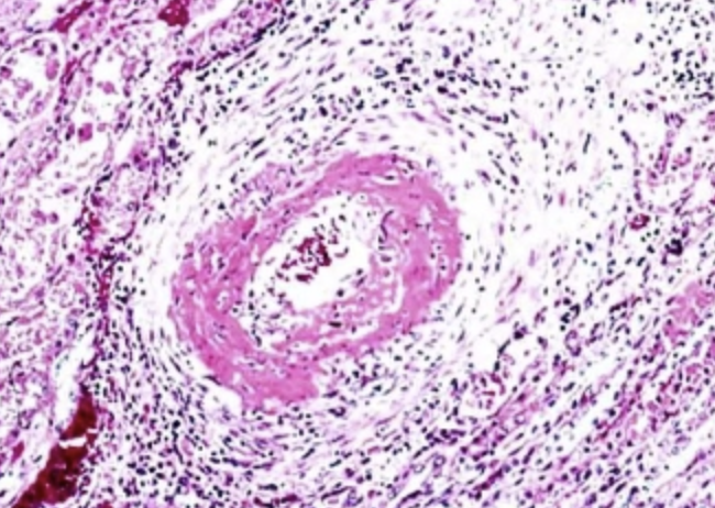
What is this? (horse)
Pathogenesis

Strongylus vulgaris
Pathogenesis:
Migration of larvae in blood vessels causes localised arteritis of the cranial mesenteric artery
Aneurysm → Rare rupture
Thrombosis → Terminal arterial embolism and intestinal infarction
4 DDx of autoimmune vasculitis
Beagle Pain Syndrome = Inflammation caused by auto-Ab against blood vessels in the meninges of the brain and spinal cord → Neck and back pain in dogs
SLE
Immune-mediated polyarteritis
Drug-induced hypersensitivity
What is this? (umbilicus)
Pathogenesis
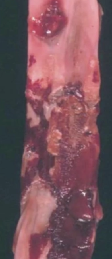
Omphalophlebitis = Only venous inflammation (phlebitis) common in vet
Pathogenesis:
Neonate does not receive adequate colostrum which allows bacteria to infect the umbilical vein
Septic embolic develop from the umbilical vein which travel to the liver to cause hepatic abscesses
Bacteraemia with septic inflammation of the joints, meninges and kidneys
3 Lymphatic diseases and examples of each
Congenital malformation of lymphatics = Rare in dogs, cats and calves resulting in chronically swollen limb with marked pitting oedema
Bacterial lymphangitis (eg. Johne's Disease)
Damaged thoracic duct of cats → Chylothorax
Hamartoma (2)
Definition: Non-neoplastic developmental defect resulting in localised collection of normal-appearing blood vessels
Signalment: Most common in horses
Treatment: Spontaneous regression or curative surgical excision
Myocytes
Structure
4 Organelles
3 Types
Metabolism
Appearance
Example muscle group
Structure: Long and narrow muscle cells which can be as long as the muscle itself (eg. LA myocytes are > 1m)
Functional unit of the muscle → Many myocytes arranged in bundles (fascicle) which are separated by fibrous tissue
Organelles:
Sarcolemma = Cell membrane of the myocyte containing supporting cells (macrophages, satellite cells and fibroblasts)
Multiple peripheral nuclei throughout the length
Many mitochondria to generate significant energy required for contraction
Many myofibrils = Contractile unit/organelle of muscle which contain actin and myosin
Many myofibrils within ONE myocyte
Types:
Type I fibres (slow twitch cells)
Metabolism: Oxidative metabolism ONLY
Appearance: Red due to high vascularisation to supply muscles with O2 for slow and sustained contraction
Example: Bovine masseter muscle
Type IIa fibres (fast twitch cells)
Metabolism: Combination of oxidative and glycolytic metabolism
Type IIb fibres (fast twitch cells)
Metabolism: Glycolytic metabolism ONLY
Appearance: White (less vascularised) as designed to generate rapid, short movement
Example: Chicken pectoral muscles
Why are damaged myocytes observed in clusters on histology?
Muscle contains more than one type of fibre, and a disease process typically affects one type more severely than the other

THREE Responses of Muscle to Injury
Cause
Pathogenesis
ONE: Easy Repair and Complete Regeneration
Cause: Sarcolemma remains intact
Pathogenesis:
Macrophages from sarcolemma enter the cell to remove necrotic debris
Satellite cells enter the myocyte to produce replacement myofibrils which bridge the defect
After repair, satellite cells return to the sarcolemma
TWO: Complete Regeneration OR Fibrosis
Cause: Sarcolemma disrupted
Pathogenesis:
Sarcolemma disruption causes loss of macrophages and satellite cells
Each end of the myocyte bulges TWO possible outcomes
If two ends make contact, sarcolemma is restored to heal myocyte by complete regeneration
If the defect is too extensive, it is bridged by fibrous tissue → Loss of muscle strength and elasticity
THREE: Cell Death
Cause: Damage to ENTIRE cell
Pathogenesis:
Cell death resulting in mineralisation
Defect created by lost myocyte is filled with fibrous tissue
Interpreting colour change in muscle on PM
Necrosis: Pale as damaged myocytes swell which squeezes blood from the tissue
Caution:
Muscle with high proportion of type IIb fibres (eg. chicken pectoral muscle) is normally paler than muscles with high proportion of type I fibres (eg. bovine masseter muscle)
Rigor mortis results in areas of pallor due to skeletal muscle contraction BUT is typically less well-demarcated than true necrosis
Describe TWO ways to classify muscle injury (+ DDx)
Distribution
Monofocal = Lesion at ONE site within the body and consistent with a local process (eg. trauma or injection site reaction)
Multifocal = Lesions at MULTIPLE site within the body and consistent with a systemic process (eg. toxicity or VitE/Se deficiency)
Time
Monophasic = Lesions that develop during a SINGLE damaging incident → Myocytes ALL at the same stage of degeneration and repair on histology (eg. single fatal toxin ingestion or capture myopathy)
Multiphasic = Lesions caused by REPEATED episodes of muscle damage → Myocytes at different stages of degeneration on histology as cell die at different times (eg. muscular dystrophy, vitE/Se deficiency, canine polymyositis)
eg. Normal, recent death = swollen, invaded by macrophages and satellite cells, mineralisation, fibrosis
FIVE Types of changes in muscle size
As myocytes cannot divide, changes in muscle size are limited to atrophy or hypertrophy (no hyperplasia)
Denervation atrophy (4)
Disuse atrophy (3)
Atrophy due to cachexia (3)
Physiological hypertrophy (4)
Pathological hypertrophy (4)
What is this? (horse larynx)
Definition
Pathogenesis
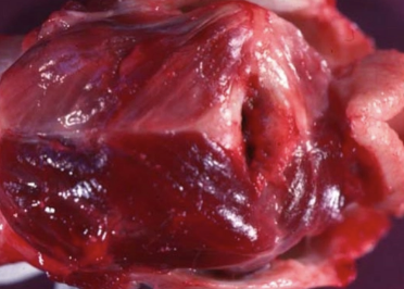
Denervation Atrophy (4) = Laryngeal hemiplegia (horse and dog)
Definition: Denervation of a significant proportion of myocytes → Rapid and marked decrease in myocyte size (vs. disuse atrophy)
Usually entire muscle atrophies as it is supplied by ONE nerve
Pathogenesis: Long-nerve degeneration
Progressive degeneration of the left recurrent laryngeal nerve (longest nerve in body)
Denervation of left intrinsic muscles of the larynx
Laryngeal hemiplegia and marked atrophy of the LEFT dorsal cricoarytenoid muscle
Vocal folds collapse resulting in roaring during exercise
Pathogenesis and characteristics of disuse atrophy (3)
Muscle immobilisation (eg. casting) → Less severe and longer period of atrophy development (vs. denervation atrophy)
Readily reversible and rarely clinically significant
Atrophy due to Cachexia (3)
Pathogenesis
Muscles LEAST affected
Pathogenesis:
Marked malnutrition/neoplasia → Cachexia
Muscle protein catabolism and atrophy
Least Affected: Postural muscles
Muscle Hypertrophy (4)
Pathogenesis
2 Types
Pathogenesis: Addition of myofibrils within the myocyte
Extreme = Additional intracellular fibrous tissue → splitting of myocyte
Types:
Physiological Hypertrophy = Normal reaction of muscle to increased workload (eg. racehorses in training)
Pathological Hypertrophy = Increased muscle mass due to disease process resulting in increased workload of remaining cells (eg. hypertrophy of contralateral limbs or remaining myocytes within the muscle)
List FOUR biochemistry/urinalysis changes associated with skeletal disease
Creatine kinase (CK)
Aspartate aminotransferase (AST)
Myoglobinuria/myoglobinaemia
Hyperkalaemia
Creatine Kinase (CK) vs. Aspartate Aminotransferase (AST)
Advantages
Disadvantages
Creatine Kinase (CK)
+ve: Specific for skeletal and cardiac muscle
-ve:
Artefactual increase in haemolysed samples or hyperbilirubinaemia
Short half-life (~4hr) → Monophasic muscle necrosis results in return of CK to normal within 24 - 48hr
Aspartate Aminotransferase (AST)
+ve: Longer half-life than CK (measure in conjunction with CK to assess severity and progression of muscle disease ± prognosis)
-ve: NOT specific for muscle (released by liver and muscle cell damage
Pathogenesis of Mb leakage with muscle necrosis
Feature of SEVERE and active muscle necrosis/degeneration
Mb released from damaged and necrotic muscle cells (myoglobinaemia = brown serum)
Mb readily filtered by renal glomeruli into urine due to low molecular weight and lack of serum binding (myoglobinuria = brown urine)
Mb is toxic to renal tubular cells resulting in acute renal failure (myoglobinuric nephrosis)
Pathogenesis of hyperkalaemia caused by muscle necrosis
Hyperkalaemia may occur in severe, necrotising muscle disease due to widespread release of intracellular K+ and exacerbated by acute renal failure caused by myoglobinuric nephrosis
Muscle Diseases
4 Congenital/genetic DDx
3 Environmental/physical DDx
5 Infectious DDx
3 Immune-mediated DDx
3 Nutritional/Toxicity DDx
Congenital/Genetic DDx:
Congenital muscular hypertrophy (4)
Splayleg in pigs and swimmer puppies (3)
Malignant hyperthermia (3)
Storage diseases (3)
Environmental/Physical DDx:
Exertional rhabdomyolysis (5)
Capture myopathy (3)
Trauma (5)
Infectious DDx:
Blackleg (4)
Gas gangrene (3)
Woody tongue (4)
Cysticercosis (3)
Sarcocystosis (4)
Immune-Mediated DDx:
Polymyositis (3)
Masticatory muscle myositis (3)
Extraocular myositis
Nutritional/Toxicity DDx:
VitE/Se deficiency (4)
Iron deficiency
Monensin toxicity (3)
Myotonia (1)
Definition: Group of rare inherited diseases causing sustained involuntary contraction of a muscle group
Signalment: Young (6 - 12w) horses, dogs and cats OR goats
Subtypes:
Necrotising Subtype (horses, dogs and cats)
Pathogenesis:
Often caused by dysfunctional membrane ion channels
Allows sustained muscle contraction
INITIAL: Results in muscle swelling due to hypertrophy and foci of muscle necrosis
LATER: Continue muscle necrosis → Visible muscle atrophy and fibrosis
Clinical Signs: Stiff gait, remarkable exercise intolerance, swelling → atrophied muscles
Histology: Mild - moderate multifocal and multiphasic necrosis and regeneration
Benign Subtype (goats)
Clinical Sign: Sudden collapse in response to voluntary effort (eg. due to stress) "Fainting goats"
Significance: NONE!
Congenital Muscular Hypertrophy (4)
Signalment
Pathogenesis
Signalment: Calves (eg. Double-muscle and Belgian Blue) = Selected trait
Pathogenesis:
Developmental defect in myostatin = Protein that inhibits muscle growth
Results in increased muscle size due to myocyte swelling and reduced IM adipose (≤ 60%)
No impaired function BUT issues with dystocia common
Steatosis (2)
Signalment: Incidental condition identified in slaughtered cattle
Pathogenesis: Improper development of myocytes results in replacement with adipose tissue
Histology: Increased adipose separating the myocytes
Difficult to differentiate from chronic myocyte necrosis


Muscular Dystrophy (1)
Definition: Group of rare genetic defects that develop within cytoskeletal proteins of the myocytes
eg. Duchenne-type muscular dystrophy #1 due to dystrophin defect which is encoded for by the largest gene in the body
Histology: Multifocal and multiplastic muscle necrosis
Signalment: Rarely seen in several dog breeds (Labrador) and DSH
ONLY males as x-linked
Pathogenesis:
Congenital defect in dystrophin = protein which anchors the myofibrils to the cell membrane
Results in excessive movement between the cell membrane and internal parts of the cell each time the muscle contracts
Causes cell damage, progressive necrosis and fibrosis of myocytes within muscles throughout the body
Clinical Signs:
Stiff gait and exercise intolerance by 10w
Muscles initially large due to swelling caused by necrosis → Marked muscle atrophy due to loss of myocytes
Aspiration pneumonia due to dysphagia
Death due to heart failure (disease affects cardiac myocytes too)
Splayleg in Pigs and Swimmer puppies
Pathogenesis
Clinical sign
Prognosis
Pathogenesis: Weakened adductor muscles (congenital predisposition?)
Clinical Sign: Animal is unable to place legs under their bodies
Prognosis: Spontaneous recovery as the animal ages
What is this? (pig muscle)
Pathogenesis
Signalment
Clinical sign
PM
Diagnosis

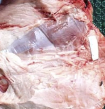
Malignant Hyperthermia (3) aka. Porcine stress syndrome
Signalment: Pig and human
Pathogenesis:
Genetic defect in ryanodine receptor results in loss of normal excitation-contraction coupling
Disease triggered by stress or exposure to halothane anaesthetic
Large influx of Ca2+ into the myocyte
Results in sustained muscle contraction
Excessive heat and intracellular lactic acid is produced resulting in myocyte necrosis
Death due to hyperthermia or acidosis
PM: Muscles of back, shoulder and thigh are swollen, pale and "cooked"
Histology: Multifocal and monophasic muscle necrosis
Diagnosis: Genetic testing (well-defined)

Storage Disease (3)
Definition
Signalment
Pathogenesis
Histology
Diagnosis
Definition: Variety of storage diseases develop due to a variety of inherited enzyme deficiencies
eg. Polysaccharide storage myopathy of horses #1
Signalment: Up to 50% of horses have varying severity of this disease
Draft-type horses #1 as bred for more CHO = increased work duration
Pathogenesis:
Increased CHO within myocytes allows muscles to contract for longer without rest
Results in excess heat and lactic acid production
Increased susceptibility to exertional rhabdomyolysis
Advanced cases of recurrent exertional rhabdomyolysis results in muscle atrophy and weakness due to necrosis
Histology: Multifocal and multiphasic muscle necrosis
Consistent with exertional rhabdomyolysis with large quantities of CHO within intact myocytes
Diagnosis: Biopsy

What is this? (horse muscle and kidney)
Signalment
Pathogenesis
2 Risk factors
3 Clinical signs
PM
Prevention

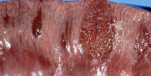
Exertional Rhabdomyolysis (5) aka. Tying up, Monday morning disease, sit-fast, azoturia
Signalment: Horse (± greyhound and sled dog)
Pathogenesis:
Horse accumulates high levels of IM glycogen
Allows oxidation of excessive glycogen in myocytes
Results in excessive production of heat and lactic acid
Because there is no alarm mechanism in horse muscle to detect excessive heat and lactic acid → Continuous oxidation of glycogen
Results in muscle necrosis and increased serum CK
Risks:
Horse with PSSM
Horses fed high-quality feed and irregularly exercised
Clinical Signs:
Stiffness, pain and swelling of muscles immediately after exercise (esp. gluteal and lumbar muscles as used most during exercise)
± Myoglobinaemia (brown serum)
± Myoglobinuric nephrosis (brown urine and AKI)
PM:
Pale and swollen muscles
Dark brown kidneys
Prevention: Feed lower quality feed or exercise more regularly
What is this? (deer)
Signalment
Pathogenesis
PM
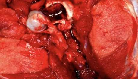
Capture Myopathy (3)
Signalment: Wild deer #1
Pathogenesis:
Capture, chase or transport results in sustained adrenaline release
Adrenaline results in continuous supply of glycogen to the muscle enabling sustained contraction (vs. exertional rhabdomyolysis which has pre-existing excess glycogen in muscle)
Sustain contraction allows accumulation of lactic acid and heat in the muscle
Results in myocyte necrosis with TWO outcomes Outcomes
Immediate death due to metabolic acidosis
Survival of acidosis → AKI due to myoglobinaemia
PM:
Swollen, pale thigh and back muscles
± Ruptured thigh muscles
± Mb-stained kidneys with survival of acidosis
± Multifocal muscle calcification with survival of acidosis
Downer Cow (5)
Pathogenesis
Diagnosis/Prognosis
Pathogenesis:
Cow goes down due to 1˚ disease (eg. hypocalcaemia or hypomagneaemia)
Weight of animal on trapped limb occludes blood flow resulting in hypoxia and muscle necrosis
Necrotic muscle swelling resulting in further hypoxia and necrosis
Resultant muscle damage may prevent cow from standing even after 1˚ problem is resolved
Diagnosis/Prognosis: Predictive scale developed to predict which cattle suffered extensive muscle damage and unlikely to ever stand again
Muscle enzymes (AST)
Number of days recumbent
Urea
Downer cows have reduced renal perfusion due to dehydration
2 Exotic viral diseases causing muscle disease
Blue-tongue (orbivirus)
Foot and Mouth Virus (picornavirus)
2 reasons why bacterial colonisation of muscle is RARE (even with severe bacteraemia)
Myocytes wrapped in tough cytoskeleton that is hard for bacteria to infiltrate
Muscle normally has rich blood supply = Lots of O2 and neutrophils
What is this? (cow muscle)
Agent
Signalment
Pathogenesis
PM
Histology
Diagnosis

Blackleg
Agent: Clostridium chauvoei
Signalment: Cattle ONLY
~1yr coming through gates OR bulling injuries
Pathogenesis: Acute necrotising myositis
Clostridium chauvoei spores ingested with pasture and enter the bloodstream to travel to the skeletal muscle
Spores lie dormant within skeletal muscle as they cannot germinate in healthy, highly oxygenated skeletal muscle
Traumatic injury results in muscle hypoxia enabling bacterial germination
Bacteria produce toxins causing vasoconstriction
Vasoconstriction results in ischaemia and necrosis of surrounding tissue which allows local extension of infection (ample new hypoxic substrate)
Vasoconstriction also prevents neutrophils from reaching bacteria (no resolution of infection and impossible to treat with antibiotics)
Rapid death within 24hr due to endotoxaemia (release of bacterial toxins and toxic metabolites of necrotic tissue)
PM:
Black discolouration of muscle due to venous pooling
Emphysematous muscle due to bacterial gas production = Gas bubbles which is palpable as crepitus under the skin
Characteristic rancid butter smell from gas produced by bacteria
Advanced autolysis due to high fever antemortem
Histology: Monofocal and monophasic necrosis
No neutrophils (vasoconstrictive toxins)
Clear spaces between myocytes (gas)
± Large G+ bacilli
Diagnosis: History of no vaccination AND histology for definitive diagnosis
Culture is NOT useful (dead animal → germination of spores)
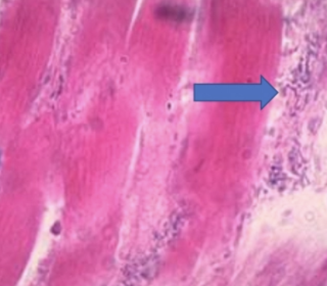
What is this? (arrow = penetrating wound)
Signalment
Agent
Pathogenesis
PM

Gas Gangrene aka. Malignant Oedema
Signalment: Herbivores more susceptible than carnivores
Big head = Ram head butting → Gas gangrene of head
Agent: Clostridium septicum/perfringens/chauvoei/novyi
Pathogenesis:
Deep penetrating wounds become infected with clostridium spores from the environment
Wounds form an anaerobic environment via hypoxia
Critical determinant of disease occurrence depends on speed which the body mounts the immune reaction
Slow immune response → Bacterial proliferation and vasoconstrictive toxins production
Swift immune response → Macrophages and neutrophils kill bacteria before they multiply and produce toxins
Same pathogenesis as blackleg from here
PM: Affected areas range from light-coloured with massive oedema and emphysema to dark and haemorrhagic
Trichinella (1)
Agent: Trichinella spiralis
Prevalence: Reduced prevalence in NZ pigs due to extensive meat inspection and improved management practices
Pathogenesis:
Pig becomes infected by eating skeletal muscle containing encysted Trichinella spiralis larvae
Larvae develop into adults and produce larvae which encyst within skeletal muscle where they are protected from the immune system
Clinical Sign: Muscle pain
Histology: Curled up larvae visible within swollen, glassy myocytes (nurse cells) surrounded by mild fibrosis and minimal lymphoplasmacytic inflammation
Occasional cyst degeneration → Exposed larvae → Stimulates acute eosinophilic or pyogranulomatous inflammatory response
Cysticercosis (3)
Agent
Signalment
PM
Histology
Agent: Cysticercus ovis
Signalment: Sheep and goats = IH (dog = DH)
Pathogenesis:
Sheep become infected with Cysticercus ovis from Taenia ovis eggs secreted by dogs in faces
Cysts develop in the heart and skeletal muscle of sheep and goats (sheep measles)
Dog becomes infected by ingesting encysted larvae in unfrozen/uncooked sheep meat
PM: Numerous small, white foci within skeletal muscle
Histology: Tapeworm larvae surrounded by mild fibrosis and small numbers of inflammatory cells
Sarcocystosis (4)
Agent
Lifecycle
Pathogenicity
Agent: Protozoa (requires TWO hosts to complete lifecycle)
Lifecycle:
Protozoan eaten by IH and enters bloodstream
After much asexual reproduction, protozoa enter skeletal muscle
DH infected by eating contaminated muscle
Pathogenicity: Asymptomatic unless IH ingests lots of protozoan which simultaneously invade myocytes
Hypersensitivity reaction to encysted protozoa → Eosinophilic myositis
Neosporosis (1)
Agent: Neospora caninum
Abortion in cattle
Neuritis and myositis in dogs
Signalment: Puppies and immunocompromised dogs
Clinical Signs: Progressive weakness of HL or rigidity
Prognosis: Poor in advanced cases
What is this?
Signalment
Pathogenesis
Clinical signs
Serum biochemistry
Histology
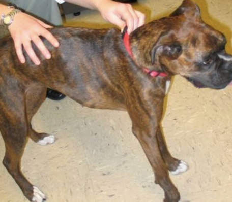
Polymyositis (3)
Signalment: Dog #1
Pathogenesis:
Body produces auto-Ab against muscle Ag
Results in necrosis and inflammation
Clinical Signs: Periodic flares
Muscle pain and swelling
Generalised atrophy and weakness due to fibrosis (± limb contracture)
Biochemistry: Chronic mild - moderate elevation in CK/AST
Histology: Multifocal and multiphasic myocyte necrosis
Regeneration and fibrosis with neutrophilic or lymphoplasmacytic inflammation
What is this?
Signalment
Pathogenesis
Histology
Treatment
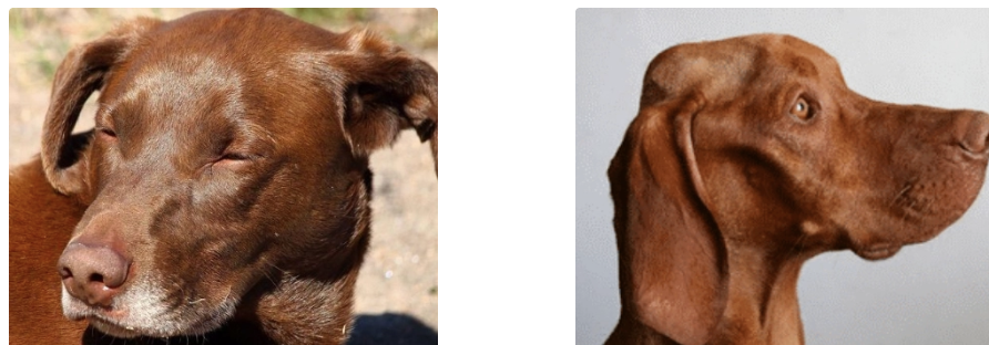
Masticatory Muscle Myositis (3) aka. Eosinophilic myositis and Atrophic myositis
Signalment: Vizsla dogs
Pathogenesis:
Body produces auto-Ab against type II masticatory myosin (unique to temporalis and masseter muscles)
INITIAL: Necrosis and eosinophilic myositis → Swelling and pain while eating
LATER: Myocytes replaced by fibrosis resulting in atrophy
Dog starves to death as they cannot open mouths wide enough to et
Histology:
ACUTE: Multiphasic myocyte necrosis with inflammation ± lots of eosinophils
CHRONIC: Loss of myocytes and replacement fibrosis
Treatment: Prompt treatment during acute muscle swelling to prevent irreversible replacement of myocytes by fibrosis