Lesson 4 - Biology of Friction Ridge Skin
1/59
Earn XP
Description and Tags
Structure, Development & Persistence of FRS
Name | Mastery | Learn | Test | Matching | Spaced | Call with Kai |
|---|
No analytics yet
Send a link to your students to track their progress
60 Terms
Weeks 8-18
Crease Formation
Major & minor creases form
Most develop concurrently with the volar pads but some develop independently
Part of the same skin structure as our ridges
8 weeks - Radial Longitudinal
9 weeks - Distal Interphalangeal & Metacarpophalangeal
10 / 18 weeks - Proximal Interphalangeal
11 weeks - Distal Transverse
11.5 weeks - 1st hand movement
13 weeks - Proximal Transverse
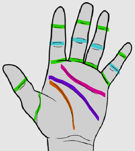
Creases vs. White Lines
Creases
Present on the dermal layer of the skin
Embryonic in nature - develop ~ 8-18 weeks EGA
Fixed shape, size & position
Ridges end at borders of a crease
Persistent & unique
White Lines
Superficial folds in the epidermal skin to the Hyalin layer (Stratum Lucidum)
Develop over time & named for the white lines they leave in inked fingerprint cards
Change shape, size & position
Ridges flow thru to the other side of a white line
NOT persistent or unique
Weeks 7-11
Volar Pad Development
Volar pads begin to swell
Palms ~6.5 weeks
Fingers ~7.5 weeks
Feet ~8 to 9 weeks
Volar pad regression = the slowing growth of volar pads & the simultaneous, more rapid, growth of the hand / feet around the pad
Regression can start ~11 weeks but is not complete until 16 weeks - right before the secondary ridges begin forming
Stratum Lucidum
“Hyalin” layer
Thin layer of cells
Clear flattened dead cells - part of the “Cornified Zone”
Found ONLY in thick skin

Differentiation
Change a cell goes through from birth until death
Loss of ability to divide
Increase in cell size & keratin while cell flattens
Internal changes including synthesis of new proteins / lipids and the addition / degeneration of organelles
External changes including changes to the plasma membrane properties, surface antigens & receptor sites
Dehydration & eventually cell death
Takes about 30-45 days
Hypodermis
Innermost layer of the skin
Composed of adipose tissue
No function in regards to friction ridge skin
Function:
Insulates the body
Serves as an energy reserve
Cushions & protects the body
Allows for skins mobility over underlying structures
Papillae Pegs
Peg-like projections from the dermal papillae that provide the template for the friction ridge arrangement
Double rows - one on either side of primary ridges
Helps increase the bond between the epidermis & dermis
Anastomoses provide additional support by acting like glue bonded to the papillae pegs
Tight Junctions
Attach the keratinocytes to each other - allow them to migrate upward in tandem
Mesenchyme
Loosely organized undifferentiated cells which give rise to numerous structures
Weeks 24-27
Dermal Papillae
Papillae pegs begin forming after the secondary ridge depth is equal to the primary ridge depth
Anastomoses appear
Adherence of the epidermis through anastomoses & the basement membrane zone
Around 24 weeks the friction ridges are fully developed & set for life
Langerhans Cells
Trigger T-cells → immune response
Phases of Scar Formation
Phase I: Inflammation
Blood flows to wound immediately
Platelets send out signals recruiting immune cells to kill bacteria & scavenge damaged cells
Fibroblasts begin to repair the dermis & endothelial cells begin to repair blood vessels
Phase II: Proliferation & Tissue Formation
Desmosomes & hemidesmosomes dissolve to allow cell movement
Pseudopodia & actin filaments help the basal keratinocytes to crawl across the wound
Dermis contracts & the ridges on the surface pucker
Mitosis & upward migration of cells in the wound continue until the proper skin thickness is obtained
Phase III: Tissue Remodeling
Fibroblasts of the dermis continue to reinforce the scar tissue for weeks or months
Scar replaces the friction ridge skin at the wounded area and remains persistent
Stratum Corneum
“Horny” layer
Outer layer of cells
Cells are fully keratinized
Can be up to 100 cells thick (making it 20 or 30 times thicker than anywhere else)
Large, flat overlapping dead cells - part of the “Cornified Zone”
Cells that “slough” off

Stratum Spinosum
“Spinous” layer
Several cell layers thick
Cells begin to shrink due to water loss & look spiny due to the abundant desmosomes attaching them together

Supra-basal layer
Located between the basal & spinous layers
Found ONLY in primary ridges
Transitional area found in thick skin
Contains transient amplifying cells → Arise from stem cells & divide a finite number of times until they become differentiated

Stratum Basale
“Basal” or “Generating” layer
Innermost layer of the epidermis
One cell thick → cells are columnar in shape
“Blueprint” for the friction ridges
Gives rise to the rest of the cells in the epidermis
Attached to the basement membrane
Stratum Granulosum
“Granular” layer
Several cell layers thick
Last living layer of the epidermis → “Malpighian”
Nuclei in various stages of degradation marking the beginning states of cell death
Cells are still alive but start to break down & flatten out

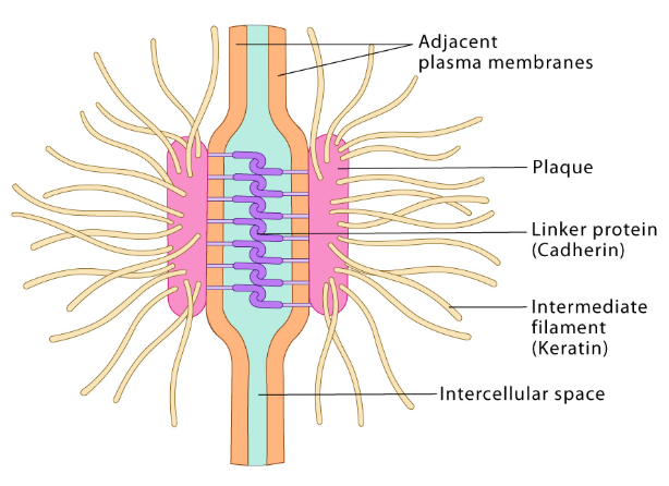
Desmosomes
Attachments between cells
Exist between cells throughout the entire epidermis
New cells are pushed up in concert from basal layer
Undergo modifications as cells progress outward from basal layer
They are broken down to allow cells to slough off as they reach the surface of the skin
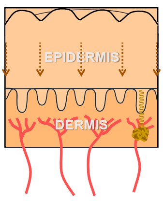
Dermis
Middle layer of the skin
Composed of collagen fibrils, elastic fibers, blood vessels, lymphatic vessels, & nerves
Almost 90% of T-cells are located within the dermis
Has two layers → Dermal Papillae & Reticular Dermis
Function:
Regulates fluids
Provides nutrients to epidermis & removes waste
Protects body from mechanical injury
Thermal regulation
Has sensory receptors
Reticular Dermis
Innermost region of the dermis
Dermal strength & resilience
Connects to hypodermis by a fibrous network
Dermal Papillae
Outermost region of the dermis
Consists of malleable rounded projections called “papillae pegs”
Weeks 5-7
Precursory Development
Paddle like hand - thick tissue
Cartilaginous bone develops from the mesenchyme
Fingers elongate & separate
Thumbs rotate
Nerve innervation occurs
Thin Skin vs Thick Skin
Thin Skin
Found on most of the body
Approx. 1.5mm thick
Contains hair follicles & hair
Contains eccrine & sebaceous glands
Thick Skin
Palmer side of hands & soles of feet
Approx. 4mm thick
Hairless
Contains eccrine glands ONLY
Contains primary & secondary ridges that form FRS
Weeks 14-16
Sweat Gland Anlagen
Precursor to sweat glands
Appear ~14 weeks where the primary ridges are forming
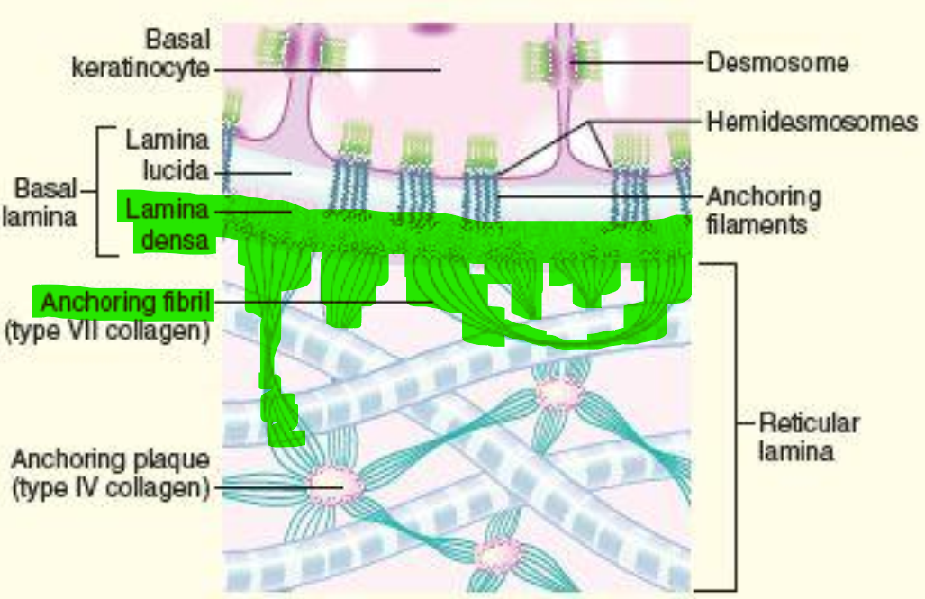
Anastomoses
Sheets of tissue that act like glue bonding to papillae pegs
Fills in around the pegs
Continues to fill in gaps as the pegs branch during aging
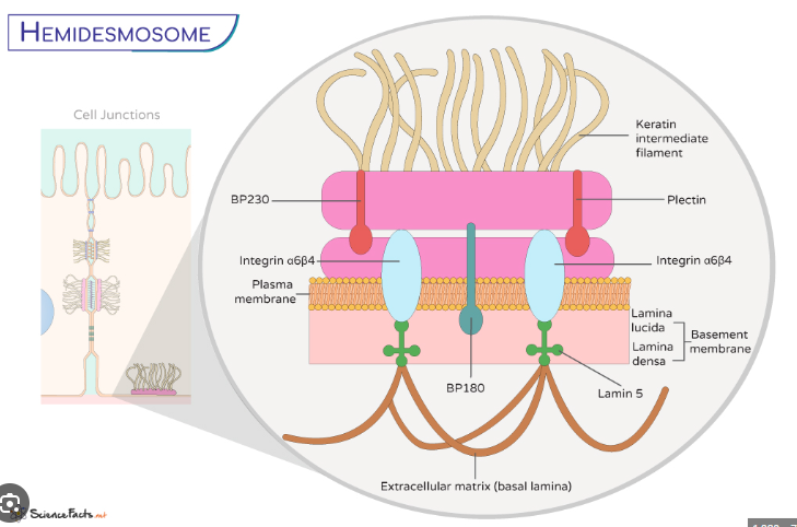
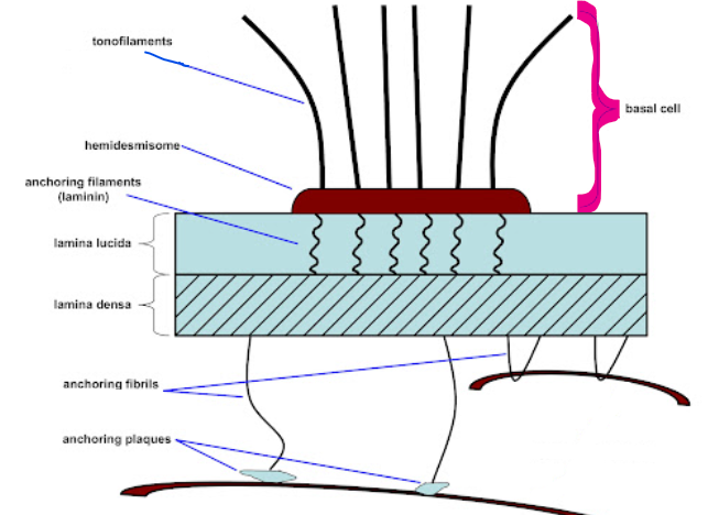
Hemidesmosmes
Located in basement membrane zone
Attachment plaques within basal cells that lock cells to the Basal Lamina
Tonofilaments secure the plaques to cells & receptor sites accept anchoring filaments from the Lamina Lucida
Merkel cells
Sensory input → extension of the body’s nervous system
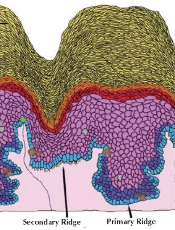
Primary Ridges vs. Secondary Ridges
Primary Ridges
Under the surface ridges
Flanked by papillae pegs
Contains supra-basal layer → transient amplifying cells
Start to form ~10.5 weeks
Secondary Ridges
Under the surface furrows
Appear between primary ridges
Penetrate as deep as the primary ridges
Lack sweat glands
Start to form ~17 weeks
Weeks 10-17
Primary Ridge Formation
Epidermis remains undifferentiated until 10-11 weeks
Around 10.5 weeks primary ridges begin to form on bottom of epidermis → “Critical Stage”
As the skin grows, it separates the existing primary ridges & new primary ridges form
15-17 weeks = Downward penetration of sweat glands & upward push of new cell growth
The entire surface is ridged by 15-17 weeks
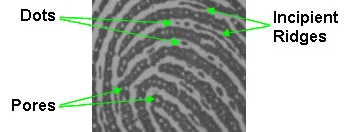
Incipient Ridge
Thin, fragmented ridges that appear in furrows between normal mature friction ridges
Immature & not fully formed
Homeostasis
Condition in which the body’s internal environment remains relatively constant within physiology limits
Achieved in the skin thru physical attachments & regulation of cell production via cell communication
Keeps everything in equilibrium → The number of cells being produced needs to match the number of cells being sloughed off
Cell Surface Receptors
Protein sites on cell membrane that accept chemical signals
Help the cells to “communicate” & signals when to start / stop cellular reproduction
Epidermis
Outermost layer of the skin
Thick Skin = 5 layers deep
Approx. 1.8mm thick
Mostly comprised of keratinocytes
Volar Pads
Transient swellings of the mesenchymal tissue that vary in symmetry & size
Palms ~6.5 weeks
Fingers ~7.5 weeks (starts with thumb and progresses to little finger → distal to proximal on each finger)
Soles of feet ~9 weeks
Regression is slowing growth of volar pad and simultaneous more rapid growth of hand/foot around pad
Gap Junctions
Connections between cell membranes that permit exchange
Keratinocytes
Primary cell of the epidermis (90-95%)
Contains keratin = providing strength
Durable protein
Organized into bundles (filaments)
Structural support
Reinforced cells so they do not break when subjected to physical stress
K9 (only in thick skin) is present in primary ridges which makes them more durable than secondary ridges
Hyperkeratosis
Overproduction of skin cells that can cause…
thickened / rough skin
clogged pores
dryness / irritation
dull / patchy / scaly
Conditions:
Psoriasis
Eczema
Corns, calluses, & warts
Causes:
Genetics
UV / sun exposure
Autoimmune diseases
Dermatitis
Pressure / rubbing of skin
Allergies
Basement Membrane Zone
Two-part fibrous zone that attaches the epidermis & dermis
Contains elements from both the epidermis & dermis
Provides structural support to the skin
Filters nutrients from the dermal blood vessels to the keratinocytes
Weeks 17-24
Secondary Ridge Formation
Second proliferation of cells into the dermis - begins ~17 weeks
All primary ridge growth STOPS
Secondary ridges appear between the primary ridges & lack eccrine glands
As the secondary ridges penetrate into the dermis they pull the already tight skin down with them which forms the surface furrows
Surface ridges spread across the finger & converge at the delta areas
Skin Regeneration
1) Skin cells slough off from the Horny layer
2) Cells communicate to maintain homeostasis
3) Basal cells replicate DNA & start mitosis
4) Additional cells replicate in the Supra-basal layer
5) Cells migrate upward in concert & differentiate
Lamina Lucida
A transparent layer containing anchoring filaments that attach to the plasma membrane of the basal keratinocytes
Top layer of the Basal Lamina
The filaments are attached below & perpendicular to the hemidesmosomes
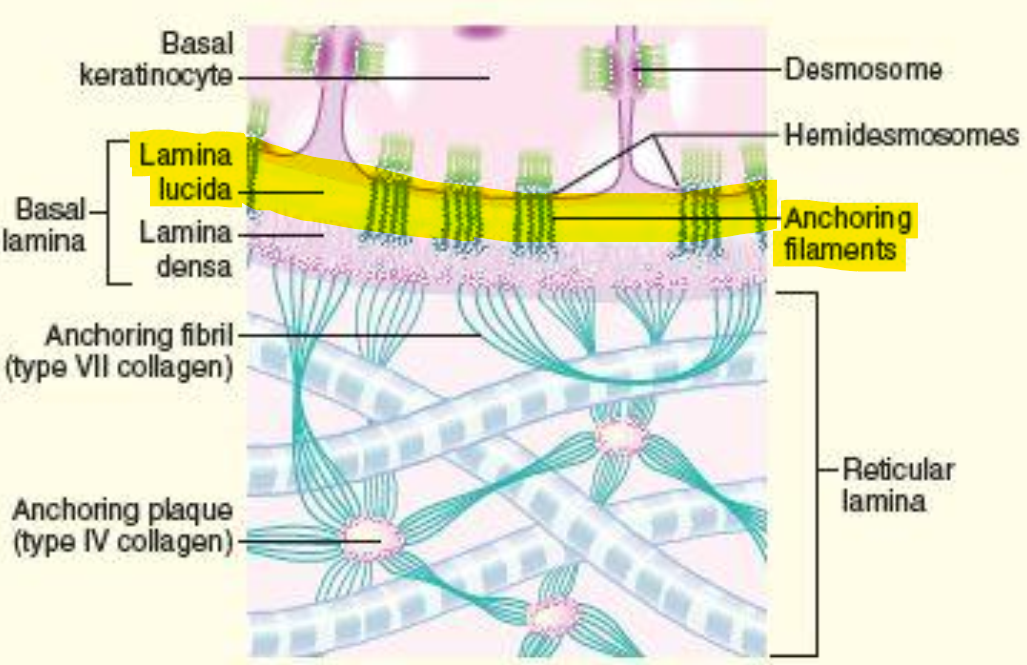
Lamina Densa
A dense layer containing anchoring fibrils that attach to collagen fibrils of the dermis
Bottom layer of the Basal Lamina
Melanocytes
Pigment cells → Protect us from UV / sun exposure
Dermal-Epidermal Junction
Multiple parts that connect the epidermis and the dermis of the skin together
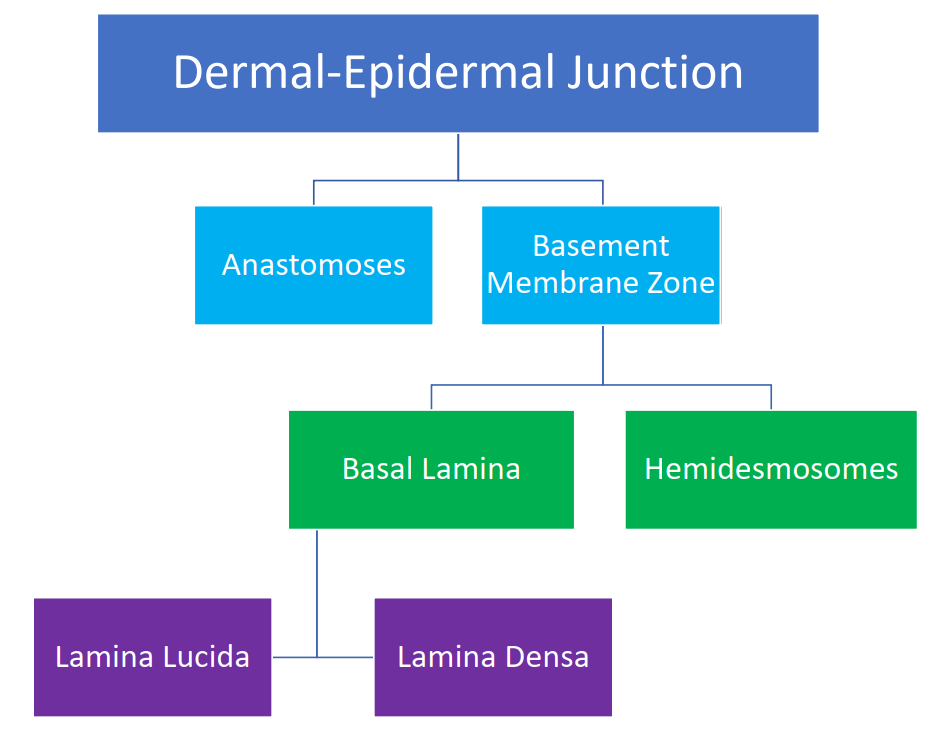
Basal Lamina
Lower half of the basement membrane zone that contains the Lamina Lucida & Lamina Densa
Helps attach the epidermis to the dermis
Causes of Variation in the Formation of our Tissues and Body Parts
Genetics
Environment
Developmental Noise
Human variation exists due to…
genotype and phenotype
What do our genetics direct?
Might direct when and where but NOT how
Pattern type is influenced by genetics but there are no genes for pattern type
Genotype
The set of chromosomes we inherit from our parents
Phenotype
Particular trait a person displays → the traits that are actually “expressed”
Ex. Hair color, eye color, etc.
Different genotypes can cause different phenotypes
Environmental Factors
Intrauterine stressors
Fetal environment
Maternal hormone levels
Maternal diet
Chemical intake, alcohol or drug use
Excess fluids
Disease
Radiation
Developmental Stability
The ability of an organism to produce a consist phenotype despite environmental and genetic variations
Growth & development of the embryo & fetus are developmentally stable
Ex. # of fingers / toes, length of bones, etc.
Developmentally Stable Growth Factors
Growth & development of fetus
Development of hands and feet
Placement of regular flexion creases
Placement of volar pads
Developmental Noise
Features that are not hardwired in genetic code and therefore are at the mercy of random formation
Exact placement of each ridge and crease is left up to chance events that take place during development
Causes different phenotypes to arise from same genotype in same environment
Source of randomness
Provides tremendous variation in the arrangement of ridges
Growth Stress
Impacts the directionality of ridges = guides direction of ridges
Differential growth establishes growth stress
Ridges grow perpendicular to growth stress
Ridge Formation
Link between growth stresses & friction ridge formation is MERKEL CELLS
Differentiate and band together in the epidermis
Perpendicular to growth stresses
Prior to ridge formation
Merkel Cell Activity
Stimulates basal cells which proliferate into dermis, beginning primary ridge development
Basal cells divide along bands of Merkle cells
Cells continue to divide & deepen primary ridges
Bands of Merkel Cells
Wherever the merkel cells band together the first primary ridges form
Longest & most continuous ridges form first with few minutiae
1st minutiae form in areas where bands of Merkel cells diverge to cover large space or converge to squeeze into a smaller space… this results in ending ridges & bifurcations
Funnel areas have less unique minutiae
Hand continues to grow in length & width
Existing primary ridges are pulled away from each other & new ridges form… they tend to be shorter in length & fill in space which adds more minutiae
Exact arrangement of ridges not dictated by DNA
Developmental Noise has its greatest impact during this process (ridge formation)
What three things are driven by growth stresses?
Patterns
Ridge counts
Minutiae densities
3 Main Growth Stresses
Growth of hands and feet - increase in length faster than width
Regular flexion creases - form prior to ridges & are sites of tension in skin
Volar pads - create localized growth stresses that redirect ridge flows in small areas