Thoracic Body Wall I - Vocabulary Flashcards
1/40
Earn XP
Description and Tags
Vocabulary-style flashcards covering thoracic wall anatomy, surface landmarks, dermatomes, osteology, and embryology concepts from Lecture 21.
Name | Mastery | Learn | Test | Matching | Spaced |
|---|
No study sessions yet.
41 Terms
Thorax
The part of the trunk between the neck and abdomen that supports the thoracic wall and houses the thoracic viscera. It is bounded by the rib cage and includes the lungs, heart, and major blood vessels.
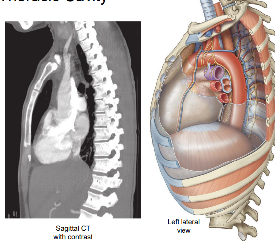
Thoracic Cavity
Contains mediastinum and left and right pulmonary cavities.
Superior thoracic aperture
The opening at the top of the thoracic cage formed by T1, first ribs, and manubrium that transmits the trachea, esophagus, nerves, and major vessels.
Inferior thoracic aperture
The bottom opening of the thorax through which structures pass between thorax and abdomen as the diaphragm forms.
Thoracic Outlet Syndrome
Crowding at the superior thoracic aperture that compromises the neurovascular supply to the upper limb.
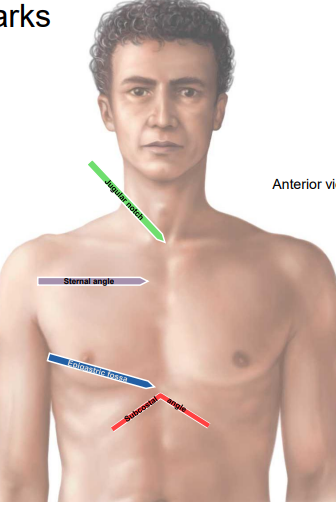
Jugular notch
The indentation at the superior border of the manubrium, just above the sternoclavicular joints. At T2 vertebral level.
Sternal angle
The palpable angle where the manubrium meets the body of the sternum; marks the level of the second rib.
Epigastric fossa
The slight indentation over the inferior end of the sternum.

Subcostal (infrasternal) angle
The angle formed by the inferior edge of the rib cage at the epigastric fossa.
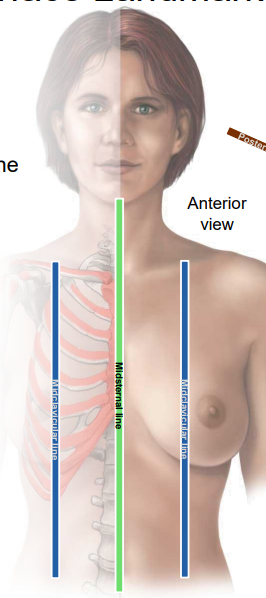
Midsternal line
A vertical surface landmark running along the midline of the sternum.

Midclavicular line
A vertical surface landmark passing through the midpoint of the clavicle, parallel to midsternal line
Anterior axillary line
A vertical line along the anterior border of the axilla, along the lateral margin of pectoralis major
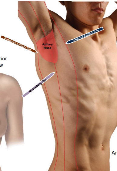
Posterior axillary line
A vertical line along the posterior border of the axilla, along the lateral margin of the latissimus dorsi

Axillary fossa
The hollow beneath the shoulder joint (armpit)

Midaxillary line
A vertical line through the midpoint of the axilla.

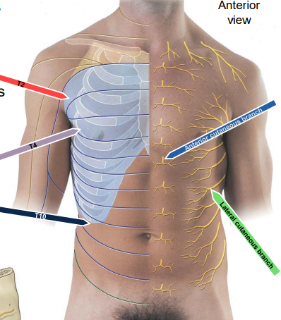
T2 dermatome
Thoracic dermatome at the axillary level.

T4 dermatome
Thoracic dermatome at the nipple level.

T10 dermatome
Thoracic dermatome at the umbilicus level.
Rib head
Posterior end of a rib with superior and inferior articular facets; ribs 1, 11, and 12 have a single facet. (light green)

Rib tubercle
Projection where the rib articulates with the transverse process of the vertebra. Not on 11 and 12 (dark blue)

Rib angle
Point of greatest curvature of a rib. (red)
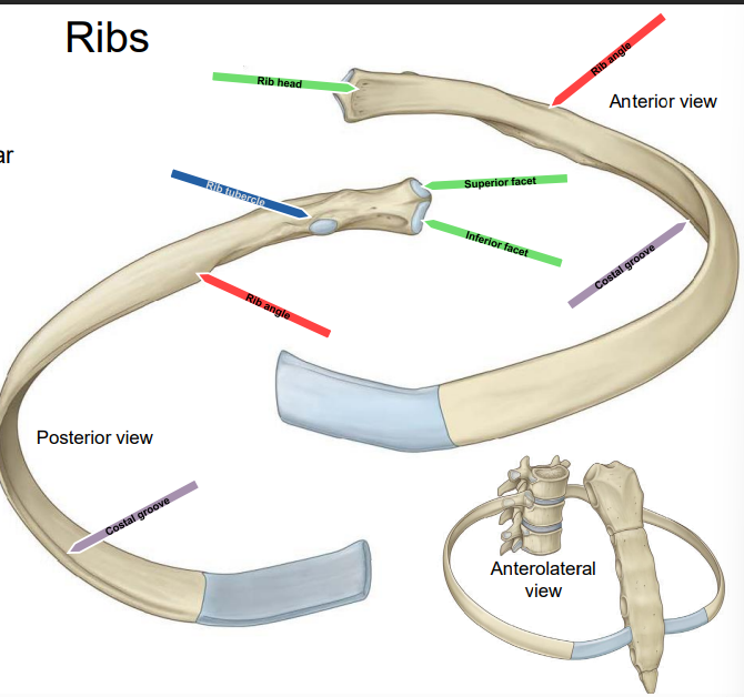
Costal groove
Groove along the inferior margin of a rib that houses intercostal vessels and nerve. (purple)

Costovertebral joints
Joints where rib heads articulate with the vertebral body of the same number and the body of the next superior vertebrae; tubercles articulate with transverse processes (except ribs 1, 11, 12- only articulate with vertebral body of same number).
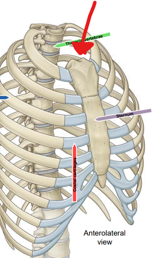
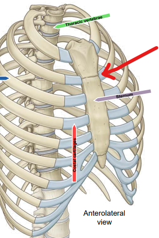
Manubrium
Upper part of the sternum; articulates with the clavicle and first two costal cartilages; contains the jugular notch.

Body (sternum)
The central part of the sternum; articulates with the manubrium at the sternal angle and with ribs 2–7 via costal cartilages.

Xiphoid process
Inferior end; articulates with sternal body at xiphisternal joint, articulates with 7th costal cartilage.
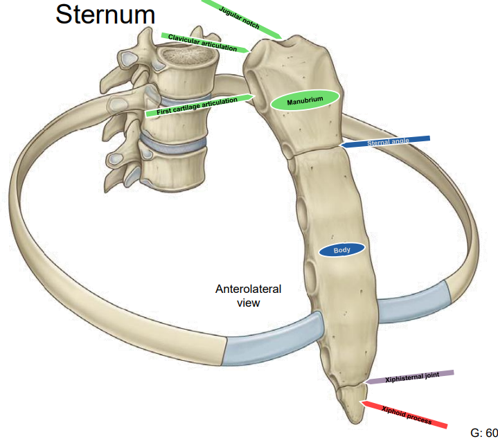
Xiphisternal joint
The joint between the xiphoid process and the body of the sternum. at T9 level.

True ribs
Ribs 1–7 whose costal cartilages attach directly to the sternum.

False ribs
Ribs 8–10 whose costal cartilages attach to the cartilage of the rib above.
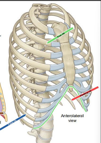
Floating ribs
Ribs 11–12 whose cartilages do not articulate with the sternum or other cartilages.

Septum transversum
Embryonic structure that contributes to the central tendon of the diaphragm and divides the pericardial and peritoneal cavities.
Pleuropericardial membranes
Membranes that grow from the lateral walls toward the lungs to isolate the pericardial and pleural cavities; remnants form the fibrous pericardium.
Pleuroperitoneal membranes
Membranes that surround the septum transversum and close off communications between pleural and peritoneal cavities.
Pericardial cavity
The space within the pericardium surrounding the heart.
Pleural cavities
The right and left serous cavities between the parietal and visceral pleura surrounding the lungs.
Peritoneal cavity
The serous cavity within the abdomen derived from the intraembryonic coelom.
Pericardioperitoneal canals
Channels between the developing pericardial and peritoneal cavities that contribute to separating the cavities.
Diaphragm (central tendon)
Musculotendinous partition separating thoracic and abdominal cavities; central tendon from septum transversum; muscular portions from C3–C5 myoblasts.
Dermatome- lateral cutaneous branch
emerges at midaxillary line and innervates most of dermatome
Dermatome- anterior cutaneous branch
emerges lateral to sternum and innervates most medial part of dermatome
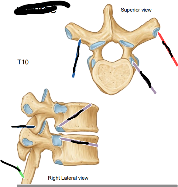
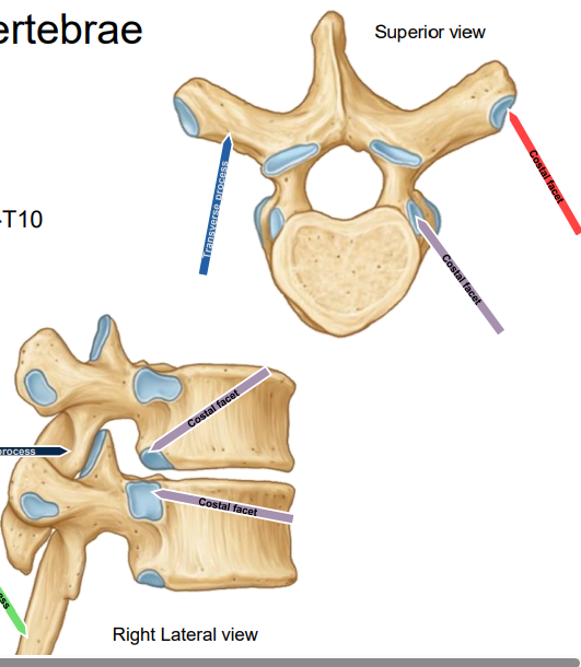
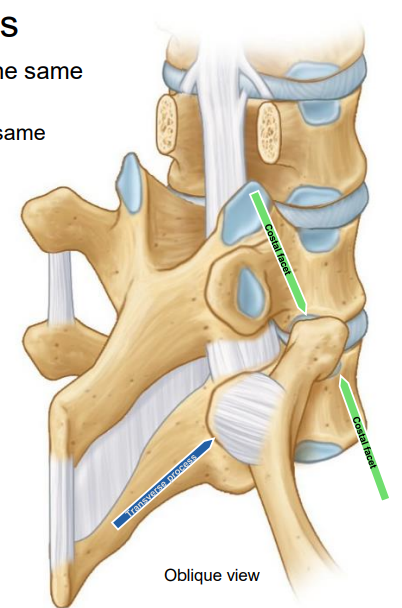
ID all and name
Thoracic vertebrae- Costal facets on all, facets on transverse processes of T1-10;
Superior view:
light blue- transverse process
purple- costal facet
red- costal facet
Lateral view:
green- spinous process
purple- costal facet
dark blue- articular process