Week 2 - Lecture - PA SC Joints and Sternum Imaging - Q&A Flashcards
1/15
Earn XP
Description and Tags
A set of Q&A flashcards covering PA SC joints, sternum RAO vs. PA oblique positioning, and lateral sternum imaging considerations.
Name | Mastery | Learn | Test | Matching | Spaced | Call with Kai |
|---|
No analytics yet
Send a link to your students to track their progress
16 Terms
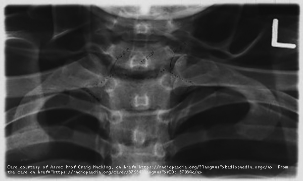
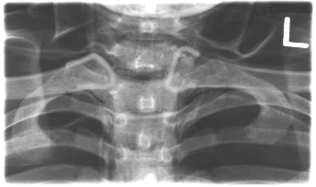
What structures are best demonstrated in the PA SC joint projection?
Lateral manubrium and medial ends of clavicles.
Which projections are used to image the sternum?
RAO (PA oblique) and Lateral.

How do you position a patient for PA SC joints?
PA erect or recumbent, arms at sides/overhead, no rotation or tilt, CR perpendicular at T3.
What rotation demonstrates the right SC joint open?
RAO.
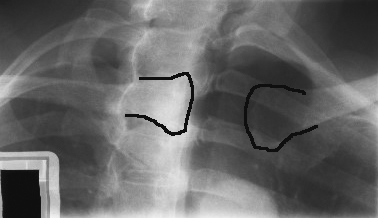
What rotation demonstrates the left SC joint open?
LAO.
What indicates correct positioning on PA SC joints?
Equal distance of SC joints from vertebral column.
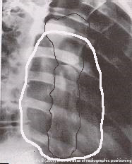
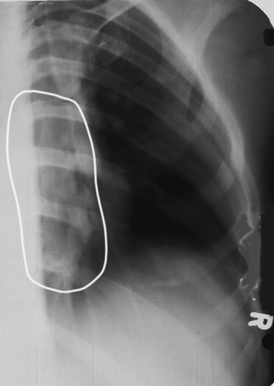
Why is RAO preferred for sternum imaging?
Places sternum over heart shadow, reducing lung/rib superimposition.
How much rotation is used for RAO sternum?
15–20° (15° hypersthenic, 20° asthenic).
Where is the CR directed for RAO sternum?
At T7, 2.5 cm left of midline.
What breathing technique helps visualize the sternum?
Shallow breathing with low mA and long exposure (2–4 sec).
What does under-rotation look like in RAO sternum?
Sternum still superimposes spine.
What does over-rotation look like in RAO sternum?
Sternum lateral to heart shadow, foreshortened.
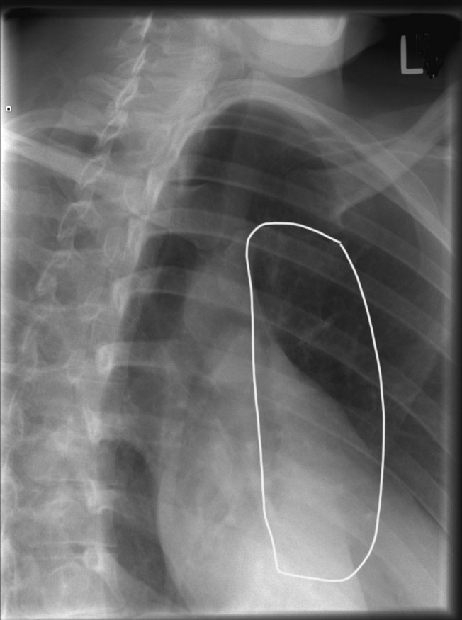
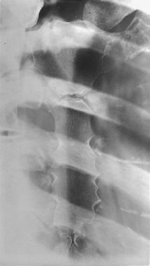
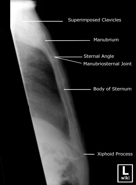
How is the patient positioned for a lateral sternum (erect)?
Arms and shoulders drawn back, chest out; mid-coronal plane perpendicular to IR.
Where is the CR for lateral sternum?
Perpendicular to mid-sternum, midway between jugular notch & xiphoid.
Why use increased SID (152–183 cm) for lateral sternum?
To reduce magnification caused by large OID.
What indicates correct positioning in lateral sternum?
Sternum free of rib/shoulder overlap, sharp bony detail, superimposed anterior ribs.
