Lower limb Connective tissue and joints
 Call Kai
 Learn
Learn Practice Test
 Spaced Repetition
 Match
Match Flashcards
 Knowt Play
1/29

There's no tags or description
Looks like no tags are added yet.
Name | Mastery | Learn | Test | Matching | Spaced | Call with Kai | Chat |
|---|
No analytics yet
Send a link to your students to track their progress
30 Terms

Knee Complex
Includes three joints — Tibiofemoral (distal femur + proximal tibia), Patellofemoral (patella + femur), and Superior Tibiofibular (proximal tibia + fibula).
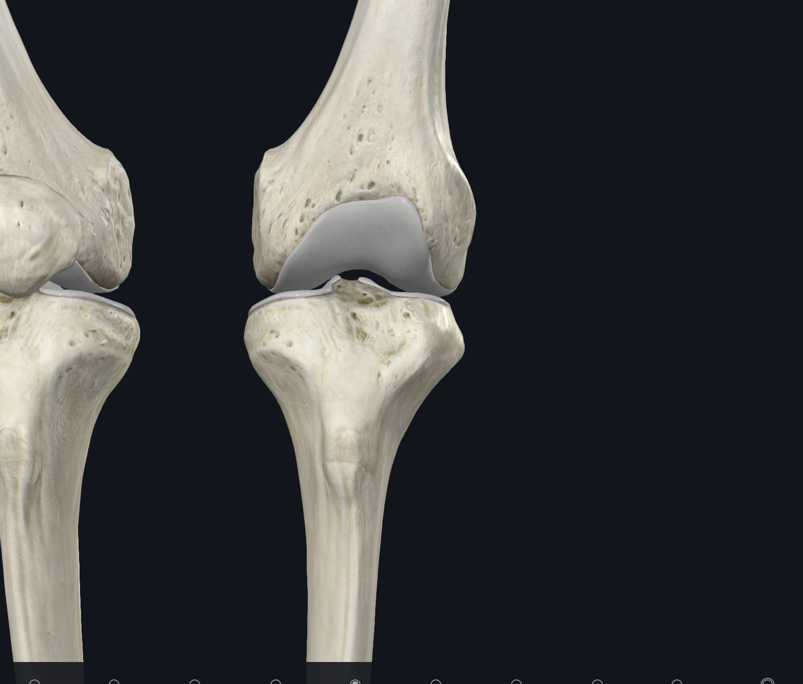
Anterior View
Joint
Name: Tibiofemoral Joint
Description: Hinge joint. Monoaxial
Intermediate joint of the lower limb. Primarily a hinge-type synovial joint with one degree of freedom (flexion/extension). In non-weight bearing, allows functional shortening of the limb for limb advancement during the swing phase of gait. Medial/lateral tibial plateau articulates with medial/lateral femoral condyles. Relatively weak mechanically since the tibial plateau facets are shallow (poor bony congruence); menisci increase congruence. All articular surfaces covered in hyaline cartilage. Stability depends on muscle/tendon strength (especially quadriceps femoris) and ligaments.
Genu Valgus vs. Genu Varum
Description: The medial femoral condyle is angled and extends farther out than the lateral condyle, producing a typical genu valgus posture at the knee (knock-knee). Genu varum = bowleg.
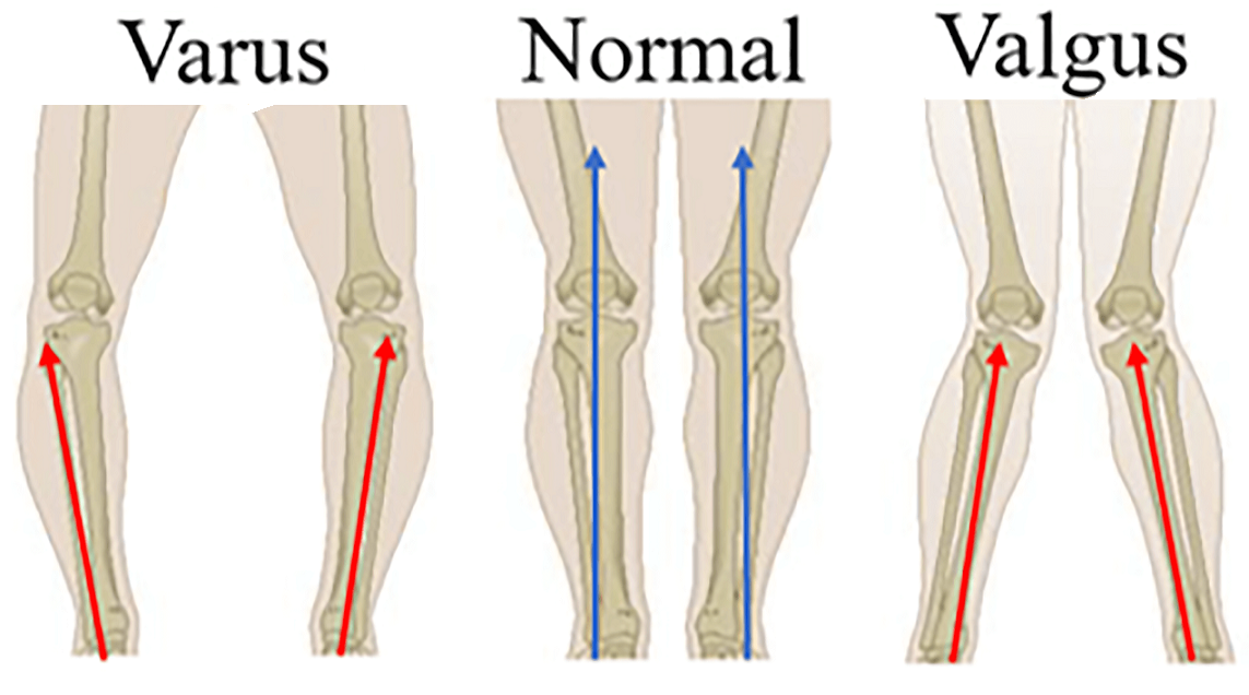
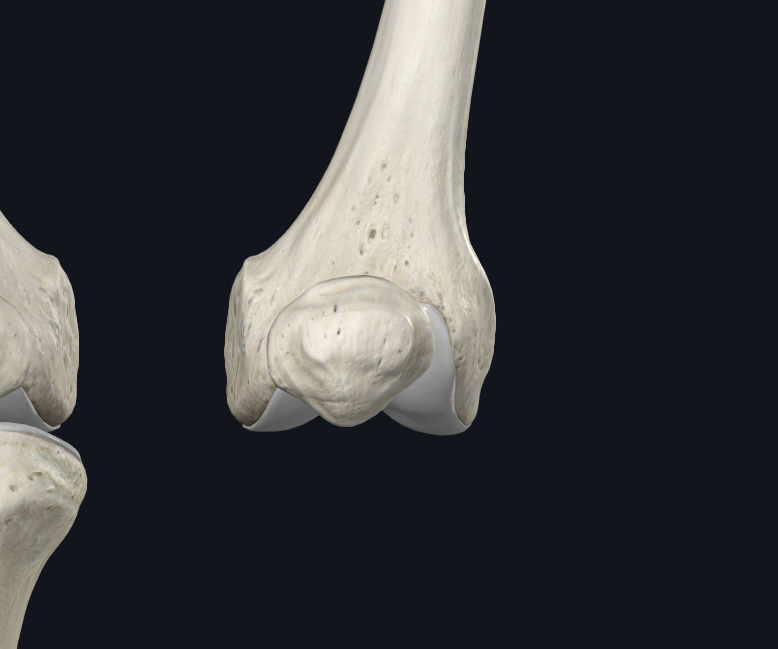
Anterior View
Joint
Name: Patellofemoral Joint
Description: A plane gliding joint. Nonaxial
The patella's three facets and intervening medial ridge articulate with the patellar surface (trochlear groove) of the femur, which acts as a gutter for the posterior patellar surface. Patella glides inferiorly with flexion, superiorly with extension. Contact area migrates superiorly with knee flexion.

Anterior View
joint
name: Proximal Tibiofibular Joint
Description: Gliding nonaxial
Synovial joint between the head of the fibula and the lateral condyle of the tibia. A flat facet on the fibular head articulates with a similar facet located posterolaterally on the lateral tibial condyle. Surrounded by a fibrous articular capsule attached to the margins of both bones' articular surfaces.

Anterior View
Connective tissue
Name: Knee Joint Articular Capsule & Synovial Membrane
Description: Fibrous capsule attaches to the femur superiorly (just proximal to the condylar margins), posteriorly to the intercondylar fossa, and inferiorly to the tibial articular margin. Absent anteriorly, allowing the synovial membrane to pouch superiorly and form the suprapatellar bursa. The synovial membrane lines the internal aspect of the fibrous capsule; this synovial cavity is the largest joint space in the body.

Anterior View
Connective tissue
Name: Patellar Ligament (Tendon)
Description: Distal continuation of the quadriceps tendon. A strong, thick, fibrous band passing from the apex of the patella to the tibial tuberosity. Separated from the knee's synovial cavity by the infrapatellar fat pad. Blends with the medial and lateral patellar retinacula; also called the patellar tendon.

Anterior view
connective tissue
Name: Patellar Retinaculum
Description: Holds the patella on track. Tissue expansions of the vastus medialis and vastus lateralis that provide support to the articular capsule.

Lateral sagitttal view
Connective tissue
Name: Fibular Collateral Ligament (LCL)
Description: Also called the Lateral Collateral Ligament. Rounded, cordlike, and strong. Extends inferiorly from the lateral epicondyle of the femur to the lateral surface of the fibular head.
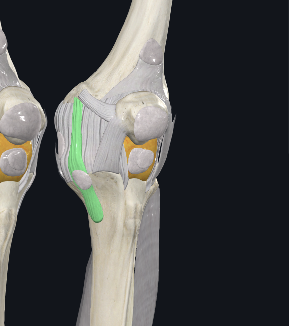
Sagital anterior view
Connective tissue
Name: Tibial Collateral Ligament (MCL)
Description: Also called the Medial Collateral Ligament. A strong, flat band — weaker than the LCL. Extends from the medial epicondyle of the femur to the medial condyle and superior medial surface of the tibia. Deep fibers attach to the medial meniscus at the midpoint. Often injured together with the LCL in contact sports.

Posterior View
connective tissue
Name: Oblique Popliteal Ligament
Description: An expansion of the semimembranosus that strengthens the knee capsule posteriorly. Arises posterior to the medial tibial condyle and passes superolaterally to attach to the central posterior fibrous capsule.

Posterior view
Connective tissue
Name: Arcuate Popliteal Ligament
Description: Strengthens the fibrous capsule posteriorly. Arises from the posterior aspect of the fibular head, passes superomedially over the popliteus tendon, and spreads over the posterior surface of the knee joint.

Anterior view
Connective tissue
Name: Anterior Tibiofibular Ligament
Description: Two or three broad, flat bands passing superomedially from the anterior aspect of the fibular head to the anterior part of the lateral tibial condyle.

Posterior view
connective tissue
Name: Posterior Tibiofibular Ligament
description: A single broad band passing superomedially from the posterior aspect of the fibular head to the posterior part of the lateral tibial condyle.

Anterior superior view
Connective tissue
Name: Cruciate Ligaments (overview)
Description: ACL and PCL join the femur and tibia, crisscrossing within the articular capsule like an "X" in the center of the joint. Both provide stability to the knee.
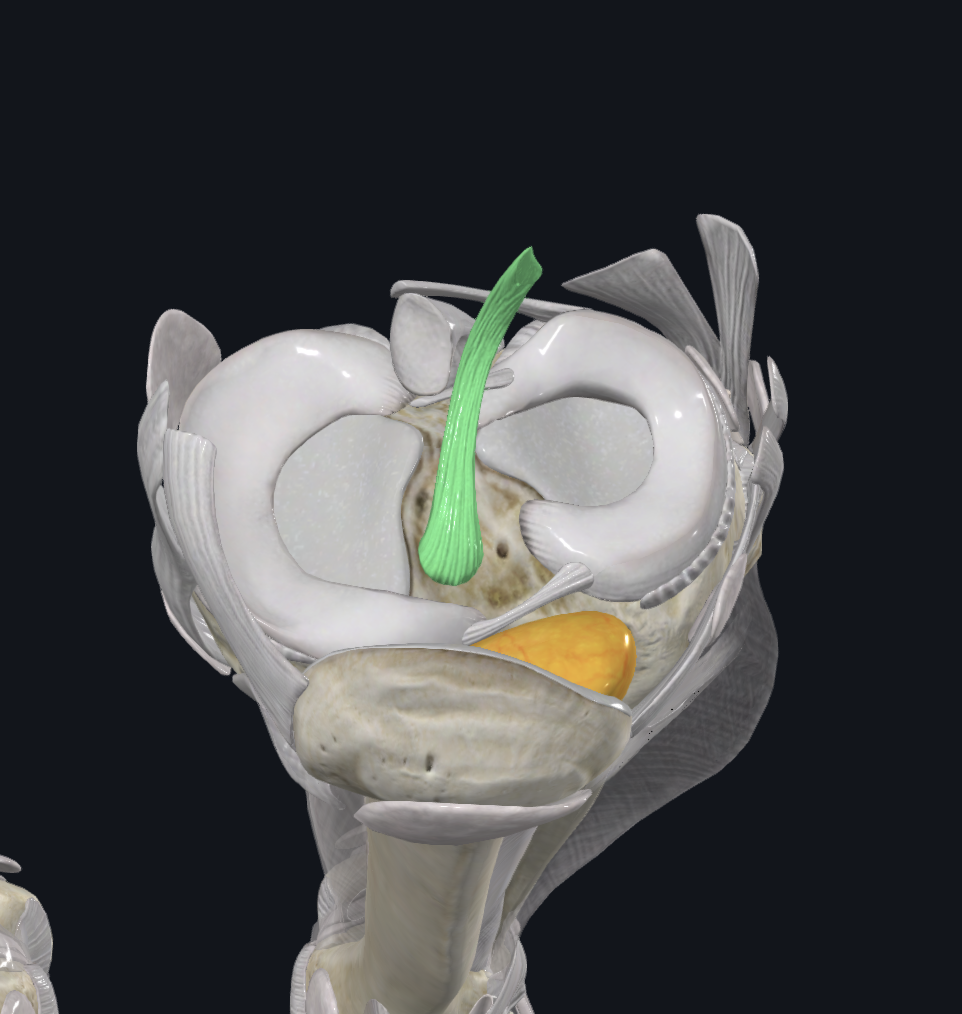
Anterior Superior view
Connective tissue
Name: Anterior Cruciate Ligament (ACL)
Description: The weaker of the two cruciate ligaments. Arises from the anterior intercondylar area of the tibia and extends superiorly, posteriorly, and laterally to attach to the medial border of the lateral femoral condyle. Relatively poor blood supply. Slack when the knee is flexed, taut when fully extended. Prevents posterior displacement of the femur on the tibia (or anterior displacement of the tibia on the femur).

Posterior view
Connenctive tissue
Name: Posterior Cruciate Ligament (PCL)
Description: The stronger of the two cruciate ligaments. Arises from the posterior intercondylar area of the tibia and passes superiorly and anteriorly on the medial side of the ACL to attach to the lateral surface of the medial femoral condyle. Tightens during knee flexion. Prevents anterior displacement of the femur on the tibia (or posterior displacement of the tibia on the femur).
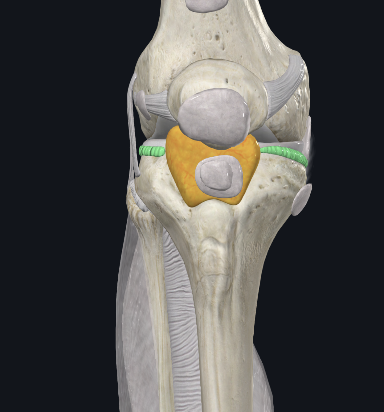
Anterior View
Name: Coronary Ligaments
Description: Capsular fibers attaching the margins of the menisci to the tibial condyles.

Anterior view
Connective tissue
Name: Transverse Ligament
Description: Joins the anterior edges of the menisci, permitting the menisci to move together during knee movements.

Posterior view
Connective tissue
Name:Posterior Meniscofemoral Ligament
Description: Joins the lateral meniscus to the PCL and the medial femoral condyle.

Anterior view
Connective tissue
Name: Menisci (overview)
Description: From the Greek "meniskos" = crescent. Fibrocartilaginous discs (one medial, one lateral) on the articular surface of the tibia. Deepen the joint surface, add to joint congruity, and act as shock absorbers for the knee. Thicker at their external margins, tapering to thin, unattached inner edges. Firmly attached at their anterior/posterior ends (horns) to the intercondylar area of the tibia; external margins attach to the fibrous capsule of the knee joint.

Anterior superior transverse view
Connective tissue
Name: Medial Meniscus
Description: "C" shaped; broader posteriorly than anteriorly. Anterior horn attaches to the anterior intercondylar area of the tibia, anterior to the ACL. Posterior horn attaches to the posterior intercondylar area, anterior to the PCL attachment. Firmly adheres to the deep surface of the MCL.

Anterior superior transverse view
Connective tissue
Name: Lateral Meniscus
Description: Nearly circular, smaller, and more freely moveable than the medial meniscus. The tendon of the popliteus separates the lateral meniscus from the LCL.
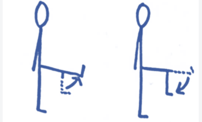
Actions of the knee
Action: Knee Flexion & Extension
The primary motions at the knee; some medial and lateral rotation also occurs. Flexion: hamstrings are the primary movers, with the gastrocnemius assisting. Extension: mostly the quadriceps.

Knee action
Action: Knee Rotation
Description: Medial rotation is produced by the popliteus and semitendinosus (slightly assisted by semimembranosus) and is checked by the cruciate ligaments. Lateral rotation is produced by the biceps femoris and is checked by the collateral ligaments.
What muscle and bones aids in locking/Unlocking of the Knee
Name: Popliteus
When fully extended, the knee "locks" due to medial rotation of the femur on the tibia, which aids weight bearing. To "unlock" the knee, the popliteus contracts, rotating the femur laterally so flexion can occur.

Bursae of the Knee
12 or more bursae surround the knee. Subcutaneous bursae: prepatellar, infrapatellar. Bursae in the synovial cavity: suprapatellar. Other bursae: popliteal, pes anserine, gastrocnemius.
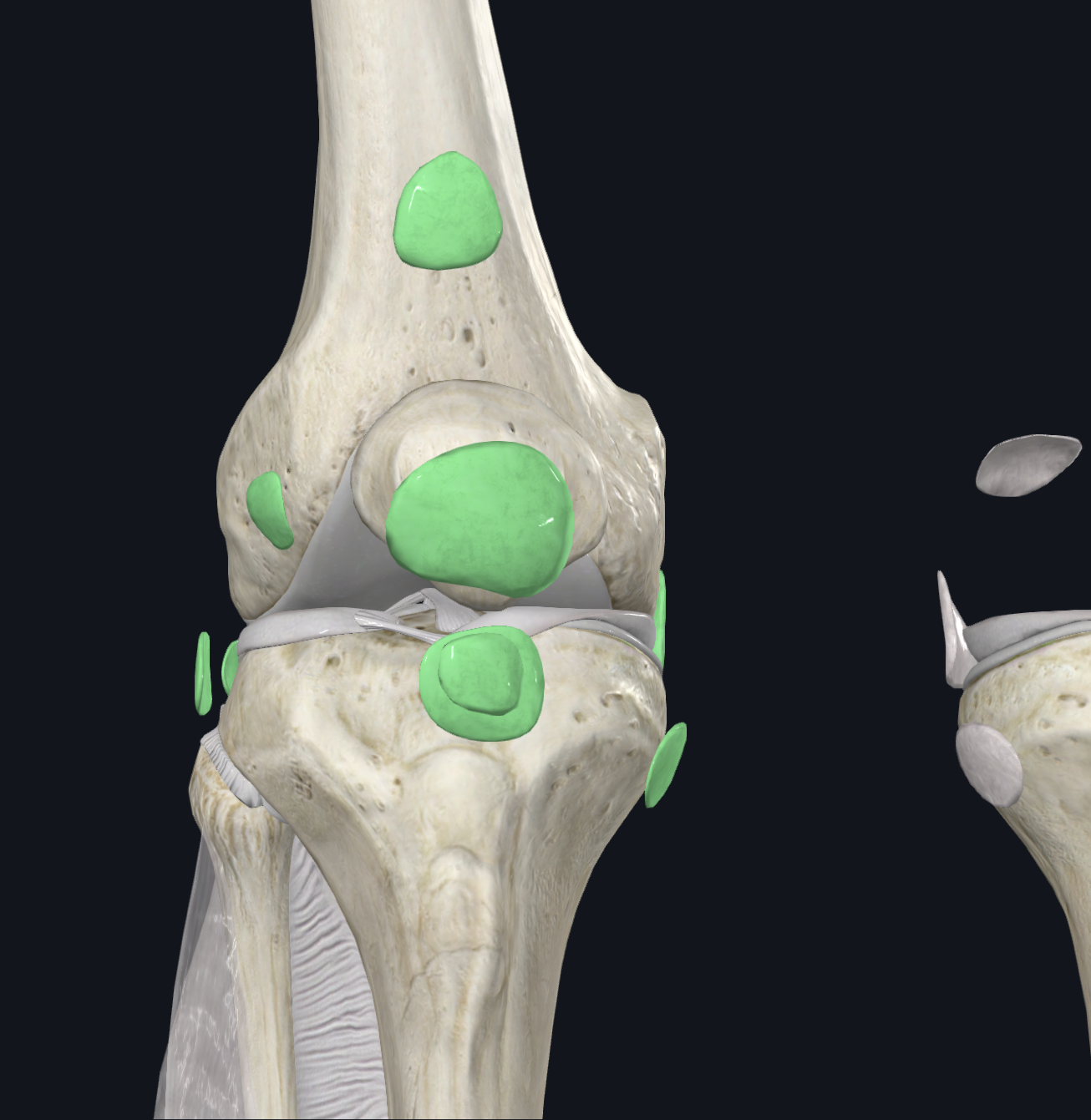
Conjunct Rotation Overview
Description: The medial femoral condyle's articular surface is longer and larger than the lateral condyle. During the last 10–15° of knee extension, the medial femoral condyle mainly articulates with the medial tibial condyle

Screw Home Mechanism
Description: The rotation occurring at the medial condyle during terminal extension. Non-weightbearing: lateral tibial rotation occurs during the last 10-15° of extension. Weightbearing: medial femoral rotation occurs during the last 10-15° of extension.

Knee Nerve Supply
Description: The knee receives innervation from branches of the obturator, femoral, tibial, and common fibular (peroneal) nerves.