RT204 - Beam Restrictors and Film Processing
 Call Kai
 Learn
Learn Practice Test
 Spaced Repetition
 Match
Match Flashcards
 Knowt Play
1/51

There's no tags or description
Looks like no tags are added yet.
Name | Mastery | Learn | Test | Matching | Spaced | Call with Kai | Chat |
|---|
No analytics yet
Send a link to your students to track their progress
52 Terms
Wetting
Swells the emulsion to permit subsequent chemical penetration
Manual: 15s
Automatic: —
Developing
Produces a visible image from the latent image
Manual: 5mins
Automatic: 22s
Rinsing in stop bath
Terminates development and removes excess chemical from emulsion
Manual: 30s
Automatic: —
Fixing
Removes remaining silver halide from the emulsion and hardens gelatin
Manual: 15min
Automatic: 22s
Washing
Removes excess chemicals
Manual: 20mins
Automatic: 20s
Drying
Manual: 30mins
Automatic: 26s
Removes water and prepares radiograph for viewing
Sequence of events in processing a radiograph
Wetting
Developing
Rinsing in stop bath
Fixing
Washing
Drying
components of the fixer and their functions
Activator
Fixing agent
Hardener
Preservative
Buffer sequestering agent
Solvent
Activator
Acetic acid
Neutralizes the developer and stops its action
Fixing agent
Ammonium thiosulfate
Removes underdeveloped silver bromine emulsion
Hardener
Potassium alum
Stiffens and shrinks emulsion
Preservative
Sodium sulfite
Maintains Chemical Balance
Buffer sequestering agent
Acetate boric acids and salts
maintains proper pH removes aluminum ions
Solvent
Water
Dissolves other components
Radiopaque
White
Radiolucent
Black
Transmission or Attenuation
X-ray photons, when interacting with matter, result in
absorbed or scattered
Scatter radiation
These are produced during a Compton interaction
It places exposures on the image receptor that are unrelated to patient anatomy
kVp and Volume of irradiated tissue
Two major factors affect the amount and energy of scatter radiation
exiting the patient
Beam Restriction
Minimizing the amount of scatter radiation can be accomplished by ____ ______ and by using grids.
Proper beam restriction
Tissue compression will keep the total amount of tissue irradiation to a minimum
BEAM-RESTRICTING DEVICES (3)
Aperture diaphragm
Cones/Cylinders
Collimators
Collimator
The foremost method of restricting the primary beam field size is the use of a
device known as a
Aperture diaphragms and cones/cylinders
are also devices that were used to restrict the beam, but are not in common use today
APERTURE DIAPHRAGM
It is the simplest of all beam-restricting devices
It is basically lead or lead-lined metal diaphragm that is attached to the X-ray tube head
Different diaphragms are needed to accomodate different receptor sizes and distances
CONES AND CYLINDERS
Radiographic extension _____ and ________ are considered modifications of the aperture diaphragm
One difficulty with using _____ is alignment
_____ are routinely used in dental radiography
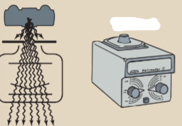
COLLIMATORS
is the most commonly employed beam restrictor in radiography
It consists of sets of lead shutters at right angles to one another that move in opposing pairs, and moves symmetrically from the center of the field
Reduces the patient radiation dose and improves contrast resolution
positive beam-limiting device, or simply, positive beam limitation (PBL)
A type of Automatic collimators
Automatic Collimator
Limits the size and the shape of the primary beam to the size and shape of the image receptor
upper shutters
are responsible for reducing the amount of off-focus radiation
Off-focus radiation
originates within the x-ray tube but not at the focal spot of the tube
Penumbra
It is a type of geometric unsharpness around the periphery of the image (edge unsharpness)
Bottom shutters
reduce penumbra along the periphery of the beam
Increase Collimation
Decrease filed size
Decrease scatter radiation
Increase contrast resolution
Decrease exposure reaching the IR
Decrease patient dose
TRUE
X-ray source > Patient > Latent image > Development > Fixing
Latent Image
is the unobservable image stored in the film; it is made manifest by processing
Film processing: Development
It is a stage of processing during which the latent image is converted to a visible image
Film Processing: Fixing
The use of Silver halide that was not exposed to radiation is the process of clearing it from the emulsion and hardening the emulsion to preserve the image
Xray tube
what is the name of this?

Primary beam
what is this?

Image receptor
what is this?
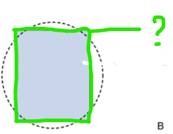
Large field size
Increased patient thickness
what is this?
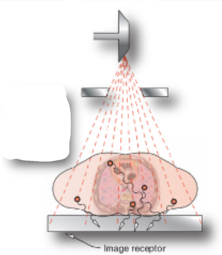
what is this?

Beam Restriction and Patient dose
As the _____ _______ or the collimation increases, the field size and patient dose decrease. As the ______ _______ or collimation decreases, the field size and patient dose increase
Appropriate Beam Restriction
In performing a radiographic examination, the radiographer should be aware of the anatomic area of interest and limit the x-ray field size to just beyond this area. Collimating to the appropriate field size is a basic method for protecting patients from unnecessary exposure
Increases
when scatter radiation _______ as the xray beam field size inceases
Diaphragm
what image is this?
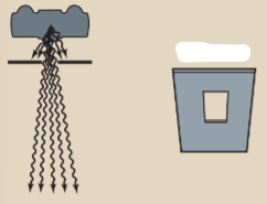
Cylinder
what image is this?

Collimator
what image is this?
Relationship: Collimation and Exposure to the IR
Collimation increases, exposure to the IR decreases: as the collimation decreases, while exposure to the IR increases
Relationship: Collimation and Radiographic Contrast
As collimation increases, the quantity of scatter radiation decreases and radiographic contrast increases: as collimation decreases the quantity of scatter radiation increases and radiographic contrast decreases
Relationship: Collimation and Scatter Radiation
As the collimation increases, the field size and quantity of scatter radiation decreases, as collimation decreases the field size and quantity of scatter radiation increases