Structure Week 2 LO's
 Call Kai
 Learn
Learn Practice Test
 Spaced Repetition
 Match
Match Flashcards
 Knowt Play
1/67

Earn XP
Description and Tags
Normal Pulmonary Anatomy, including Unique Vasculature of Lung & Pathology and Imaging of Pulmonary Embolism
Name | Mastery | Learn | Test | Matching | Spaced | Call with Kai | Chat |
|---|
No analytics yet
Send a link to your students to track their progress
68 Terms
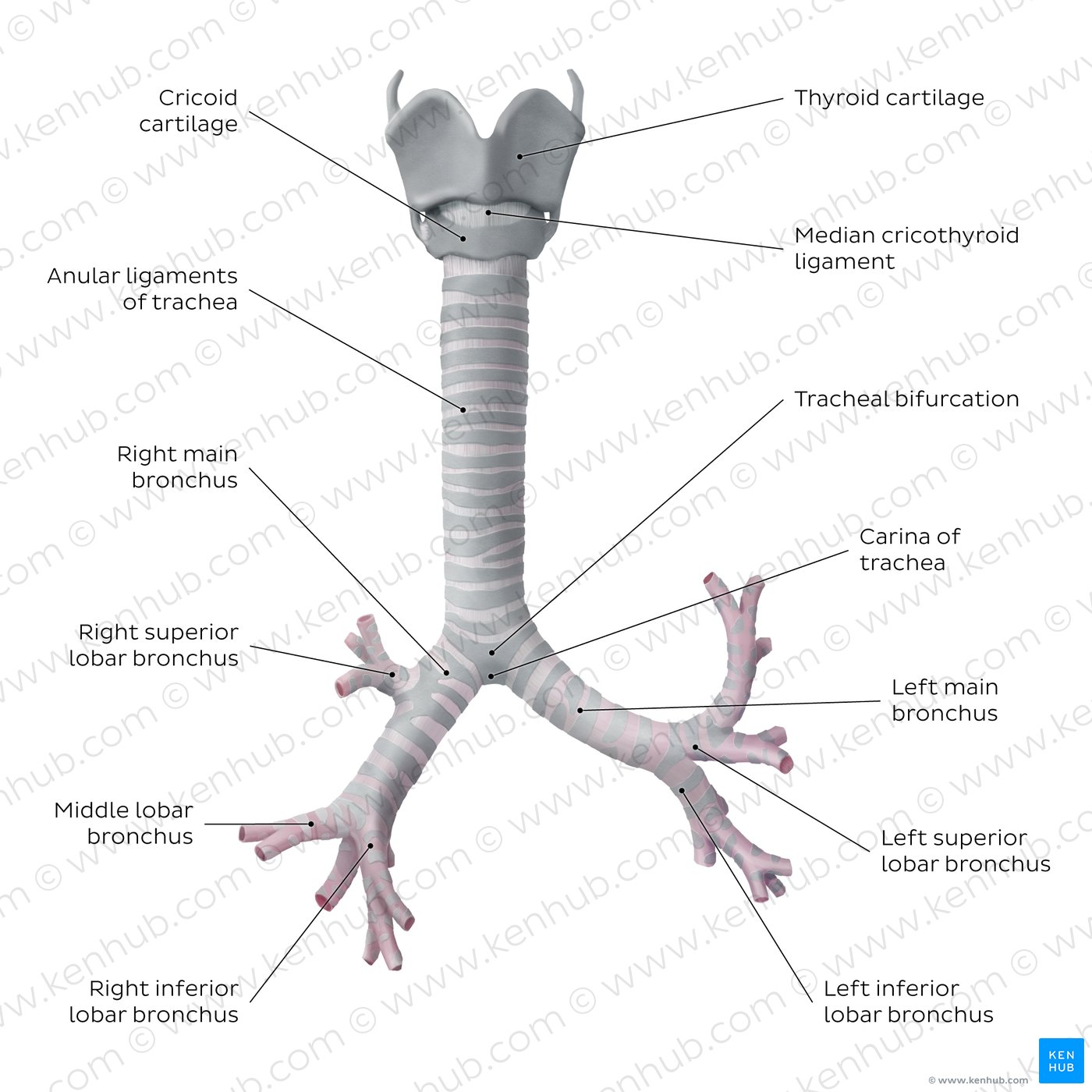
LO 1: what does the trachea split (bifurcate) into where & what T level & what is a mneuomnic
right & left mainstem bronchi
at the carina
T4/T5
Trachea bifourcates at T4/T5
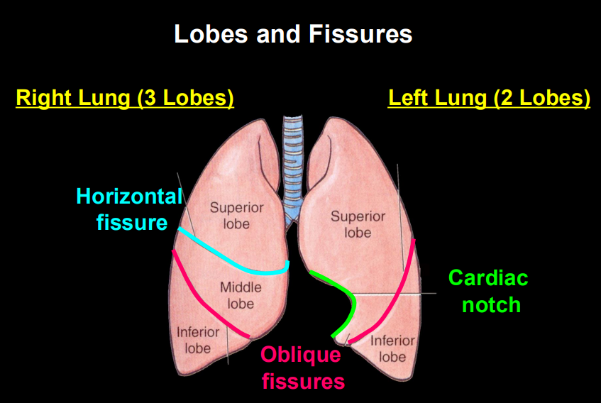
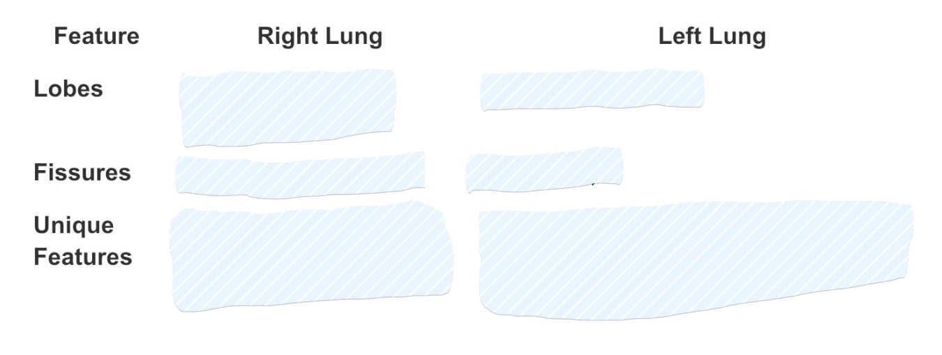
LO 1: what 2 the lobar division of the lung (right and left: size & why, how many lobes & what they are called, how many fissures, describe the fissures)
Lobar Division:
Right Lung: Larger and wider. It has 3 lobes (Superior, Middle, Inferior) separated by two fissures: the Horizontal (separating superior/middle) and Oblique (separating middle/inferior).
Left Lung: Smaller due to the heart. It has 2 lobes (Superior, Inferior) separated by only one fissure: the Oblique.
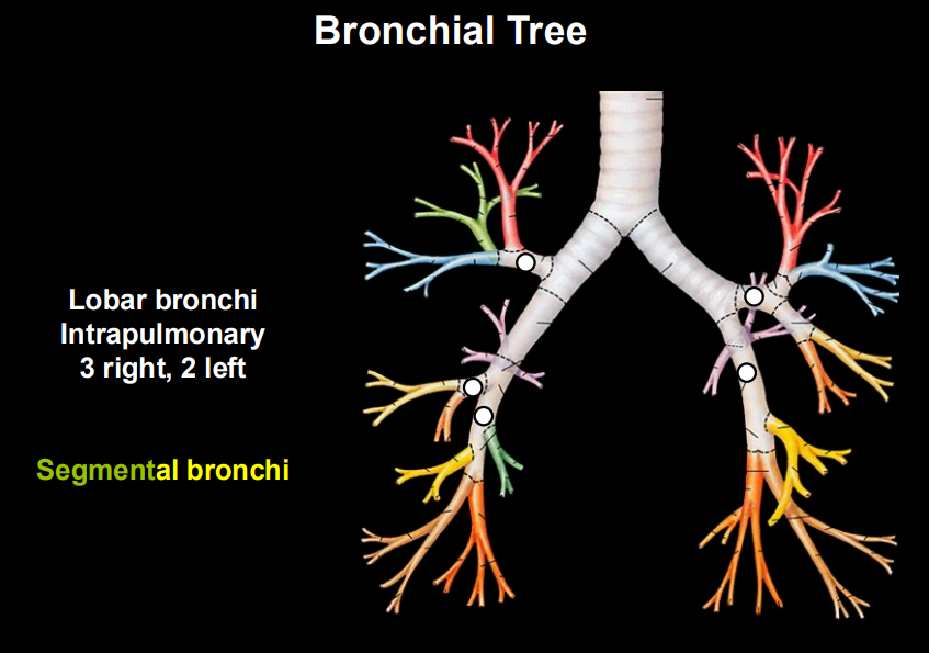
LO 1: segmental anatomy of the lungs (what is each lung divided into, describe the function of the segments & what do they contain)
Segmental Anatomy: Each lung is further divided into 10 bronchopulmonary segments.
LO 1: describe bronchopulmonary segments how they function and why & how many per lung
Each segment is functionally independent because it has its own dedicated segmental bronchus and segmental pulmonary artery.
10 per lung
Part | Function |
|---|---|
Segmental bronchus | Brings air into that segment |
Segmental pulmonary artery | Brings deoxygenated blood to that segment for gas exchange |
LO 1: lobar bronchi right vs left how many
right: 3
left: 2
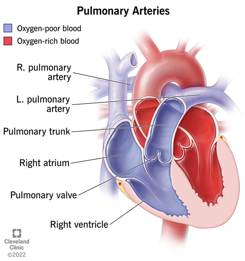
LO 1: pulmonary arterial supply where does the pulmonary trunk exit and what does it carry, what does it bifurcate into and where
The Pulmonary Trunk exits the right ventricle carrying oxygen-poor blood (deoxygenated)
It bifurcates into the Right and Left Pulmonary Arteries at the level of the sternal angle.
LO 1: what do the pulmonary arteries branch into
These arteries branch exactly like the bronchial tree: Main → Lobar → Segmental.
Bronchi (Airways) | Pulmonary Arteries (Blood Vessels) |
|---|---|
Main bronchus | Main pulmonary artery |
Lobar bronchus | Lobar pulmonary artery |
Segmental bronchus | Segmental pulmonary artery |
LO 1: where are the pulmonary arteries located in comparison with pulmonary veins
Structure | Location |
|---|---|
Segmental bronchus | Inside (intrasegmental) the bronchopulmonary segment |
Segmental pulmonary artery | Inside (intrasegmental) the bronchopulmonary segment |
Pulmonary veins | Between (intersegmental) the bronchopulmonary segments |
LO 1:
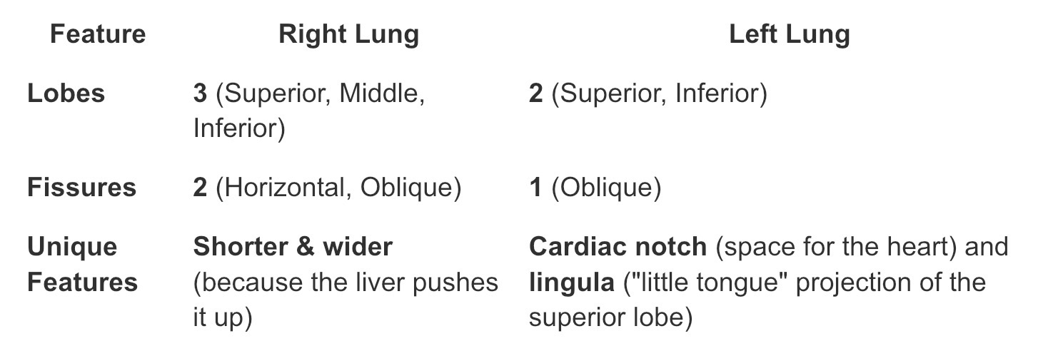
LO 1: if a patient aspirates & accidentally inhales a foreign object (like a peanut), which bronchus is it most likely to lodge and why
Aspiration: If a patient accidentally inhales a foreign object (like a peanut), it is most likely to lodge in the Right Main Bronchus because it is wider, shorter, and more vertical than the left.
LO 1: why can surgeons perform segmentectomy’s to remove a localized tumor while spearing healthy lung tissue
Segmentectomy: Because each segment is separated by connective tissue septa and has its own blood/air supply, surgeons can perform a segmentectomy to remove a localized tumor while sparing healthy lung tissue.
LO 1: The pulmonary arteries carry ________blood and follow the branching of the bronchi.
The right lung has ____ lobes; the left has ____lobes.
There are _____ bronchopulmonary segments per lung.
The pulmonary arteries carry deoxygenated blood and follow the branching of the bronchi.
The right lung has 3 lobes; the left has 2 lobes.
There are 10 bronchopulmonary segments per lung.
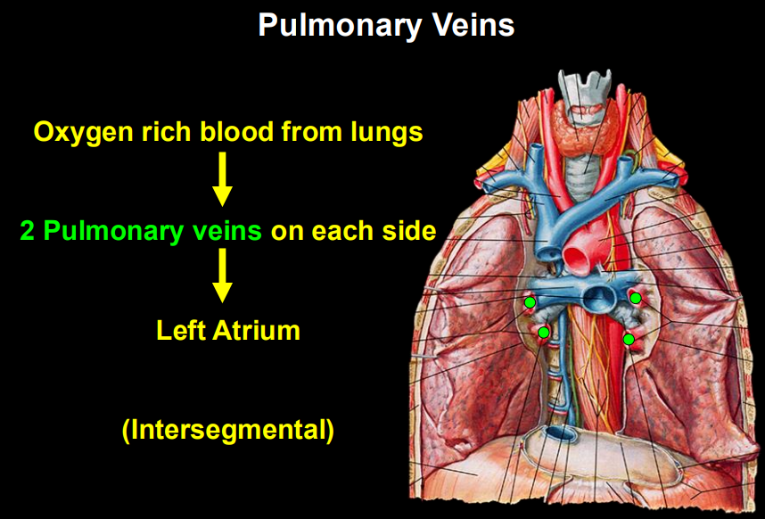
LO 2: what the pulmonary veins collect
Collection: Small veins collect oxygenated blood from the capillaries surrounding the alveoli.
LO 2: where are the pulmonary veins located in comparison to the pulmonary arteries
Course: These veins travel intersegmentally (in the connective tissue between bronchopulmonary segments) rather than running alongside the bronchi (pulmonary arteries).
LO 2: what do the pulmonary veins merge into and where is their destination
Exit: They eventually merge into four main pulmonary veins—two from each lung (Superior and Inferior Pulmonary Veins).
Destination: All four veins empty directly into the Left Atrium of the heart.
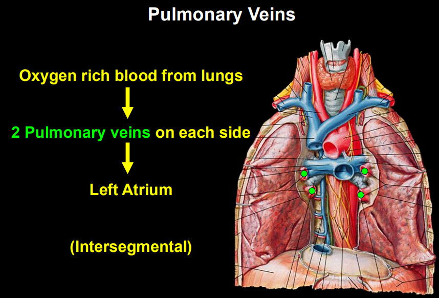
LO 2: why are pulmonary veins helpful in surgery
Clinical Correlation In surgery, because the veins run between segments, they serve as helpful landmarks for identifying the boundaries of a bronchopulmonary segment during a resection.
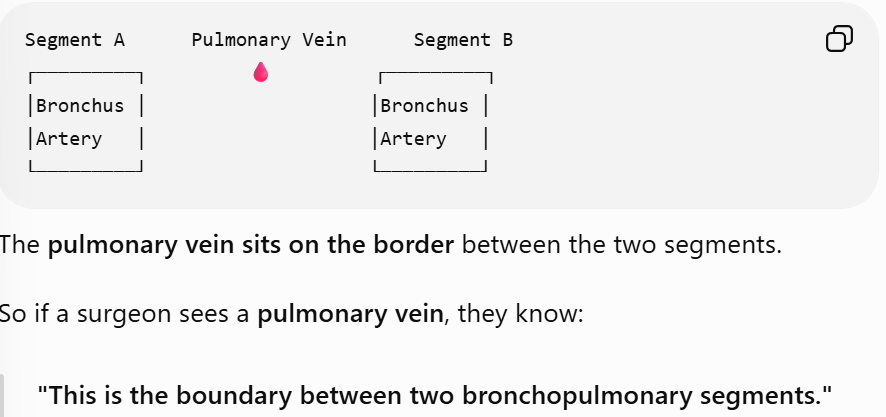
LO 2:
There are ___ pulmonary veins total (___ left, __ right).
Pulmonary veins are ________, meaning they do not travel with the bronchi.
They carry _________ blood to the left atrium.
There are 4 pulmonary veins total (2 left, 2 right).
Pulmonary veins are intersegmental, meaning they do not travel with the bronchi.
They carry oxygenated blood to the left atrium.
LO 3: what is the trachea, what is it supported by, where is it palpable
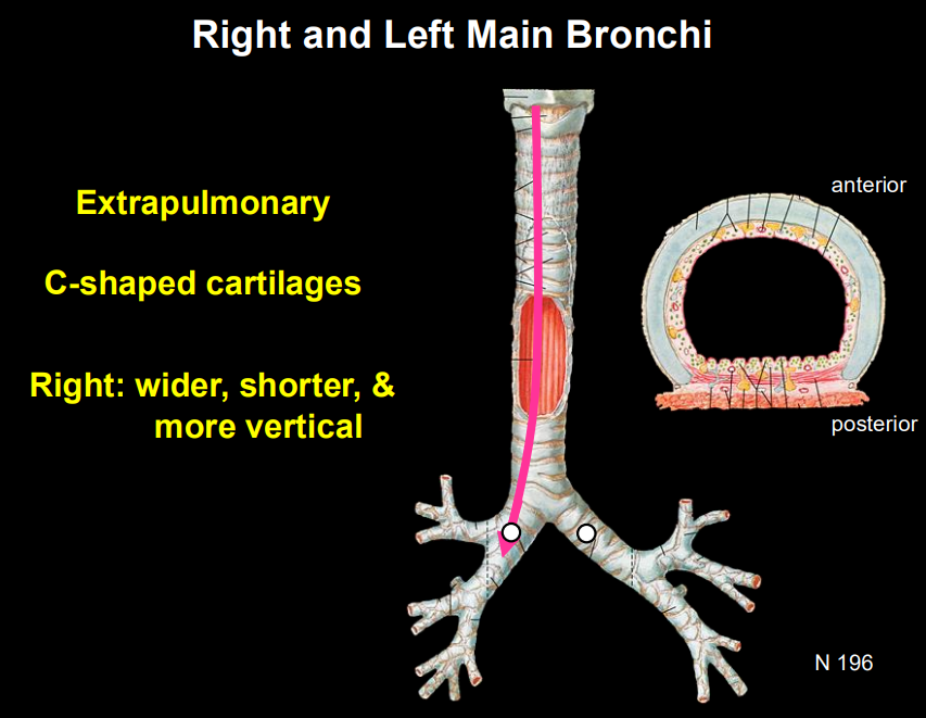
Trachea: A rigid tube supported by C-shaped hyaline cartilage rings.
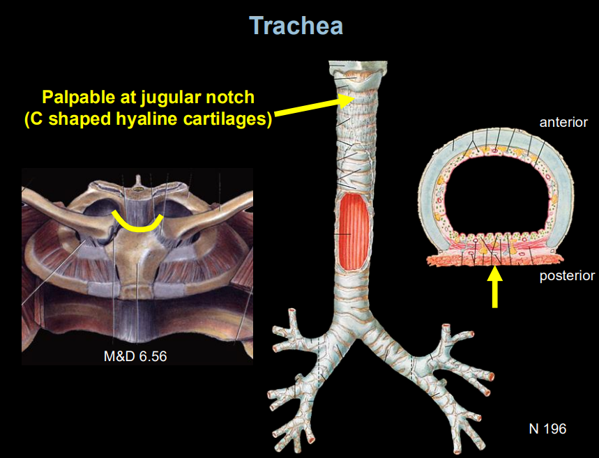
LO 3: where does the trachea bifurcate and into what
Bifurcation: It splits at the carina (level of the sternal angle, T4/T5) into the Right and Left Main Bronchi.
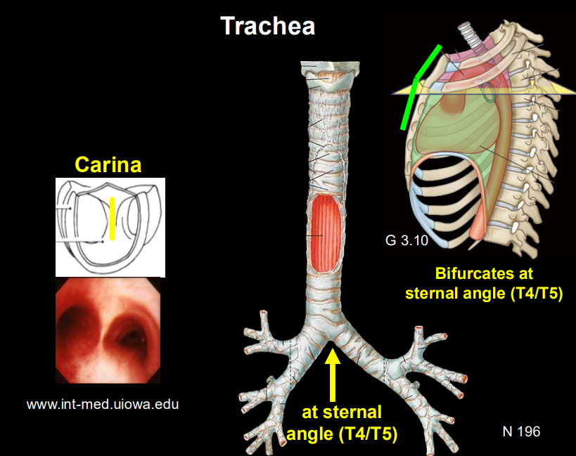
LO 3: what do the bronchi further branch into
Branching: Main Bronchi → Lobar (Secondary) Bronchi (3 right, 2 left) → Segmental (Tertiary) Bronchi.
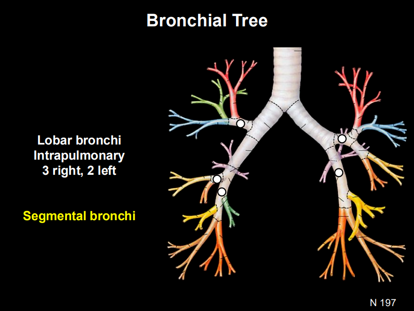
LO 3: describe the right mainstem bronchi
wider, shorter, more vertical
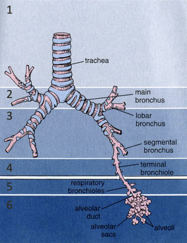
LO 3: what are the 2 types of bronchioles & what respiratory zones are they associated with
Bronchioles: Eventually, the tubes lose cartilage entirely and become bronchioles (less than 1mm diameter).
Terminal Bronchioles: The end of the "conducting zone" (no gas exchange).
Respiratory Bronchioles: The start of the "respiratory zone" (have tiny alveoli for gas exchange).
LO 3: describe the bronchial supply of the lung parenchyma what supplies it & from where
Bronchial Supply : The lung tissue itself needs (oxygenated blood). This is provided by the Bronchial Arteries, which typically arise from the Aorta.
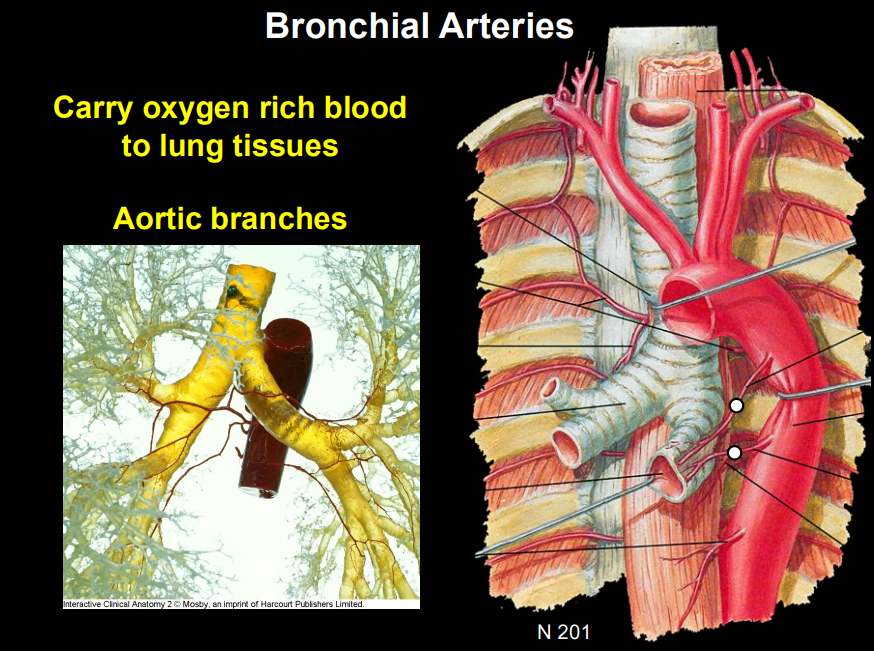
LO 3: what do bronchial arteries do
carry oxygen rich blood to lung tissues
LO 3: what do bronchial veins do
return deoxygenates blood from lung tissues to azygous or intercostal veins
LO 3:
_______/_______: Have cartilage support.
___________: Have no cartilage but have smooth muscle.
__________: Provide the "nutritive" blood supply to the lung tissue itself (not for gas exchange).
The lungs have a "dual circulation": one for work (_______ arteries) and one for nourishment (________arteries)
Trachea/Bronchi: Have cartilage support.
Bronchioles: Have no cartilage but have smooth muscle.
Bronchial Arteries: Provide the "nutritive" blood supply to the lung tissue itself (not for gas exchange).
The lungs have a "dual circulation": one for work (pulmonary) and one for nourishment (bronchial)
LO 4: what is the blood supply of the trachea
Trachea: Supplied by branches of the inferior thyroid arteries and bronchial arteries.
LO 4: Bronchial Tree & Parenchyma blood supply (nutritive)
Parenchyma means the functional tissue of an organ
Bronchial Arteries (usually 1 right, 2 left) arise from the Aorta to feed the lung tissue.
LO 4: Lung Parenchyma blood supply (Functional)
Pulmonary Arteries bring deoxygenated blood for gas exchange.
LO 4: what are the 2 types of autonomic innervations of the lung
parasympathetic
sympathetic
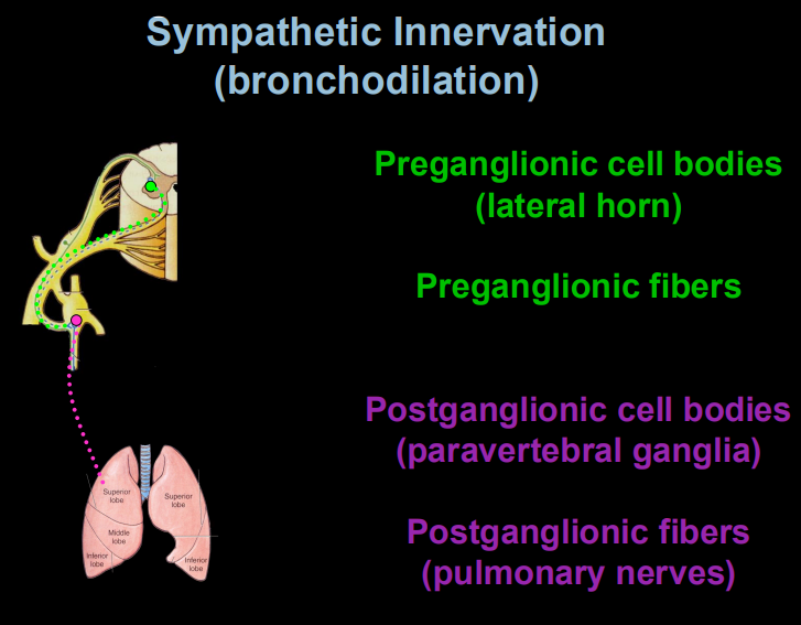
LO 4: sympathetic innervation of the lungs what does it cause & what receptors are involved , & sympathetic pathway
Sympathetic: Causes bronchodilation (widening) via beta-2 receptors—essential for "fight or flight".
L → P → P → L
Lateral horn
Preganglionic fiber
Paravertebral ganglion (postganglion cell bodies)
Lung (via pulmonary nerves) (postganglionic fibers)
LO 4: parasympathetic innervation of the lungs what are the innervation nerves, what does it cause & parasympathetic pathway
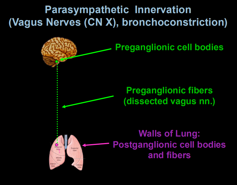
Parasympathetic (Vagus Nerve - CN X cranial nerve 10): Causes bronchoconstriction (narrowing) and increased mucus secretion.
Preganglionic cell body
↓
Preganglionic fiber (Vagus nerve)
↓
Postganglionic cell body (lung wall)
↓
Postganglionic fiber
↓
Bronchoconstriction
LO 5: where is the diaphragm located
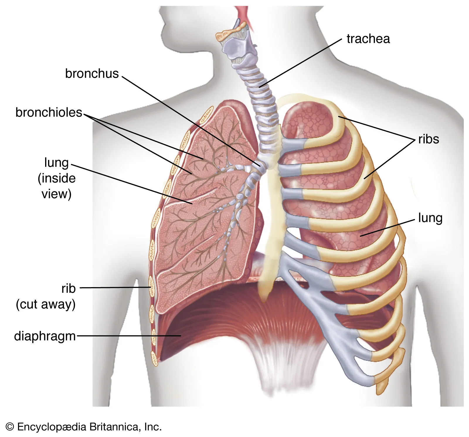
directly below the lungs and heart, right at the base of the rib cage
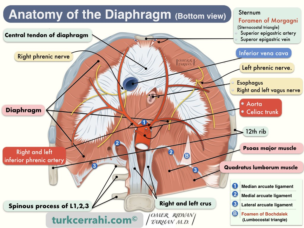
LO 5: what are 3 superior surface components of the diaphragm
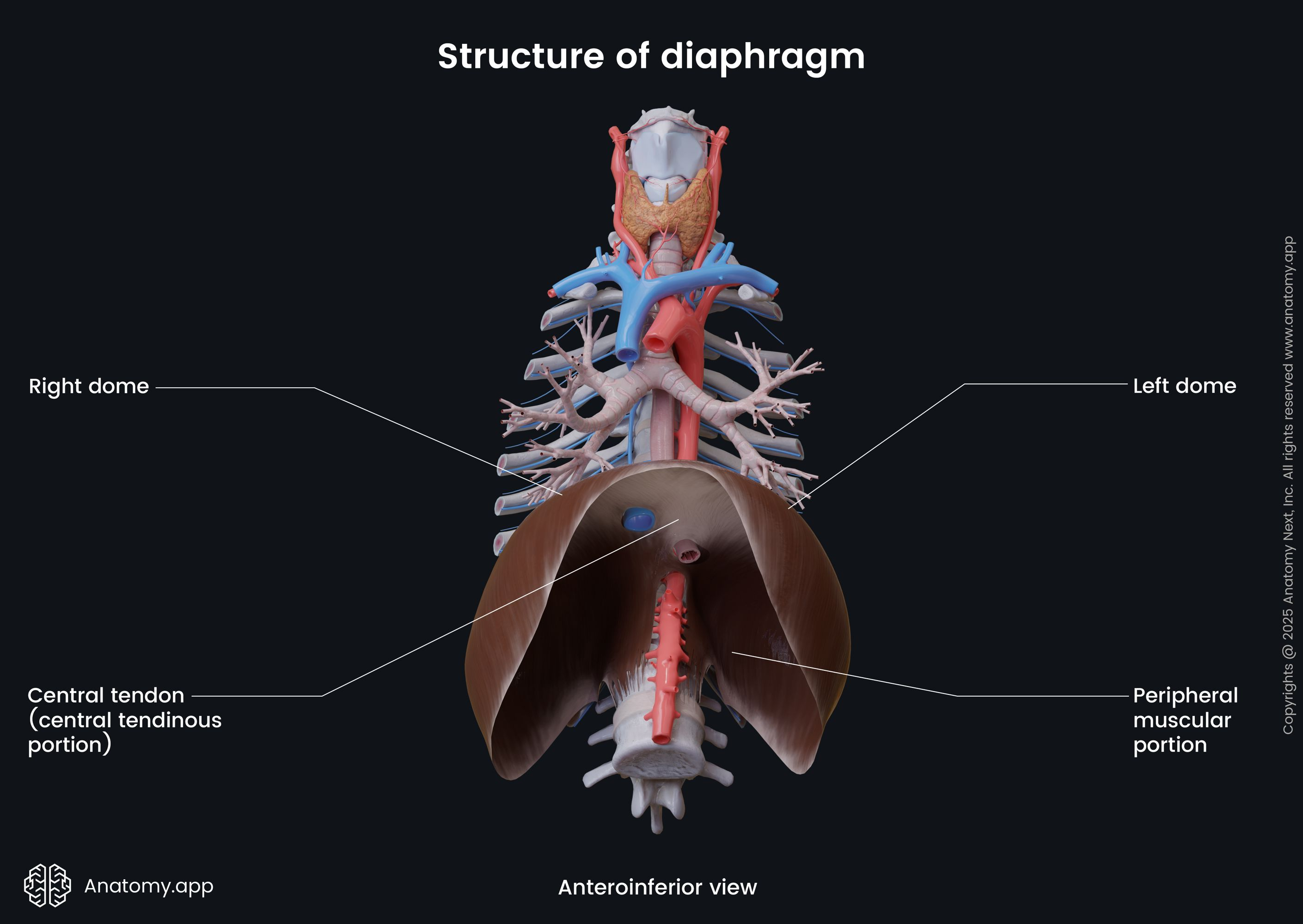
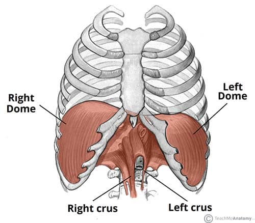
2 domes (right is higher due to the liver)
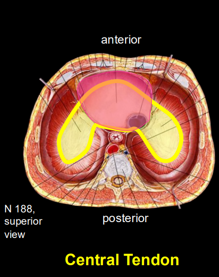
central tendon
LO 5: what are diaphragm domes
curved tops of the diaphragm
LO 5: what is the central tendon
strong, flat center of the diaphragm
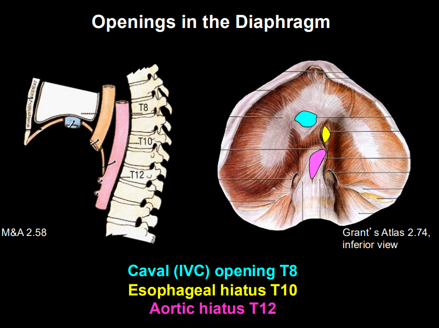
LO 5: what are the 3 openings in the diaphragm & what passes through each include vertebral level
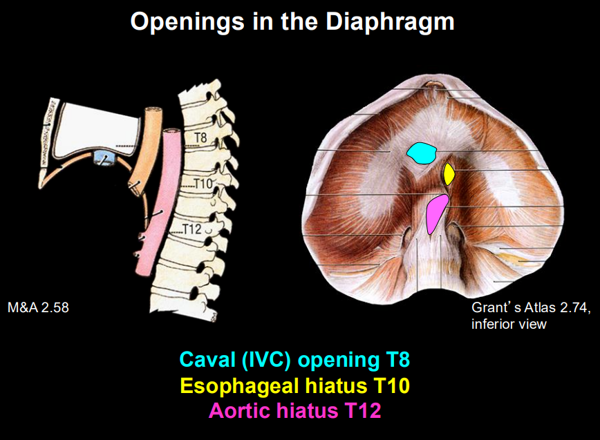
T8: Caval Opening → Inferior Vena Cava passes through.
T10: Esophageal Hiatus → Esophagus and Vagus Nerves pass through.
T12: Aortic Hiatus → Aorta, Thoracic Duct, and Azygos Vein pass through.
LO 5: mneoumonic for openings in the diaphragm
"I 8, 10 Eggs At 12."
LO 5: what is the vascular supply of the diaphragm
Supply:
Phrenic arteries (superior/inferior) → supply diaphragm
pericardiophrenic arteries → supplies central diaphragm travels with phrenic nerve
LO 5: what is the motor innervation of the diaphragm in include vertebral levels & how are the domes innervated
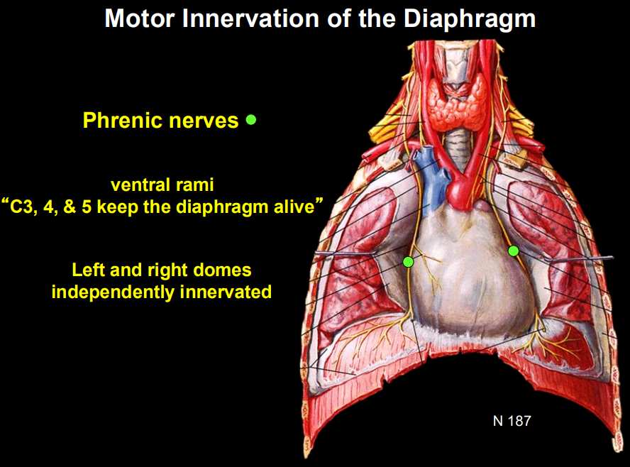
Motor Innervation: Phrenic Nerve (C3, C4, C5).
left & right domes independently innervated
LO 5: what are components of the sensory innervation (central vs peripheral) of the diaphragm nerves involved, location, and what happens if irritated
Sensory Innervation:
Central: Phrenic nerve.
Peripheral: intercostal nerves
Part of Diaphragm
Location
Sensory Nerve
If Irritated...
Central
Middle of the diaphragm
Phrenic nerve (C3–C5)
Referred pain to the shoulder
Peripheral
Outer edge (rim) of the diaphragm
Intercostal nerves
Pain in the lower chest wall or upper abdomen
LO 5: mneumonic for phrenic nerves & what are they also called
ventral rami “C3, C4, & C5 keep the diaphragm alive
LO 5: if there is a phrenic nerve injury what will happen to the diaphragm & how will it appear on x-ray & explain why
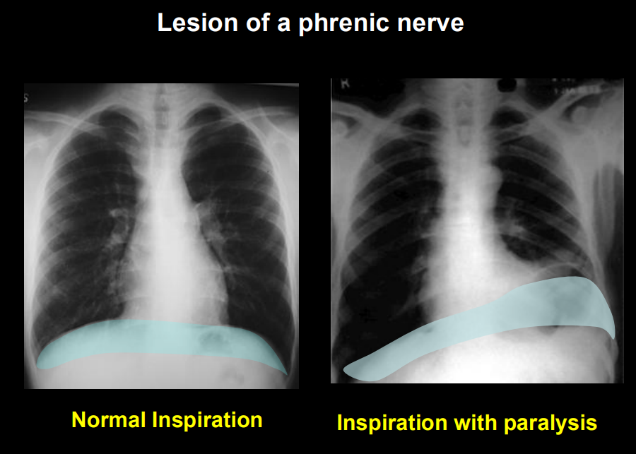
Phrenic Nerve Injury:
If a phrenic nerve is damaged (e.g., by a tumor), that half of the diaphragm becomes paralyzed.
On a chest X-ray during inspiration, the paralyzed side will rise (paradoxical movement) because it is pushed up by abdominal pressure instead of contracting downward.
what muscles are used during forceful inspiration vs forceful expiration
forceful inspiration: pectoralis major
forceful expiration: oblique muscles & rectus abdominis
LO 5:
Openings: _______, ________, ________.
Motor innervation is solely the________ Nerve.
Openings: IVC (T8), Esophagus (T10), Aorta (T12).
Motor innervation is solely the Phrenic Nerve.
LO 6: conducting zone function & histologic components & their functions
location: trachea → terminal bronchioles
function: Conduct, warm, humidify, and filter air
Histologic Components
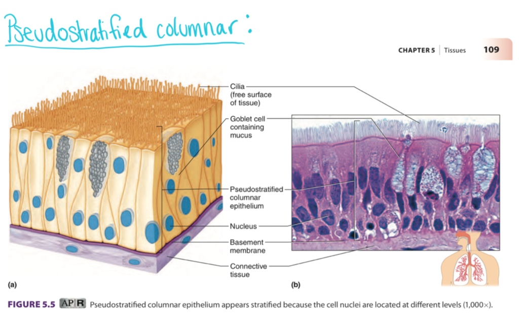
Conducting Zone (Trachea/Bronchi): Lined with Pseudostratified Ciliated Columnar Epithelium (Respiratory Epithelium).
Goblet Cells: Secrete sticky mucus to trap dust.
Cilia: Hair-like projections that move mucus up (the Mucociliary Escalator).
LO 6: bronchioles function & histologic components & their function
function: deliver air to the alveoli, regulate airflow, and protect the lungs
Histological Components
Bronchioles: Epithelium becomes simpler (cuboidal).
Club (Clara) Cells: Replace goblet cells; they secrete protective fluids and can act as stem cells to repair the lining.
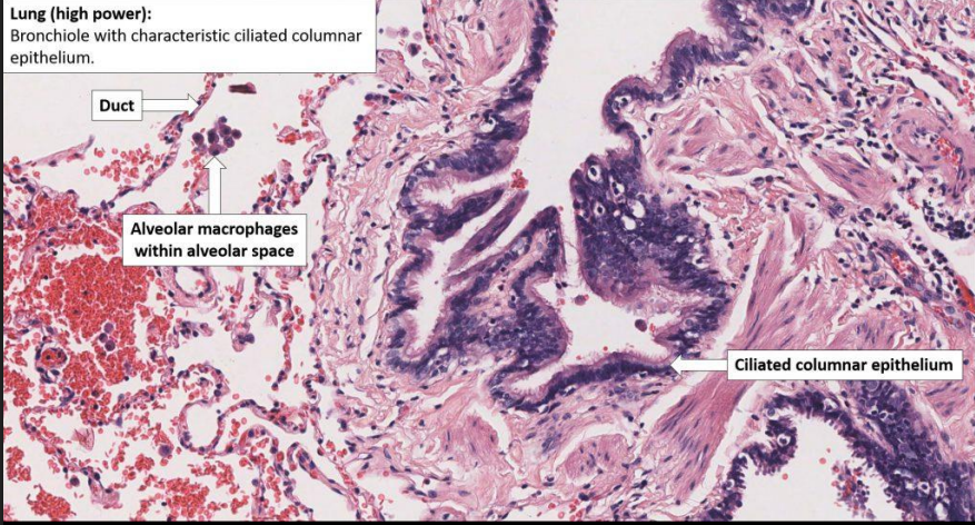
LO 6: alveoli function, 2 types of pneumocytes & their functions, macrophages & histological components
function: gas exchange
Type I Pneumocytes: Extremely thin, flat cells that cover 95% of the surface area. This is where gas exchange happens.
Type II Pneumocytes: Cuboidal cells that secrete surfactant (reduces surface tension so alveoli don't collapse) and repair Type I cells.
Alveolar Macrophages (Dust Cells): "Janitors" that gobble up any particles that made it past the mucus.
LO 6:
Type I Pneumocytes: shape → function.
Type II Pneumocytes: shape→ function.
Club Cells: location & function
Type I Pneumocytes: Thin/Flat → Gas exchange.
Type II Pneumocytes: Cuboidal → Surfactant.
Club Cells: Found in bronchioles; produce protection/repair.
LO 7: what is an x-ray result in a PE patient & what do you need to see the clot
A standard Chest X-ray (CXR) is often normal in a patient with a Pulmonary Embolism (PE).
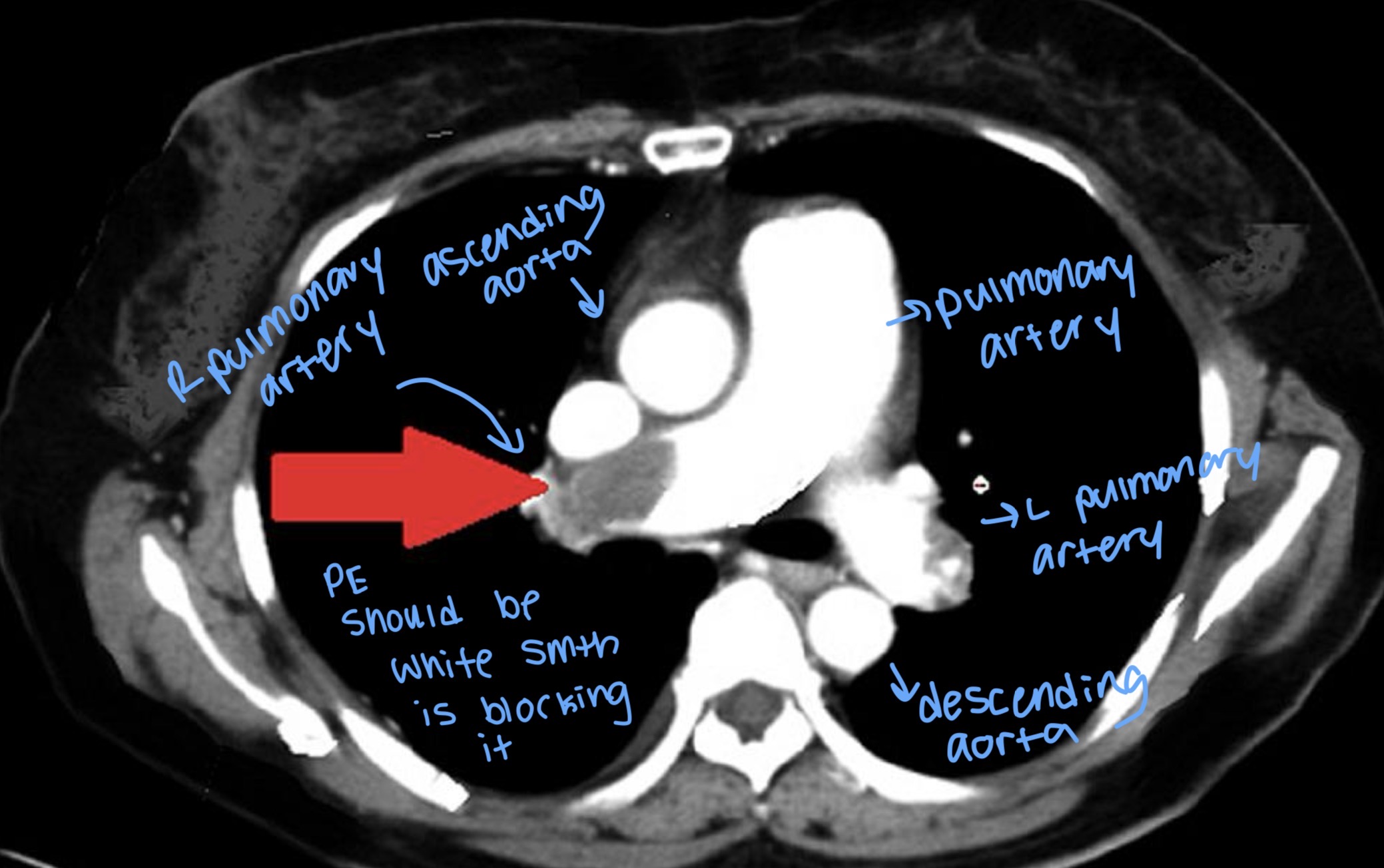
To actually see the clot, you need more specialized imaging that uses dye to highlight the blood vessels.
LO 7: what is the gold standard test for a PE
The "Gold Standard": The CT Pulmonary Angiogram (CTPA) is the preferred test because it is fast and precisely shows where the clot is located.
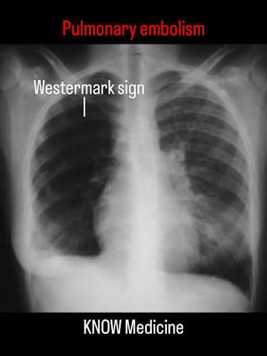
LO 7: what is Westernmark’s sign
Dark lung region due to decreased blood (oligemia) flow from a pulmonary embolus.
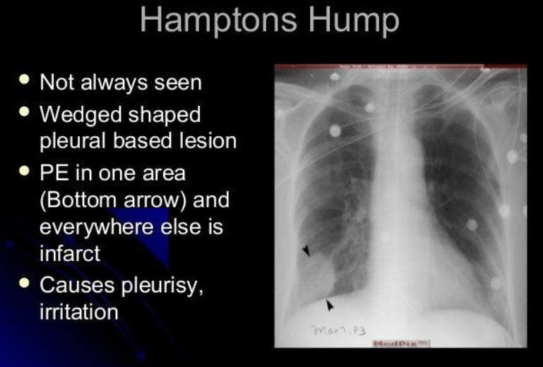
LO 7: what is Hampton’s Hump
Hampton’s Hump: Wedge-shaped opacity indicating lung tissue death (infarct).
won’t always see bc dual blood supply that’s why need CT angiogram
LO 7:
_______ is often normal in PE.
_______: Decreased blood vessel markings (oligemia).
_________: Wedge-shaped opacity indicating lung tissue death (infarct).
CXR is often normal in PE.
Westermark’s sign: Decreased blood vessel markings (oligemia).
Hampton’s Hump: Wedge-shaped opacity indicating lung tissue death (infarct).
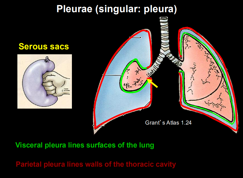
LO 8: what do the visceral & parietal pleura line
Visceral pleura lines surfaces of the lung
Parietal pleura lines walls of the thoracic cavity
LO 7: if someone feels pain which pleura is affected
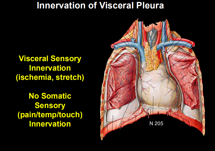
The lung tissue itself (parenchyma) and the inner "shrink-wrap" (visceral pleura) cannot feel pain.
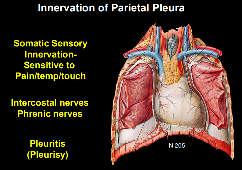
Pain only happens when the outer lining of the chest wall (parietal pleura) gets irritated.
LO 7: innervation of visceral pleura
visceral sensory innervation (ischemia aka reduced O2 & blood flow and stretch)
no somatic sensory (pain, temp, touch) innervation
LO 7: innervation of parietal pleura
somatic sensory innervation (sensitive to pain, temp, touch)
somatic nerves (intercostal nerves & phrenic nerves)
LO 7: when would one feel pain from a PE
A PE only causes pain if it leads to a Pulmonary Infarction near the surface of the lung.
The resulting inflammation "rubs" against the sensitive parietal pleura, causing pleuritic chest pain (sharp pain when breathing in).
LO 9: what is the D-dimer test, sensitivity vs specificity, what is it used to do
blood test that looks for evidence that the body has recently made and broken down a blood clot
What it is: A breakdown product of cross-linked fibrin.
Sensitivity: Very high (80-85%). If the test is negative, you can almost certainly rule out a PE (93-100% negative predictive value).
Specificity: Very low. Many things cause a high D-Dimer: pregnancy, surgery, infection, age over 80, or even a simple bruise.
LO 9: sensitivity vs specificity (what it looks for, what a high value means, good for, negative result, positive)
Feature | Sensitivity | Specificity |
|---|---|---|
Looks for | Sick people | Healthy people |
High value means | Few false negatives | Few false positives |
Good for | Screening | Confirming |
Negative result | Rules OUT disease | Not as helpful |
Positive result | Not as helpful | Rules IN disease |
LO 10: what is a PE vs Infarct
Pulmonary embolism = intravascular obstruction of pulmonary arteries by thrombus or other material. Examples include thromboembolism, air embolism, fat embolism, amniotic fluid embolism
Pulmonary infarction = ischemic necrosis of lung parenchyma distal to an obstructed vessel.
LO 10: describe the blood supply of the lungs & what are the components
The Lung Has Dual Blood Supply
Pulmonary arteries: carry deoxygenated blood for gas exchange and is the vessel that gets occluded in PE
Bronchial arteries : arise from the aorta, provide oxygenated blood to lung tissue itself
why do most emboli not produce ischemic necrosis
Because bronchial arteries continue to perfuse the lung parenchyma even when a pulmonary artery is blocked, most emboli do not produce ischemic necrosis (bc the lungs have dual blood supply)
LO 10: pulmonary embolism gross and histopathologic features
Gross: A solid mass inside the artery. A "Saddle Embolus" sits right where the main artery splits.
Histology: Lines of Zahn. These are alternating pale layers (platelets/fibrin) and dark layers (red blood cells) that prove the clot formed while the patient was alive. (diagnostic of antemortem clot)
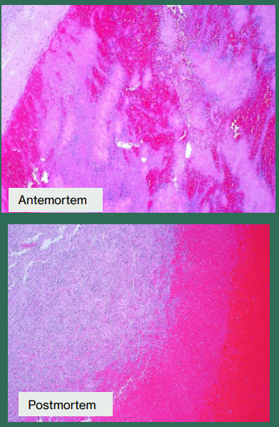
LO 10: what is antemortem vs postmortem
antemortem means "before death"
postmortem means "after death"
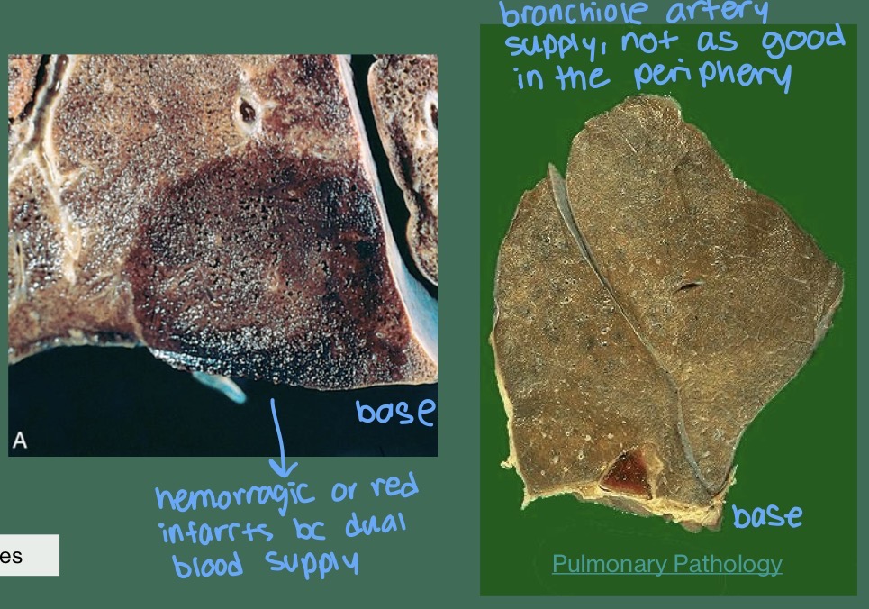
LO 10: pulmonary infarction (tissue death) gross and histopathologic features
Gross: Wedge-shaped lesion with the base pointing toward the outer edge (pleura).
Type: Red (Hemorrhagic) Infarct. Because the bronchial arteries are still pumping some blood into the area, the dead tissue gets soaked in blood.
Histology: Necrosis (death) of the alveolar walls and red blood cells filling the air spaces.
LO 10:
Lines of Zahn:
Wedge-shaped & Red:
Lines of Zahn: Diagnostic of an antemortem thrombus (clot formed before death).
Wedge-shaped & Red: Classic appearance of a pulmonary infarct.