NEUR420 Exam 2
 Call Kai
 Learn
Learn Practice Test
 Spaced Repetition
 Match
Match Flashcards
 Knowt Play
1/25

There's no tags or description
Looks like no tags are added yet.
Name | Mastery | Learn | Test | Matching | Spaced | Call with Kai | Chat |
|---|
No analytics yet
Send a link to your students to track their progress
26 Terms
What is the purpose of performing histology or immunohistochemistry on brain tissue?
To differentiate between brain structures; find specific antigens of interest; examine arrangement of cells
What is the difference between a retrograde and anterograde tracer? Give an example of each.
Retrograde: going from the axon terminal back to cell body; HRP
Anterograde: going from the cell body to the axon terminal: viral tracers
What types of tracers are most commonly used now in Neuroscience research and why?
Viral tracers are most commonly used now because they are able to jump the synapse and provide more exact connections (monosynaptic, for example).
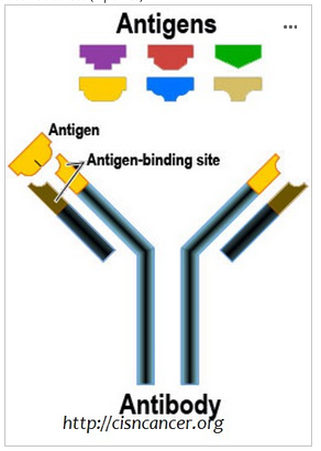
A) Pictured below is a diagram of an antibody with multiple antigens. What are antibodies used for? (2 points)
B) What is the diagram attempting to depict about antibodies? (2 points)
A) To label tissue for a certain antigen of interest.
B) Specificity
Experimental Neuroanatomy: Choose 1 below to answer. Indicate in your answer which option you chose.
Option B: You are a student rotating in Dr. Mitrano’s neuroanatomy lab for the summer. You have been given the task to determine whether the serotonin2A receptor is found presynaptically (in axon terminals) or postsynaptically (in dendrites or spines) in the amygdala. Describe an immunohistochemical technique to do this and how you would examine the amygdala to determine this (including what type of microscope you would use to observe your findings).
Obtain antibody for the 5HT2A receptor. Euthanize animal and obtain brain tissue; slice and obtain sections that contain the amygdala. Using the antibody and immunoperoxidase or nanogold secondary. Use electron microscope to see if the labeling is pre- or post-synaptic.
A CT scan measures the density of _______________________ , while an MRI measures the density or intensity of __________________.
A .protons; x-rays
B. blood flow; protons
C. tissue; protons
D. x-rays; blood flow
C. tissue; protons
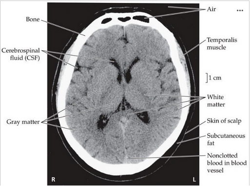
A) What type of brain scan is pictured below (CT or MRI)? (2 points)
B) What would be an advantage of using this type of scan with contrast? What information would it provide? (3 points)
A: CT
B: CT with contrast allows for the visualization of blood flow /perfusion in the brain, particualry if a hemorhage or blokage occured
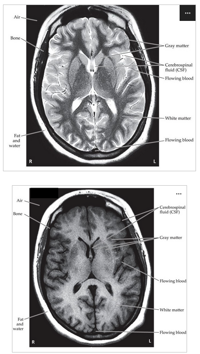
1) What type of brain scans are pictured above?
2) Which one is T1-weighted? Which one is T2-weighted?
1) MRIs
2) A is T2-weighted; B is T1-weighted
Why are both types of MRI imaging done (i.e., what information does each provide)? (2 points)
T1: ______________________________________________________________________
T2:______________________________________________________________________
T1: good for identifying anatomy
T2: good for identifying pathological changes
What does DT-MRI allow us to image?
white matter
You have just started rotations in medical school and were placed in the ER for the next 4 weeks. Late one night a patient arrives and the EMTs inform you that the individual was rescued from a car accident, is breathing but unconscious, and may have sustained a head injury. Your supervising resident asks you to determine the course of action in determining the extent of the injuries.
First, what are three most common types of neuroimaging? (3 points)
CT, CT with contrast, , xray, MRI
You have just started rotations in medical school and were placed in the ER for the next 4 weeks. Late one night a patient arrives and the EMTs inform you that the individual was rescued from a car accident, is breathing but unconscious, and may have sustained a head injury. Your supervising resident asks you to determine the course of action in determining the extent of the injuries.
A) What imaging technique would you order first? (2 points)
B) Explain why you would choose that technique in terms of what the technique measures and the advantages of it compared to other techniques. (3 points)
C) If that test failed to give a conclusive diagnosis what test would you order next and why (3 points).
A) CT scan
B) quick, trying to find major injuries
C) MRI, get more detailed image of the brain.
Name two techniques that are able to look at the activity of the human brain (aka two examples of functional neuroimaging).
EEG and BOLD fMRI
When would you want to use the immunoperoxidase technique versus the immunogold technique for examining tissue using an electron microscope? (2 points)
Immunoperoxidase- subcellular level
Immunogold- subsynaptic level
“An Anatomic Study of the Autonomic Nervous System”
Which of the following would NOT be regulated by the autonomic nervous system? (1 point)
A - Dilation of pupils
B - Breathing rate
C. Driving a car
D - Monitoring changes in blood pressure
C. Driving a car
“An Anatomic Study of the Autonomic Nervous System”
Which of the following would be controlled by the parasympathetic division of the autonomic nervous system? (2 points)
A - Increased heart rate:
B - Increased saliva production
C Dilation of pupils
D - Bronchodilation
B - Increased saliva production
In the case, we are introduced to a condition called FD or familial dysautonomia.
Name 3 symptoms of FD.
inability to feel pain, inability to produce tears, Constipation
In the case, we are introduced to a condition called FD or familial dysautonomia.
Explain why someone with familial dysautonomia may have difficulty moving from a dimly lit room into a room that is very bright.
Since pupils change size when moving from dark to light, and pupil dilation/constriction is controlled by the autonomic nervous system, someone with FD may have trouble if their pupils do not dilate correctly.
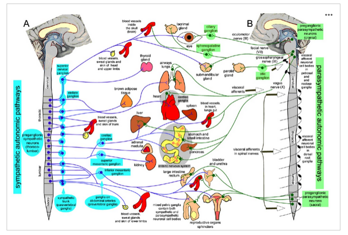
In the figure above, the nerve pathways and target organs of the sympathetic (left) and parasympathetic (right) divisions of the ANS are depicted. Why is the sympathetic division sometimes also called the “thoracolumbar” division, and why is the parasympathetic division often referred to as the “craniosacral” division? (4 points)
All neurons that form the sympathetic pathway originate from either the thoracic or lumbar areas of the spinal cord; hence why it is also known as thoracolumbar.
All neurons that form the parasympathtic pathway originate from either the cranial or sacral nerves, hence the name craniosacral.
Sympathetic innervation to the heart exits the spinal cord in the _______ (thoracic/lumbar) region. Electrical signals first travel down a/an __________ (cholinergic/adrenergic) preganglionic neuron that synapses with the ganglionic neuron in the sympathetic trunk ganglia. At the synapse _________ (ACh/ NE) is released and binds to _________ (nicotinic/muscarinic) receptors on the ganglionic neuron. This __________ (excites/inhibits) the __________ (cholinergic/adrenergic) ganglionic neuron. At the heart, the ganglionic neuron releases ___________ (ACh/NE), which binds to ____________ (alpha/beta) receptors on the heart, _______________ (stimulating/inhibiting) contraction of the heart.
Sympathetic innervation to the heart exits the spinal cord in the thoracic_______ (thoracic/lumbar) region. Electrical signals first travel down a/an ___cholinergic________ (cholinergic/adrenergic) preganglionic neuron that synapses with the ganglionic neuron in the sympathetic trunk ganglia. At the synapse _____ACh______ (ACh/ NE) is released and binds to _____nicotinic______ (nicotinic/muscarinic) receptors on the ganglionic neuron. This ____excites________ (excites/inhibits) the ___adrenergic__________ (cholinergic/adrenergic) ganglionic neuron. At the heart, the ganglionic neuron releases _____NE________ (ACh/NE), which binds to ____beta__________ (alpha/beta) receptors on the heart, _____stimulating___________ (stimulating/inhibiting) contraction of the heart.
In reference to the activity we did exploring brain regions using the Allen Institute mouse brain atlas:
A) What is a brain atlas? (2 points)
B) What do we use a brain atlas for? (2 points)
A) A brain atlas maps out all the known brain structures in a specific species.
B) We use brain atlas to find a region of interest when examining the brain that has been stained, or to determine stereotaxic coordinates for brain surgery, to inject a tracer or some other drug, etc.
In reference to the activity we did exploring brain regions using the Allen Institute mouse brain atlas:
A) What planes did we view in the mouse brain in? (coronal, sagittal, horizontal) (2 points)
B) Which plane generally included more brain regions per image? (2 points)
A) sagittal and coronal
B) sagittal
In reference to the Allen Institute Activity on Neuropathology and Alzheimer's disease (AD):
In this activity we looked at tissue from the SEA-AD project. In order to conclusively diagnose Alzheimer’s disease post-mortem, scientists look for biomarkers in brain tissue. What are the two primary biomarkers for AD? (2 points)
Beta-amyloid plaques
Neurofibrillary tangles (tau)
In reference to the Allen Institute Activity on Neuropathology and Alzheimer's disease (AD):
What experimental technique did the neuroscientists use to find those biomarkers? (2 points)
Immunohistochemistry
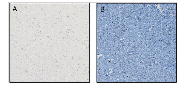
Below are two images from two different brain donors to the SEA-AD study. The images depict pTau (AT8) and p-TDP43 in the middle temporal gyrus.
Based on what we learned about Braak stages, which is the extent of anatomical distribution of neurofibrillary tangles, which image would you say has a higher Braak stage, A or B, and WHY? (2 points)
Based on what we learned about the ADNC score (the overall Alzheimer’s disease neuropathologic change score), which patient would you say has a LOW ADNC score and WHY? (2 points)
B, as it contains far more tau tangles (in blue)
A as they have a much lower concentration of tau tangles, which correlates to a lower progressed stage of alzheimers
In reference to the Allen Institute Activity on Neuropathology and Alzheimer's disease (AD):
What does looking at AD patients’ brain tissue post-mortem help the biomedical community do? (2 points)
Develop treatments that are aimed at treating and removing the protein buildup from plaques and tangles. In class example, Kisunla.