Cell structure and division
1/108
There's no tags or description
Looks like no tags are added yet.
Name | Mastery | Learn | Test | Matching | Spaced | Call with Kai |
|---|
No analytics yet
Send a link to your students to track their progress
109 Terms
What are the four kingdoms from eukaryotic cells?
Animalia, Plantae, Fungi, Protista
What are the two kingdoms from prokaryotic cells?
Archaebacteria archaea, Bacteria monera eubacteria
What is the structure of a nucleus?
Nuclear envelope
Nuclear pores
Nucleoplasm
Chromosomes
Nucleolus
what is the nuclear envelope?
double membrane, the outer membrane has a rough ER which controls the entry/exit nucleus and its where reactions occur
what are nuclear pores?
they allow the passage of large molecules out of the nucleus
what are nucleoplasm’s?
jelly-like material that makes up the nucleus
what are chromosomes?
contain the DNA
what are nucleolus?
small region in the nucleoplasm that makes RNA
what is the function of the nucleus?
controls the centre of the cell
produces mRNA and tRNA
contains genetic material
makes ribosomes
where are ribosomes found
cytoplasm
what are the two types of ribosomes
80S, which are found in the eukaryatic cells
70S, which are found in prokaryotic, mitochondria and chloroplasts.
what are the two sub-units in ribosomes
one is larger and one is smaller, they each have RNA and protein
what is the function of ribosomes
protein synthesis
what is the endoplasmic reticulum
a system of sheet-like membranes that encloses a network of tubules and flattened sacs called cisternae.
cells that have more carbohydrates, proteins and lipids have extensive ER
what is the rough ER
has ribosomes on the outer surface
has a large surface area for synthesis of protein and glycoproteins
makes a pathway for transport of materials
what is the smooth ER
has no ribosomes and is tubular
synthesises, stores and transports lipids and carbohydrates
what is the structure of the golgi apparatus
has cisternae and small round structures called vesicles
what is the function of the golgi apparatus
proteins and lipids produced by ER are passed through the golgi apparatus in strict sequences
the golgi modifies the proteins, often adding other components and correctly labels them
the modified molecules are transported by the golgi vesicles to the outside
adds carbohydrates to protein to make glycoproteins
produces secretory enzymes
secretes carbohydrates
transports, modifies and stores lipids
forms lysosomes
what is the structure of lysosomes
contains enzymes such as proteas, lipase and lysozymes
lysozymes hydrolyse the cell walls of certain bacteria
what is the function of lysosomes
hydrolyse material ingested by phagocytic cells
releases enzymes to destroy material
digest worn out organelles
completely break down dead cells
what is the structure of cell walls
Consists of microfibrils of cellulose embedded in a matrix. Microfibrils have considerable strength.
They have a thin layer, called the middle lamella, which marks the boundary between adjacent cell walls and cements adjacent cells together.
what is the function of cell walls
Provide strength so that cells don’t burst from osmosis
Allow water to pass along it
what is the structure of vacuoles
A fluid-filled sac bounded by a single membrane
The single membrane around it is called the tonoplast
A plant vacuole contains a solution of mineral salts, sugars, amino acids, wastes and sometimes pigments such as anthocyanins
what is the function of vacuoles
Supports herbaceous plants by making cells turgid
Sugars and amino acids may act as a temporary food store
The pigments may colour petals to attract pollinating insects
what is the structure of mitochondria
Around it is a double membrane that controls entry and exit. The inner has two membranes that is folded to make extensions called the cristae.
Cristae provide a large surface area to attach enzymes and other proteins for respiration.
The matrix makes up the remainder. It has proteins, lipids, ribosomes and DNA that allows the mitochondria to make some proteins. Site of many enzymes.
what is the function of mitochondria
Sites of aerobic respiration
Make ATP as they have a high metabolic rate
what is the structure of chloroplasts
chloroplast envelope
granum
chlorophyll
stroma
DNA
what is the chloroplast envelope
double plasma membrane which surrounds the organelles, it decides what enters and leaves
what is the granum/grana
stacks of disc like structures called thylakoids, these have the photosynthetic pigments (chlorophyll)
the granal membranes provide a large surface area for the attachment of chlorophyll
what is the stroma
fluid-filled matrix where the second stage of photosynthesis takes place
fluid from the stroma possesses all enzymes needed to make sugars
what is the function of chloroplasts
carry out photosynthesis
What are multicellular organism
Organisms that are composed of multiple cells
In an an embryo cell, what happens when it matures?
each cell takes on its won individual characteristics to suit its own function
how are all cells in an organism produced?
by mitotic divisions from the fertilised egg.
how many genes do each cell have in an organism
all of them, but some are switched off
what do all different cells have?
different shapes and different numbers of organelles
what makes an organism efficient?
when all the cells have evolved to become suited to a specialised function
what do epithelial tissues consist of?
Sheets of cells
what is the function of epithelial tissue?
line the surface of organs and have a protective and secretory function and can be where diffusion happens (on alveoli)
what is the xylem made of and what is its function?
Made of similar cell types
Transports water and mineral ions throughout the plant.
Gives mechanical support.
what is the stomach made of?
muscle tissue to churn the contents, epithelium to protect the wall and produce secretions and connective tissue to hold together the other tissue.
what is the digestive system made of and whats its function
The digestive system digests food and is made of salivary glands, oesophagus, stomach, duodenum, pancreas and liver.
what is the respitory system and what is it made of
used for breathing and gas exchange, its made of the trachea, bronchi and lungs and more
what is the circulatory system and whats it made of?
pumps and circulates blood and is made of the heart, arteries and veins.
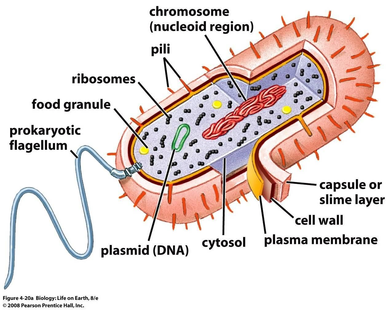
what is the structure of bacteria?
They are versatile and adaptable
They are successful due to their small size
Simple cellular structure
what is the role of the cell wall in a bacteria
Physical barrier that excludes certain substances and protects against mechanical damage and osmotic lysis.
what is the role of a capsule in bacteria
Protects bacteria from other cells and helps groups of bacteria to stick together for further protection as they secrete mucilaginous slime.
what is the role of cell-surface membranes in bacteria
Inside the cell wall where the cytoplasm is. It has 70S ribosomes. Acts as a permeable layer that controls entry and exit of chemicals.
what is the role of circular DNA in bacteria
Where genetic material is for the cells to replicate. |
what is the role of plasmids in bacteria
It has genes that help the cell to survive, e.g against antibiotics.
how do bacteria replicate
binary fission
how does binary fission work
The first step of this is the circular DNA molecule replicates and both copies attach to the cell membrane.
The plasmids also replicate.
The cell membrane begins to grow between the two DNA molecules and begins to pinch inwards dividing the cytoplasm into two.
A new cell wall forms between the two molecules of DNA, dividing the cell into two identical daughter cells, each with a single copy of the circular DNA and a variable number of copies of the plasmids.
what are the parts of prokaryotic cells
No nucleus, DNA free in cytoplasm
DNA is in the form of circular strands- plasmids
No membrane-bound organelles
No chloroplasts, only bacterial chlorophyll
70S ribosomes
Cell walls made of murein
May have a slime capsule
what are the parts of eukaryotic cells
Nucleus with nuclear envelope.
DNA is associated with proteins.
No plasmids
Membrane-bound organelles
80S
Cell wall made of cellulose
No capsule
label the bacteria cell

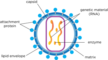
what is the structure of viruses cells
Acellular, non-living particles
Smaller than bacteria
Contain nucleic acids (DNA or RNA) as genetic material.
Only multiply in host cells
Nucleic acid is enclosed in a protein coat – capsid.
Have attachment proteins which allow the virus to identify and attach to host cells.
label the virus cells

how do viruses replicate?
They replicate by attaching to their host cell with the attachment of protein on their surface.
They inject their nucleic acid into the host cell, the genetic information on the injected viral nucleic acid then provides the instructions for the host cells metabolic processes.
what do microscopes do?
produce a magnified image of an object
what do convex glass lenses do?
acts as magnifying glass, they work more effectively if they are used in pairs.
what do long wavelengths of light mean for microscopes?
it lowers the resolution
what is the resolution of light microscopes?
0.2um (micrometres)
what is the resolution for electron microscopes
0.1nm
how can long wavelengths of light rays be overcome?
using beams of electrons rather than beams of light that have a shorter wavelengths
what is the definition of an object
the material under the microscope
what is an image
appearance of the object
whats the equation for magnification
magnification= image size/actual size
what is the resolution
the distance apart that two objects can be, so they are still seen as two separate objects.
what is the limit of resolution
up to this point increasing the magnification, increases the resolution. after this point, the resolution doesn’t increase, the image just becomes blurred.
what is cell fractionation
the process where cells are broken up and the different organelles are separated out
what are the conditions needed for cell fractionation
Cold- to reduce enzyme activity that might break down the organelles.
Same water potential- prevent osmotic activity of the cell bursting or shrinking.
Buffered- so the pH doesn’t fluctuate as any change in the pH could alter the structure of the organelles or affect enzyme's function.
what does the homogeniser do
breaks up cells in a homogeniser that opens up the cell membrane and releases the organelles from the cell. the homogenate is then filtered to remove any complete cells and cellular debris
what is ultracentrifugation
This is when the fragments in the filtered homogenate are separated in a machine called a centrifuge. This spins tubes of homogenate at a high speed to create a centrifugal force
what is the ultracentrifugation for animal cells
The tube filtrate is placed in the centrifuge and spun at a slow speed.
The heaviest organelles, the nuclei, are forced to the bottom to form the pellet.
The fluid at the top, the supernatant) is removed, leaving the pellet.
The supernatant is transferred to a different tube and then it is spun at a faster speed.
The next heaviest (density) is mitochondria, then chloroplasts, lysosomes, endoplasmic reticulum/Golgi, ribosomes.
why do light microscopes have poor resolution?
due to their long wavelengths
how can electron beams help resolution improve?
they have a short wavelength and can therefore resolve objects well (high resolving power). electrons are absorbed or deflected by the molecule in air or near-vacuum must be created for the electron microscope to work
what does a transmission electron microscope consist of?
an electron gun that produces a beam of electron that is focused onto the specimen by a condenser electromagnet.
what is a photomicrograph?
an image produced on a screen to be photographed
what are the limitations of the transmission electron microscope?
o Difficulties preparing the specimen limit the resolution (0.1nm).
o A higher energy electron beam is required, and this may destroy the specimen.
o The whole system must be in a vacuum and therefore living specimens cannot be observed.
o A complex ‘staining’ process is required and even then, the image isn’t in colour.
o The specimen must be thin to allow electrons to penetrate.
o Artefacts may interfere with the photomicrograph.
what do scanning electron microscopes do?
It directs a beam onto the surface of the specimen from above, rather than penetrating from below.
The electrons are scattered by the specimen and the pattern of this depends on the contours of the specimen.
what type of image can be built using a SEM?
3D, by the computer analysis of the scattered electrons
does the TEM or SEM have a lower resolution?
SEM (20nm)
what are the limitations of SEM?
same as TEM, but the specimens dont have to be extremely thin as electrons dont penetrate.
What is an eyepiece graticule?
measures the size of objects on a light microscope
What is a graticule?
a glass-disc placed in eyepiece of a microscope.
Why cant graticules measure objects?
it needs to be calibrated
What is a stage micrometer?
used to calibrate graticule, usually with sub-divisions of 10um
What is the cell cycle?
cells that don’t divide continuously but undergo a regular cycle of division, separated by periods of cell growth
What are the stages of the cell cycle?
o Interphase- most of the cycle, resting phase as there is no division.
o Nuclear division- nucleus divides into two or four.
o Division of cytoplasm (cytokinesis)- cytoplasm divides to form two new cells or four new cells.
What is cancer?
group of diseases caused by a growth disorder of cells, result of damaged genes that regulate mitosis and the cell cycle. This leads tumors to develop and expand.
How can the rate of mitosis be affected?
the environment of the cell and growth factors and controlled by two types of gene.
What can a mutation result in?
A mutation of a gene can result in uncontrolled mitosis, mutated cells are different shapes and sizes than normal cells. They can divide and form clones and make tumors. Malignant tumors grow rapidly and can be life-threatening, benign ones are slower and not life-threatening.
What does treatment of cancer involve?
Involves killing cells by blocking parts of the cell cycle so division is disrupted to slow cancer growth.
How does chemotherapy disrupt the cell cycle?
o Preventing DNA from replicating.
o Stopping metaphase by interfering with spindle formation.
Whats a problem with chemo?
can disrupt cell cycles of normal cells
How does chemo work?
The drugs are more effective against rapidly dividing cells, so cancer cells are damaged to a greater degree.
How do you prepare a root tip?
1. Add some 1 mole of HCl to a boiling tube, enough to cover the root tip. Put the boiling tube in a bath that has reached 60 degrees-Celsius.
2. Use a scalpel to cut 1cm from the tip of a growing root.
3. Transfer root tip into boiling tube with acid, incubate for 5 minutes.
4. Use tweezers to remove the root tip from tube and use pipette to rinse with cold water, leave to dry on paper towel.
5. Place root tip on microscope slide and cut 2mm from tip, discard the rest.
6. Use a mounted needle to break the tip and spread the cells.
7. Add a few drops of acetic orcein stain to make the chromosomes easier to see.
8. Place cover slip over the top and use a paper towel to push firmly down onto them to make the tissue thinner and allow light through.
9. Look at the stages of mitosis under an optical microscope.
How can you use an optical microscope?
o Start by clipping the slide onto the stage.
o Select the lowest-powered objective lens.
o Use the coarse adjustment knob to bring the stage up to below the objective lens.
o Look down the eyepiece and use the coarse adjustment knob to move the stage downwards, away from the objective lens until the image is in focus.
o Adjust the focus with the fine adjustment knob until you get a clear image of the object.
o You can swap to a higher power to increase the magnification.
What is the mitotic index?
The proportion of cells in a tissue sample that are undergoing mitosis allows you to see how quickly the tissue is growing.
How do you work out the mitotic index?
Mitotic index= number of cells with visible chromosomes/total number of cells observed