Chapter 35, Assessment: Cardiovascular System
 Call Kai
 Learn
Learn Practice Test
 Spaced Repetition
 Match
Match Flashcards
 Knowt Play
1/98

Earn XP
Description and Tags
Structures and Functions of Cardiovascular System
Name | Mastery | Learn | Test | Matching | Spaced | Call with Kai | Chat |
|---|
No analytics yet
Send a link to your students to track their progress
99 Terms
what are the layers of the heart? (hint: 3)
Endocardium—thin, inner lining
Myocardium—layer of muscle
Epicardium—outer layer
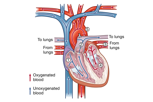
describe blood flow of the heart
1, The right atrium receives venous blood from the inferior and superior vena cava and the coronary sinus. The blood then passes through the tricuspid valve into the right ventricle. 2, With each contraction, the right ventricle pumps blood through the pulmonic valve into the pulmonary artery and to the lungs. 3, Oxygenated blood flows from the lungs to the left atrium by way of the pulmonary veins. 4, It then passes through the mitral valve and into the left ventricle. 5, As the heart contracts, blood is ejected through the aortic valve into the aorta and enters the systemic circulation.
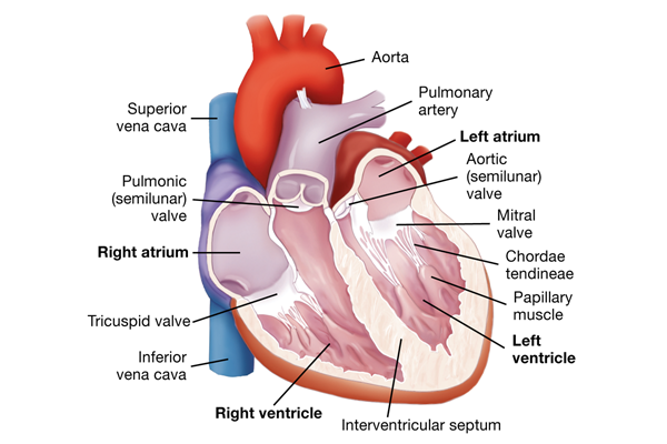
name the anatomic structures of the heart and heart valves.
how does the coronary circulation system supply the myocardium with blood?
coronary circulation supplies the myocardium with blood. blood flow into the two major coronary arteries occurs primarily during diastole.
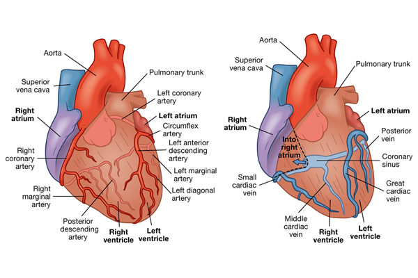
The left coronary artery arises from the aorta and divides into the left anterior descending artery and left circumflex artery. These arteries supply the:
Left atrium
Left ventricle
Interventricular septum
Part of the right ventricle
The right coronary artery arises from the aorta, and its branches supply the:
Right atrium
Right ventricle
Part of the posterior wall of the left ventricle
The right coronary artery supplies blood to the:
atrioventricular (AV) node and the bundle of His in 90% of people. blockage of this artery often causes serious defects in cardiac conduction.
coronary veins are what to the coronary arteries?
parallel
most blood from the coronary system drains and empties where?
most of the blood from the coronary system drains into the coronary sinus and empties into the right atrium.
how does an electrical impulse travel through the heart, and how does the use of an ECG monitor this impulse?
the conduction system of the heart creates and transports the electrical impulse
what starts the depolarization of heart cells?
the impulse or action potential
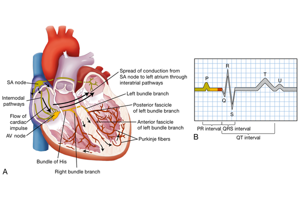
describe conduction system of the heart
AV, Atrioventricular; SA, sinoatrial. (B) The normal ECG pattern. The P wave represents depolarization of the atria. The QRS complex indicates depolarization of the ventricles. The T wave represents repolarization of the ventricles. The U wave, if present, may represent repolarization of the Purkinje fibers or be associated with hypokalemia. The PR, QRS, and QT intervals reflect the time it takes for the impulse to travel from one area of the heart to another.
what is depolarization?
The impulse normally begins in the sinoatrial (SA) node, which is the pacemaker of the heart. (The impulse is delivered within 0.12 second)
what is repolarization?
Repolarization occurs when the contractile and conduction pathways cells return to resting.
what does an electrocardiogram do?
the electrical activity of the heart can be detected using electrodes and recorded on an electrocardiogram (ECG). The letters P, QRS, T, and U are used to name the separate waveforms.
what does the p wave represent?
begins with the firing of the SA node and represents depolarization of the atria.
what does qrs represent?
qrs represents depolarization from the AV node throughout the ventricles.
how do we measure a delay of impulse through the AV node?
it is the time between the beginning of the P wave and the beginning of the QRS wave.
what does the t wave represent?
The T wave represents repolarization of the ventricles
what does a u wave represent if seen?
may represent repolarization of the Purkinje fibers and a large u wave may occur with hypokalemia.
what is systole?
contraction of the heart muscle that results in ejection of blood from the ventricles.
what is diastole?
relaxation of the heart muscle that allows for filling of the ventricles.
what is cardiac output?
the amount of blood pumped by each ventricle in 1 minute.
what is stroke volume?
the amount of blood ejected from the ventricle with each heartbeat.
how do we calculate cardiac output?
CO = SV x HR
what is the normal range of cardiac output?
4 to 8 L/min
what is HR controlled by?
the ANS (primarily)
what happens if HR is rapid?
there is less time for diastole and perfusion of coronary arteries.
how fast can the heart beat for short periods of time without harmful effects?
180 beats/min
since SV is affected by preload, contractibility, and afterload, if we were to increase these factors what would happen?
this increases the workload of heart muscle and O2 demand
what is preload?
the volume of blood stretching the ventricles at the end of diastole.
what does the Frank-Starling law state?
that the more myocardial fibers are stretched, the greater their force of contraction.
what can increase preload?
by hypertension, aortic valve disease, and hypervolemia
what can decrease preload?
rapid HR or hypovolemia, which reduces ventricular filling during diastole
what is contractility increased by?
epinephrine and norepinephrine, which are released by the sympathetic nervous system (note: increasing contractility raises SV by increasing ventricular emptying).
what is afterload?
is the peripheral resistance against which the left ventricle pumps.
what factors does afterload depend on?
size of the ventricle, wall tension, and arterial BP
when can ventricular hypertrophy occur? (an enlargement of the heart muscle)
when arterial BP rises, ventricles meet increased resistance to ejection of blood and increase the work demand.
what does the right ventricle pump against?
the afterload of pulmonary arterial resistance
what is cardiac reserve?
the ability to respond to demands by maintaining or increasing CO
Which explanation would the nurse include when teaching a patient why there can be a blockage in the left anterior descending artery, causing damage to the right ventricle?
“The left anterior descending artery supplies blood to the left side of the heart and part of the right ventricle.”
Which structure is considered the pacemaker of the heart?
Sinoatrial (SA) node
Which information about cardiac output is essential when developing the plan of care for a patient with decreased cardiac output (CO) due to heart failure?
CO is calculated by multiplying the patient’s stroke volume by the heart rate.
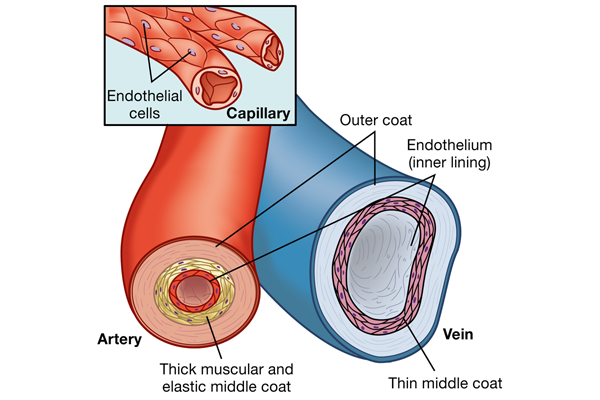
what do arteries do? (except for the pulmonary artery)
carry oxygenated blood away from the heart.
what do veins do? (except for the pulmonary veins)
carry deoxygenated blood toward the heart.
what do capillaries do?
exchange cellular nutrients and metabolic end products (contain no elastic or muscle tissue)
how does blood circulate?
blood circulates from the left side of the heart into arteries, arterioles, capillaries, venules, and veins, and then returns to the right side of the heart.
describe large arteries
the large arteries, such as the aorta and PA, have thick walls composed mainly of elastic tissue that cushion pressure from ventricular contraction.
describe arterioles
Arterioles have more smooth muscle and little elastic tissue, respond to local changes like o2 or co2 levels, and control arterial bp and distribution of blood flow.
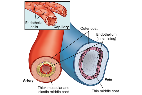
what is the endothelium?
the innermost lining of the arteries that maintains hemostasis, promotes blood flow, and, normally, inhibits blood coagulation.when disrupted, the coagulation cascade results in the formation of a fibrin clot
what do venules do?
collect blood from the capillary beds and channel it to larger veins
describe veins
large-diameter, thin-walled vessels that return blood to the right atrium.
what are the largest veins in the body?
the superior vena cava and the inferior vena cava
the venous system is…
low pressure and high volume
what are factors that affect the amount of blood in the venous system?
Arterial flow
Compression of veins by skeletal muscles
Changes in thoracic and abdominal pressure
Right atrial pressure
Which complication would the nurse expect if the endothelium is disrupted?
Fibrin clot
Which characteristic is an attribute of capillaries?
Has a thin vessel wall
Connect arteries and veins
Made up of smooth muscle tissue
Exchange of cellular nutrients
Contain valves
Has a thin vessel wall
Exchange of cellular nutrients
Which assessment finding is expected with elevation of right atrial pressure?
Neck vein distension
if the SNS is stimulated what occurs?
increases to HR, speed of impulse conduction through the AV node, and force of atrial and ventricular contractions.
how is SNS stimulation mediated?
by beta (β)-adrenergic receptors that act as receptors for norepinephrine and epinephrine.
(α1)-adrenergic receptors are found in vascular smooth muscle that help control blood vessels. what occurs if there is increased/decreased stimulation of these receptors?
Stimulation of α1-adrenergic receptors results in vasoconstriction.
Decreased stimulation to α1-adrenergic receptors causes vasodilation.
how does the PNS slow HR?
by decreasing impulses from the SA node and conduction through the AV node. the effect is mediated by the vagus nerve.
How do baroreceptors and chemoreceptors affect blood flow? (found in the aortic arch and carotid sinus).
baroreceptors are sensitive to stretch or pressure and if there is for example volume overload, it will increase influence of PNS for decreased HR and vasodilation (and vise versa). chemoreceptors respond to hypercapnia, acidosis, and hypoxia which may alter RR and BP.
Which mechanism mediates the effects of the parasympathetic system?
Vagus nerve stimulation
Which component of the autonomic nervous system controls the force of atrial and ventricular contractions?
Sympathetic system
Which outcome would the nurse attribute to baroreceptor stimulation?
Decreased heart rate
what is systolic blood pressure (SBP)?
is the peak pressure exerted against the arteries when the heart contracts.
what is diastolic blood pressure (DBP)?
is the residual pressure in the arterial system during ventricular relaxation.
how to calculate BP?
BP = CO × SVR
how can BP be measured? (invasive/noninvasive)
noninvasive: Sphygmomanometer and Stethoscope and Doppler Ultrasonic Flowmeter
invasive: inserting a catheter into an artery
what is pulse pressure?
the difference between the SBP and DBP. is normally one-third of the SBP
what is MAP?
the average pressure within the arterial system.
how do you calculate MAP?
MAP = (SBP + 2 DBP) ÷ 3
what is the average MAP?
The MAP must be greater than 60 mm Hg to perfuse the vital organs under most conditions.
CVD is the leading cause of death in adults over?
age 65
CAD is due to? (most common problem)
atherosclerosis
what causes aging of the cardiovascular system?
The aging process
Disease
Environmental factors
Lifetime health behaviors
what happens during orthostatic hypotension?
the sympathetic nerve pathway produces a reduced HR response, which can decrease BP
what is postprandial hypotension?
a decrease in BP of at least 20 mm Hg that occurs within 75 minutes after eating
blood pressure changes increase what?
fall risk in older adults
What is the mean arterial pressure (MAP) for a patient whose blood pressure is 102/78 mm Hg?
86
Which condition results in a heart murmur?
Turbulent blood flow across a heart valve
Which anatomic feature of the heart directly stimulates ventricular contractions?
Purkinje fibers
Which aspect of the heart’s action does the QRS complex on the electrocardiogram (ECG) represent?
Depolarization from atrioventricular (AV) node throughout ventricles
Which outcome would the nurse expect with a prolonged increase in arterial blood pressure?
Ventricular hypertrophy
place the structures of the heart in order of blood flow starting after the superior and inferior vena cava.
1 Right atrium
2 Right ventricle
3 Lungs
4 Left atrium
5 Mitral valve
6 Left ventricle
Which characteristic is an attribute of arteries?
Arteries branch into smaller vessels called arterioles.
Arteries are lined by endothelial tissue.
Which age-related change contributes to a blood pressure of 160/70 mm Hg in an older adult?
Loss of elasticity in arterial vessels
Which structure prevents the eversion of mitral and tricuspid valves during ventricular contraction?
Chordae tendineae
Which change in vital sign measurement would the nurse expect for a hypovolemic patient?
Decrease in pulse pressure
Which assessment finding in the older adult would the nurse attribute to kyphosis?
Distant heart sounds
Which cardiovascular response would the nurse expect in a patient with a plasma pH of 7.32?
Activation of chemoreceptors to increase respiratory rate