RHS anatomic Landmarks
 Call Kai
 Learn
Learn Practice Test
 Spaced Repetition
 Match
Match Flashcards
 Knowt Play
1/60

There's no tags or description
Looks like no tags are added yet.
Name | Mastery | Learn | Test | Matching | Spaced | Call with Kai | Chat |
|---|
No analytics yet
Send a link to your students to track their progress
61 Terms
identify the following anatomical landmarks as maxillary or mandibular:
lingual foramen
mandibular
the broad radiolucent band seen just below the internal oblique ridge is the submandibular fossa
true
all the following can appear on radiograph of the mandibular incisors EXCEPT one. which is the EXCEPTION?
hamular process
all the folowing can appear within the maxillary sinus EXCEPT one. which is the EXCEPTION?
coronoid process
a radiopaque Y in the maxillary canine region represents the junction of the border of the maxillary sinus and the
floor of the nasal cavity
The nasal septum ends inferiorly at another radiopaque landmark known as the:
anterior nasal spine
identify the thin, radiolucent line seen surrounding the cementum of the root of the tooth
periodontal ligament space
all of the following landmarks will appear radiolucent EXCEPT one. Which one
ridge
the V-shaped structure seen in the mandibular incisor area is the
mental ridge
what radiolucent anatomical structure is indicated by the arrow?
nasal fossa
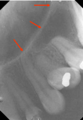
what radiolucent anatomic structure is indicated by the arrow?
mental foramen
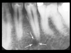
what radiopaque anatomical structure is indicated by the arrow?
floor of the maxillary sinus

what radiolucent anatomical structure is indicated by the arrow?
maxillary sinus
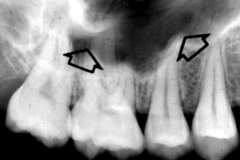
what radiopaque anatomical structure is indicated by the arrow?
nasal septum
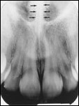
what radiolucent anatomical structure ( dark oval) is indicated by the arrow?
incisive foramen

what radiolucent anatomical structure (thin, dark line) is indicated by the line?
median palatine suture
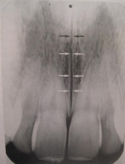
what radiopaque anatomical structure s indicated by the arrow?
genial tubercles

what is the proper method for mounting radiographs?
radiographs should be mounted as if you were facing the patient
which of these structures appears radiolucent?
dental pulp
which of these structures appears radiopaque?
maxillary tuberosity
the inverted-Y landmark is composed of which two structures?
inferior border of the nasal cavity and anterior border of maxillary sinus
which of the following best describes the appearance of bone on a radiograph?
cortical bone appears radiopaque, cancellous bone appears radiolucent
identify the area the arrow points to on the dental patient
mandibular canal

identify the radiopaque area the arrow points to
zygoma

identify the radiopaque area the arrow points to
coronoid process
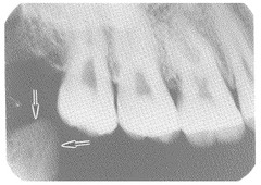
identify the radiolucent area the arrow points to
nutrient canal

what are the arrows pointing to?
lateral fossa
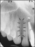
identify this landmark
mylohyoid ridge
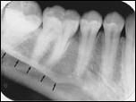
identify the landmark the arrow points to in the dental image?
incisive foramen

what is the arrow pointing to in the dental image?
mental foramen
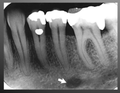
identify the landmark
maxillary sinus

identify this landmark of the dental image
maxillary tuberosity
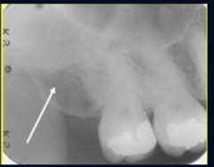
identify this landmark
nasal spine
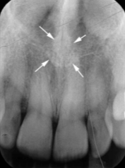
identify this landmark
mental ridge

on a dental image, the alveolar crest is typically located _________ mm below the junction of the crown and the foot surfaces
1.5 to 2.0
which of the following tooth structures is most radiolucent?
pulp
the anterior nasal spine is located at the ________ portion of the nasal cavity
anterior and interior
on a maxillary periapical image, the floor of the nasal cavity appears as a _______ band of bone ________ the maxillary incisors
radiolucent, above
on maxillary periapical image, the nasal septum may be superimposed over the
median palatal suture
the lateral walls of the nasal cavity are formed b y the ______ bone and the maxillae
ethmoid
on a dental image, the lateral fossa appears as a ______ area of _____ appears, depending on the anatomy of the individual
radiolucent, variable
the lateral fossa is located between the
Maxillary canine and lateral incisor
the median palate suture is located between the two _______ of the maxilla
palatine process
on a periapical image, the maxillary sinuses appear as radiolucencies located superior to the apices of the
maxillary molars and permolars
the superior foramina of the incisive canal are located on the ________ of the nasal cavity
floor
the superior foramina of the incisive canal are ________ tiny openings or holes in bone
two
on a ________ perapical image, the nasal fossa appears as a large _______ are located above the roots of the maxillary incisors
maxillary, radiolucent
the ______ nerve exits the maxilla through the incisive foramen
nasopalatine
This foramen is almond-shaped and is located between the apices of the maxillary central incisors.
incisive
which of the following landmarks would appear radiolucent on a dental image?
suture
an empty soace within bone is known as a
sinus
a pointed, spikelike protrusion of bone is a
spine
which of the following describes a ridge
Linear protrusion of bone
which type of bone is composed of bony trabeculae?
cancellous
cancellous bone is the soft ________ bone located between two layers if ________ cortical bone
spongy, dense
(1) cancellous bone is considered radiolucent
(2) cortical bone is considered radiolucent
both statements are false
compact bone is also called ________ bone
cortical
Where is the mental fossa and is it radiopaque or radiolucent?
Mandibular anterior, radiolucent
Where are the oblique ridges? Are they radiopaque or radiolucent? What’s difference between external and internal?
Mandibular posterior, radiopaque, external is on outer surface of mandible and internal is on lingual side
Where is mental foramen located?
Mandibular premolar region, radiolucent, circular