All Para Pracs: Pigs/Poultry/Small Animal
1/333
There's no tags or description
Looks like no tags are added yet.
Name | Mastery | Learn | Test | Matching | Spaced |
|---|
No study sessions yet.
334 Terms
Eimeria tenella
Station 1:
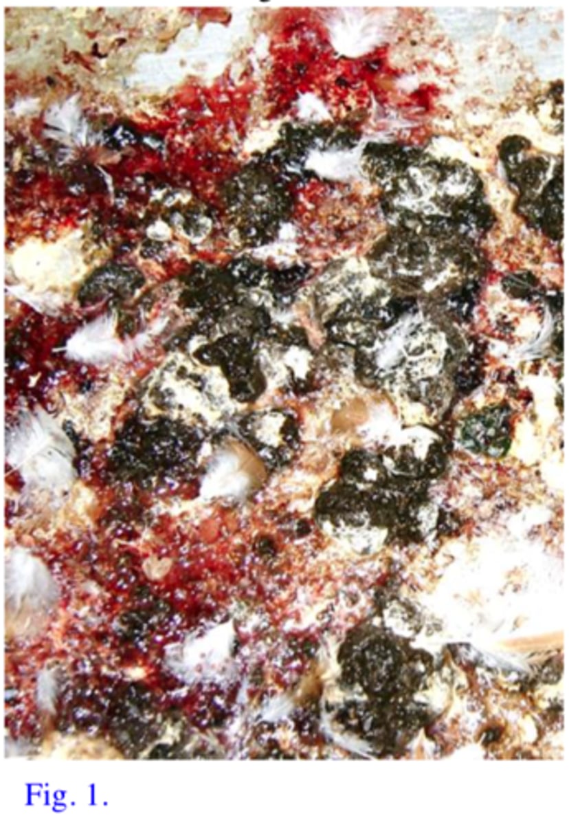
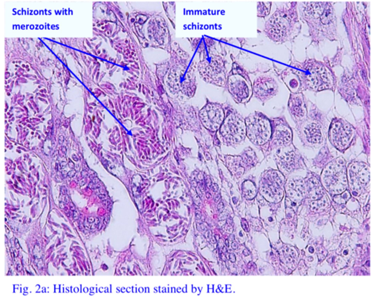
The owner of a poultry farm is telling you that most of his 4-5 week chickens that are reared on the floor show anorexia, have wings dropped and feathers ruffled and eyes closed. Most of the sick chickens present hemorrhagic diarrohea (Fig. 1) and die in large numbers. The histological section (H&E staining) on the microscope 1 is from the caeca of a chook that died with hemorrhagic diarrhoea.
Q1: Name the parasite responsible for the death of the chickens
2nd generation mature schizonts (contain banana shape stages called merozoites).
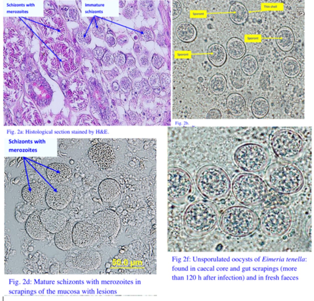
Q2: What parasite stages are associated with pathology for Eimeria tenella? Identify them on the microscope slide (microscope 1) and on Fig. 2a.
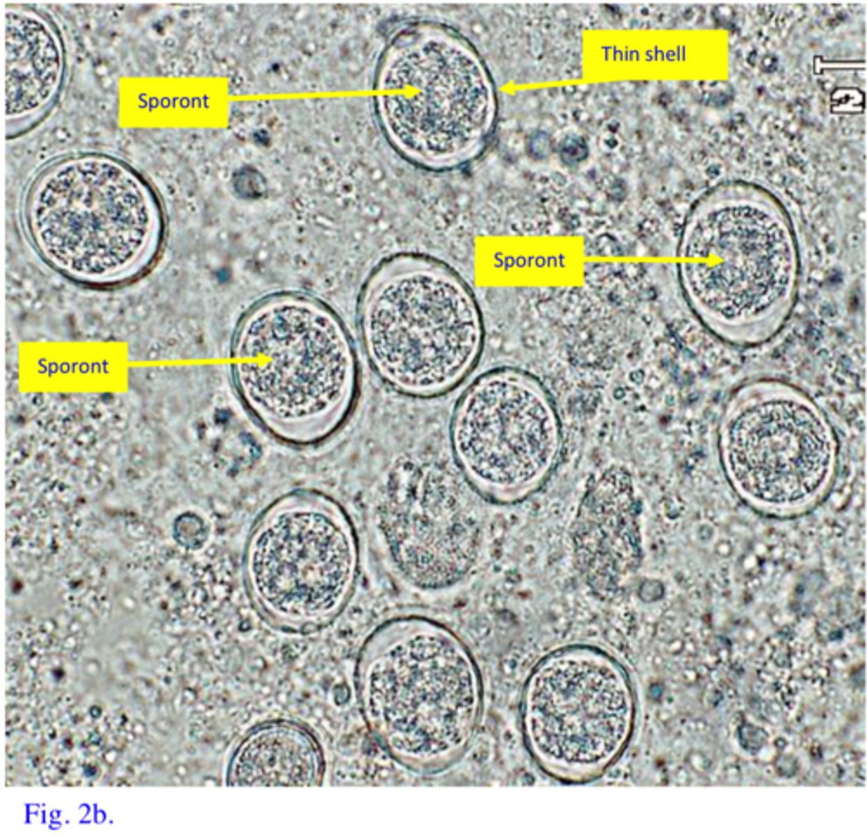
The few parasites seen in the faeces of infected birds (and on microscope 2) are unsporulated oocysts. The pathology is caused by stages (mature schizonts) that precede the development of oocysts so that is why the oocysts are in low numbers at this stage of the disease. If the birds survive large numbers of oocysts will be shed in the faeces.
Station 1:
Q3: In the faeces of the sick birds you can see few parasites as shown in Fig. 2b and on microscope no 2 (you cannot see them on the histological section). These parasites are characterized by a huge reproductive potential and you wonder why the parasites in faeces are in low numbers. Do you have an explanation why the parasites are in low numbers?
Scrapings of the mucosa and/or histological sections from the affected areas of the intestine. Depending on the stage of the disease we can see either:
1. Schizonts and merozites in the Acute form of the disease
2. Gametocytes/zygotes/oocysts in the Chronic stage of the disease
Station 1:
Q4: The owner of the farm is sending your practice chickens that died because of infection with this parasite and he is asking for a diagnosis. How would you confirm the diagnosis (methods, stages of the parasite you can find, how are you going to identify them) Eimeria tenella
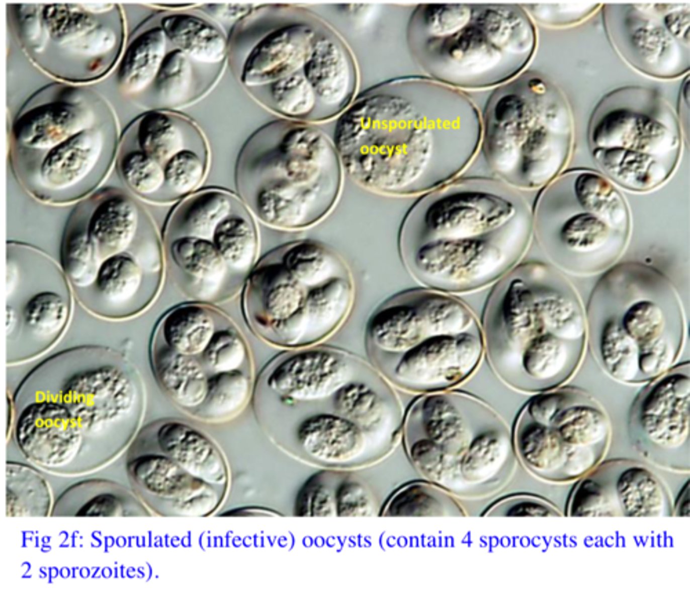
Ingestion of sporulated oocysts (fig. 2g) with food/water (it takes a couple of days for the unsporulated oocysts shed by infected birds to become sporulated/infective oocysts);Sporulated oocysts contain 4 sporocyts each with 2 sporozoites.
Station 1:
Q5: How did the birds become infected (parasite stage and way of entry into the host) with Eimeria tenella
The sporulated oocysts might survive long time in the environment and they are resistant to the common disinfectants. Furthermore they can be transported easily by rats, mice, flies, boxes for eggs, tools, boots of personal etc.
Q6: The broilers are managed ‘all-in/all-out’ with very good cleaning and disinfection of the sheds. How is it that this outbreak occurred? Eimeria tenella
Toltrazuril (Baycox) is the drug of choice to betried on these birds. Amprolium and sulfonamides might work (the chickens do not feed/drink water)!!
Station 1:
Q7: How do you control this outbreak? Eimeria tenella
1. Chemoprophylaxis (common in broilers, shuttle program better than straight program)
OR
2. vaccination using live/attenuated vaccines (common in laying hens/breeders)
3. Hygiene (try to prevent the sporulation of oocysts), prevent overcrowding of birds,proper nutrition, do not grow birds of different ages together (old birds might beresistant because of immunity but shed oocysts that can prove fatal for young birds),moisture content of the floor etc.
Station 1:
Q8. How do you prevent outbreaks of this disease? Eimeria tenella
Widespread Chemresistance
Station 1:
Q9. What is one of the biggest challenges that farmers that are using chemoprophylaxis are facing nowadays?
Station 2:
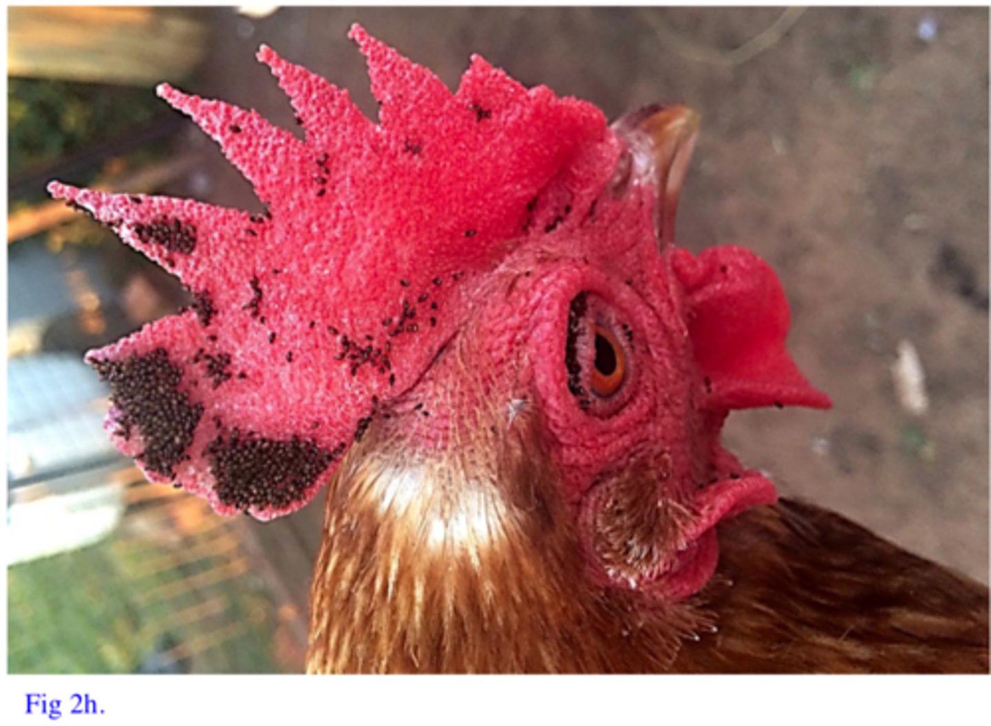
The owner of a very small poultry farm from Qld is relating you that his poultry are anaemic, irritated and they keep scratching that caused lesions around the eyes. In laying eggs the production of eggs decreased while the young birds are emaciated and started to die. On inspection of the birds you can see small parasites fixed on the head and comb (Fig. 2h) of the birds
Parasite a: Echidnophaga gallinacea.
- No ctenidia
- Angular head
- Thorax reduced dorsally
The owner of a very small poultry farm from Qld is relating you that his poultry are anaemic, irritated and they keep scratching that caused lesions around the eyes. In laying eggs the production of eggs decreased while the young birds are emaciated and started to die. On inspection of the birds you can see small parasites fixed on the head and comb (Fig. 2h) of the birds
Q1. Out of parasites in Petri dishes ‘a” and ‘b’ which one is most likely to be found attached around the eyes of the chooks as shown in Fig. 2h? Name the parasite and describe the most important morphological features used for identification.
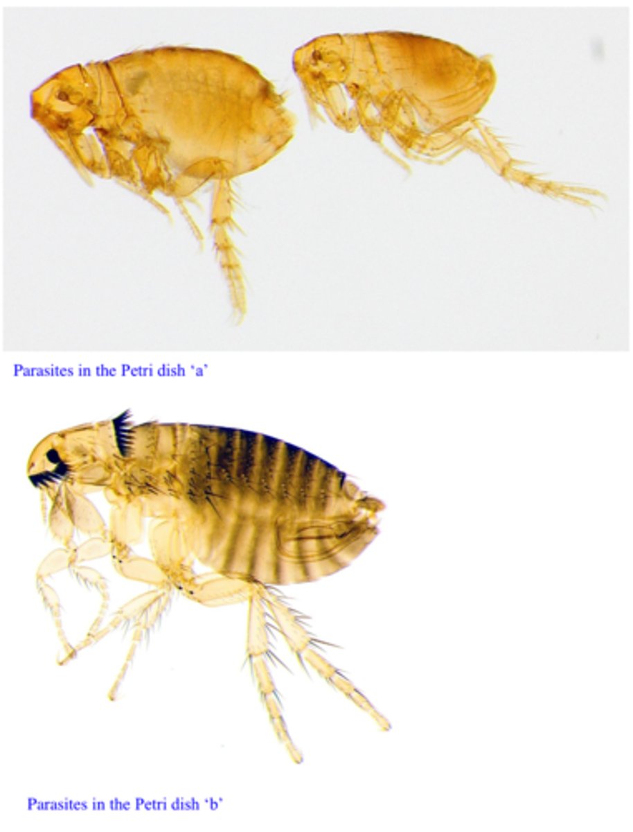
Parasite b: Ctenocephalides felis
- On dogs, cats and other species including humans, calves etc.
Station 2:
Q2. On what host species are you going to find the other parasite? Name the parasite.

From the environment (larvae and pupae/adults within cocoons are present in the environment)
Station 2:
Q3. How did the birds become infested with Echidnophaga gallinacea
Pyrethrins/Pyrethroids - check the products are registered for poultry. Treat also the environment/shed (the immature stages are in the environment).
Station 2:
Q4. How would you treat this bird? with Echidnophaga gallinacea
1. Confinement of poultry
2. Provision of impervious floor
3. Regular removal of droppings and cleaning of the sheds
4. Regular treatments of birds
Station 2:
Q5. How would you prevent further outbreaks? with Echidnophaga gallinacea
Yes
Station 2:
Q6. Can these parasites feed on humans? Echidnophaga gallinacea
Dogs, Cats, Rabbits ect.
Station 2:
Q7. On what other animal species can these parasite be found? Echidnophaga gallinacea
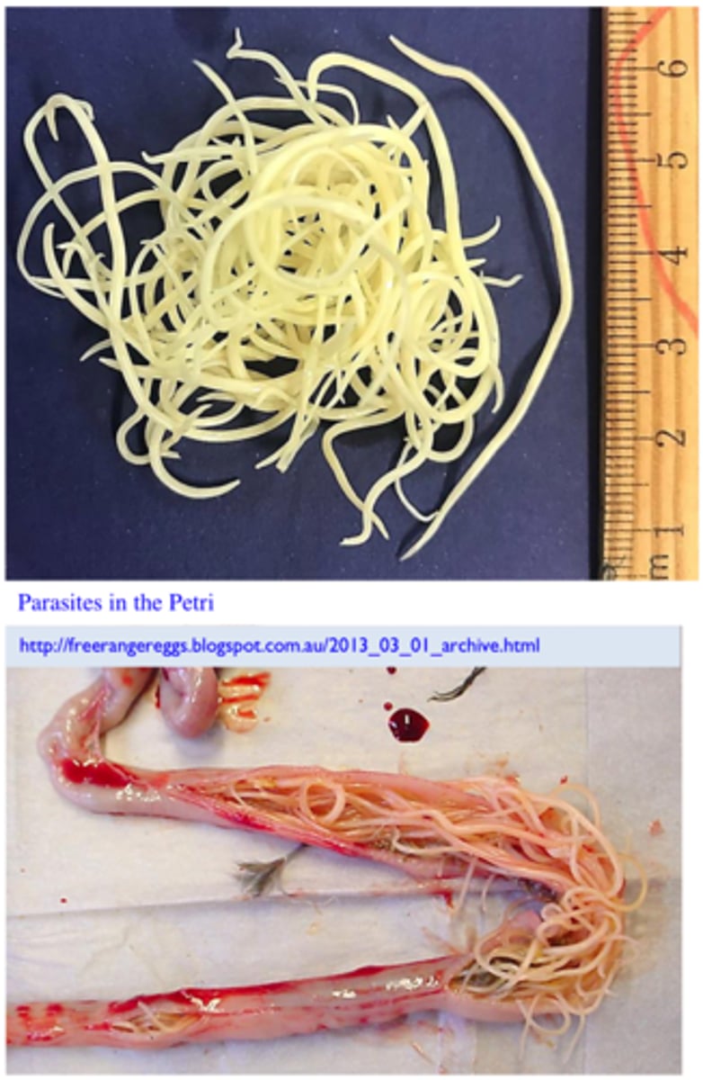
Ascaridia galli
- Long (3-12 cm), stout, whitish parasites (larges nematode of poultry);three large lips around the mouth;
Station 3:
The parasites in the Petri dish were found in the small intestine (Fig. 3) of a 2 month old chook that was showing diarrhea, anaemia, anorexia, unthrift etc to. Most of the chickens inthat particular flock manifest the same clinical signs. Q1. Name the parasite and tell how you identified it (most important morphological features)
Ingestion of embrionated eggs (eggs that contain L3) and paratenic hosts (earthworms,grasshoppers)
Station 3:
Q2. How did the birds become infected (parasite stage and way of entry into the host) with Ascaridia galli?
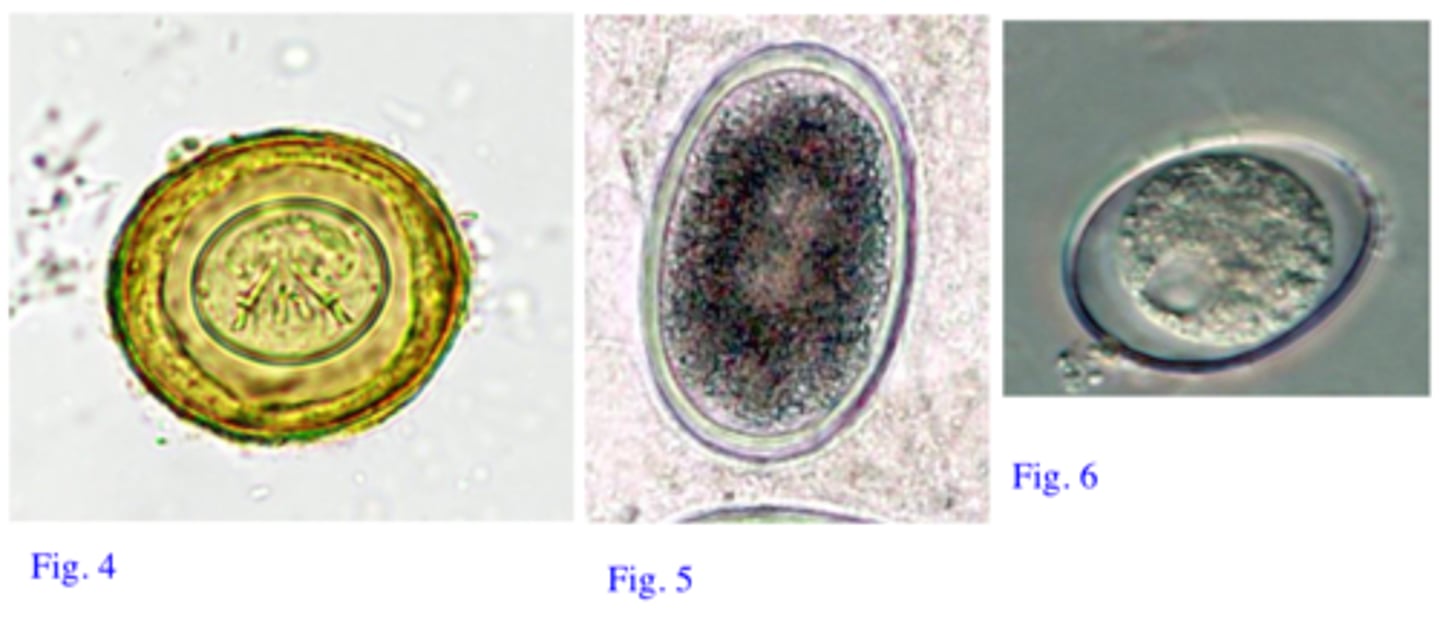
Fig. 4: Eggs of tapeworms – contain an embryo provided with 6 hooks (hexacanth);
Fig. 5: Eggs of A. galli - 75-80 μm, brown, elipsoidal, smooth, rather thick shell, one cellinside;
Fig. 6: Unsporulated oocyst of Eimeria - small size, 27-40 μm, thin shell, inside one cellcalled sporont;
Station 3:
Q3: Which of the eggs from the Fig 4-6 are likely to belong to this parasite? Identify the other twoeggs and describe their morphological features (Ascaridia galli)
Eggs (like the eggs of ascarid parasites) survive for long time in the environment (more than one year).
Station 3:
Q4: How long are the eggs of this parasite likely to survive in the environment? Any implicationsfor the prevention of other outbreaks? Ascaridia galli
Piperazine: active only against A. galli, cheap, administered in food or water.
Levamisole: given in water.
Benzimidazoles (Fenbendazole) work but they are not approved in Australia; they can beadministered in the food, active against most roundworms of chicken and some tapeworms;
Station 3:
Q5: Recommend 2 drugs for the treatment of this parasitic infection. How are you going to use them, why did you select them (price, activity against other parasites, easy to administer etc)? Ascaridia galli
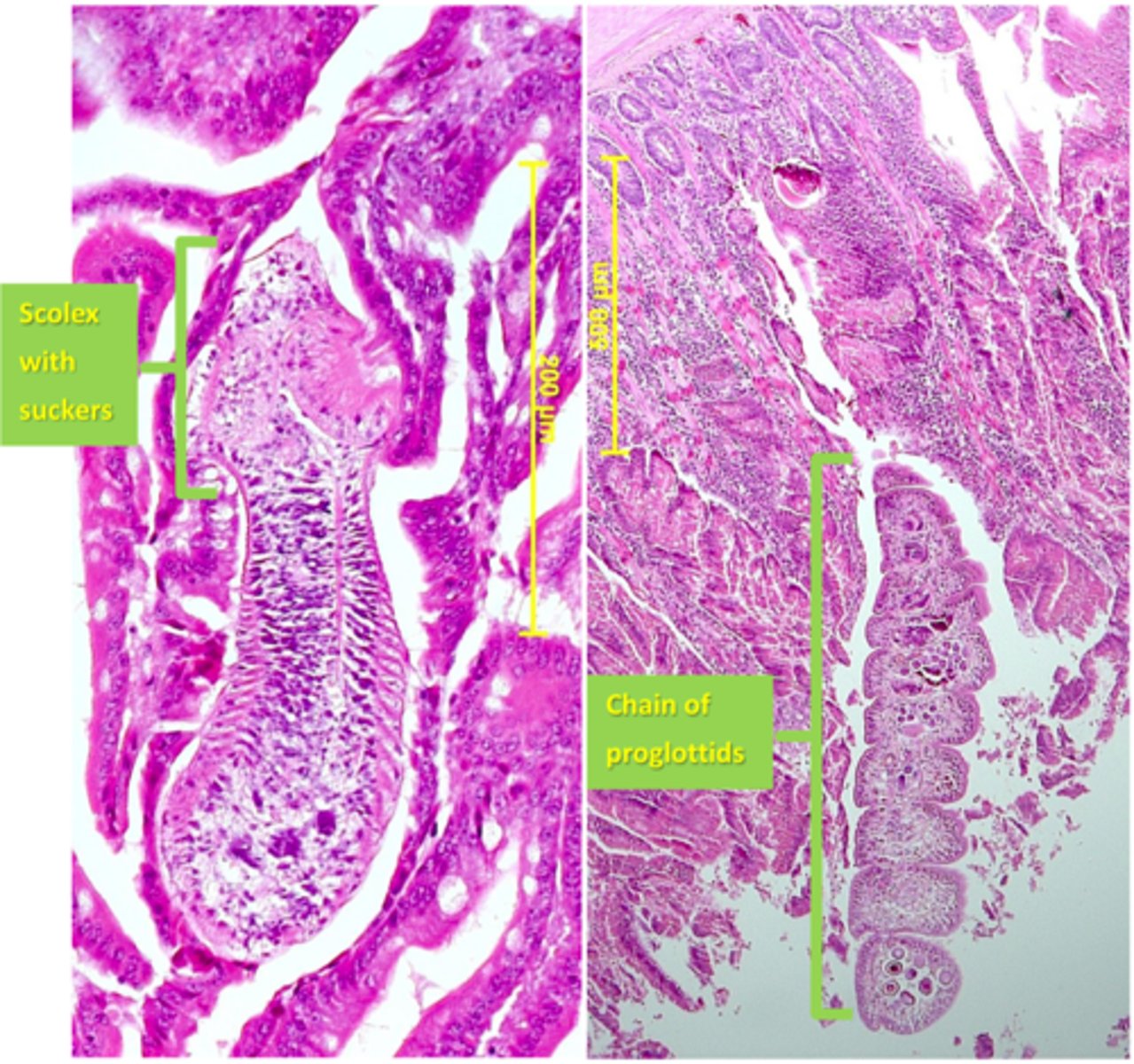
Cestoda/tapeworm, scolex and suckers fixed on the mucosa of the small intestine and chains of proglottids are easily seen.
Station 4:
The histological sections (H&E staining) on the attached microscope and shown below arefrom the small intestine of a bird that was culled because it broke one of its legs. Q1: What type of parasite can you see on the microscope?
Not all tapeworms infecting birds are pathogenic. Numbers of parasites infecting the birds are also important in pathogenesis of the disease.
Station 4:
Q2: Before culling the bird did not show any clinical signs, how do you explain this? Tapeworm
1. Davainea proglottina
2. Raillietina echinobothrida
Station 4:
Q3: Name two of the most pathogenic species of tapeworms that might infect poultry.
Praziquantel: active but not approved for poultry
Fenbendazole: active but not approved for poultry
Station 4:
Q4: Name 2 drugs that might be active against poultry tapeworms
- Outdoor/free-range chickens
- The birds become infected by ingesting intermediate hosts that contain cysticercoid larvae.
- Depending on the species of tapeworms the intermediate hosts might be represented by earthworms, slugs, flies, ants etc that normally should not occur indoors.
Station 4:
Q5: In which rearing system infections with poultry tapeworms are common and why? 1. Davainea proglottina
2. Raillietina echinobothrida
- Grow the birds indoors
- Prevent contact with IH
- Eliminate IH.
Station 4:
Q6: How do you prevent infections with these parasites? 1. Davainea proglottina
2. Raillietina echinobothrida
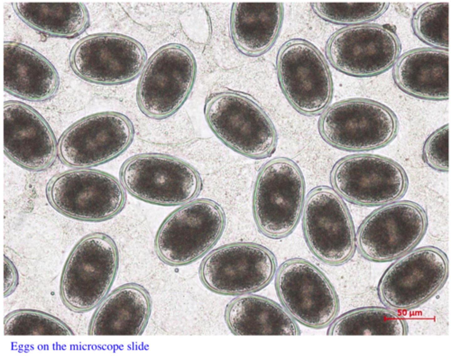
Ascaridia galli or Heterakis gallinarum
(difficult to differentiate the eggs of these two species of worms)
Station 5:
Eggs like those shown on the microscope slide were found in the faeces (floatation) of 6 month old chooks reared on the floor in an indoor farm. The birds do not show any clinical signs. Identify the parasite
Ingestion of embryonated eggs and paratenic hosts (earthworms), the latter way is more common for H. gallinarum than for A. galli
Station 5:
Q2: How could the birds become infected with Ascaridia galli or Heterakis gallinarum
Acaridia galli is pathogenic for young birds,
H. gallinarum is not pathogenic but it is the vector for Histomonas meleagridis, a very pathogenic parasite for turkeys and poultry
Station 5:
Q3: What is the significance of these two parasites for the poultry? Ascaridia galli or Heterakis gallinarum
Station 6:
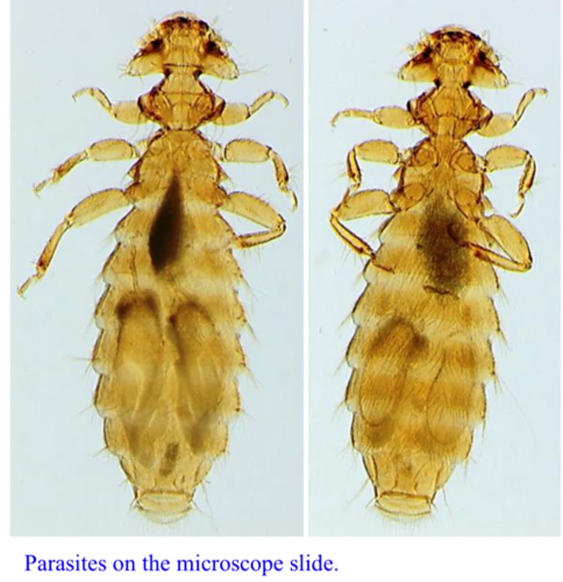
On inspection of a flock of birds you notice that birds present frequent pecking, severe irritation, erythematous and inflamed skin and scabs (Fig. 7). On close examination the formations shown in Fig 8 and in Petri dish A (stereomicroscope) were seen around the feathers, especially in the anus area. Parasites like those on the microscope were moving very quickly on the skin of the birds, some of them even transferred to you
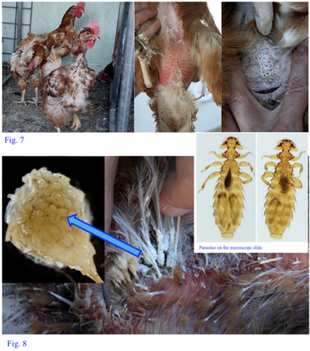
Menacanthus stramineus:
3-3.5 mm long, head triangular: two short, spine like processes on the underside of the head; abdomen with dense covering of medium size setae
Chewing lice (head wider than the thorax, body made up of 3 segments, 3 pairs of legs).
Station 6:
Q1: Name the parasite/group of parasites (and the particular species).
Most likely by direct contact
Station 6:
Q2: How can the birds become infested with this parasite?
Clusters of egg lice glued on the base of feathers
Station 6:
Q3: What are the structures seen in Fig. 8 (and in the Petri dish A) (examine them under a stereo-microscope)?
Lice are permanent parasites: all stages of the lice stay and feed on the host.
Dermanyssus gallinae hides during the day in the crevices of the shed and attacks birds only during thenight (larvae do not feed).
Station 6:
Q4: What are the main differences between the biology of this group of parasites and the biology of Dermanyssus gallinae (red poultry mite)
Maldison, Pestene
Generally insecticides are not active against eggs
Repeat treatment after 7-10 days.However, you have to follow the instructions that come with the product!! Check if the drugs are approved for poultry
Station 6:
Q5: How do you treat these birds (name two drugs and describe how are you going to use them –number of applications etc)
None but they can transfer on the body of the vets while examining/treating the birds
Station 6:
Q6: What is the significance of these parasites for humans?
Station 7:
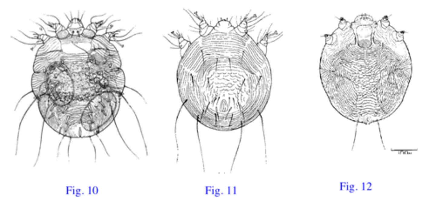
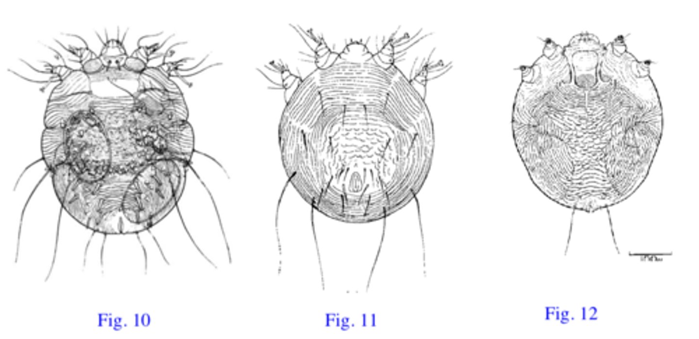
The cockatiel (Fig. 9) was presented to a local vet with white crusts around beak and eyes.The crusts are thick and deformed the beak causing difficulties in feeding and respiration.The Vet carefully collects crusts, put them on a slide, add few drops of KOH, squash the crusts, add a coverslip and examine the slide under microscope.
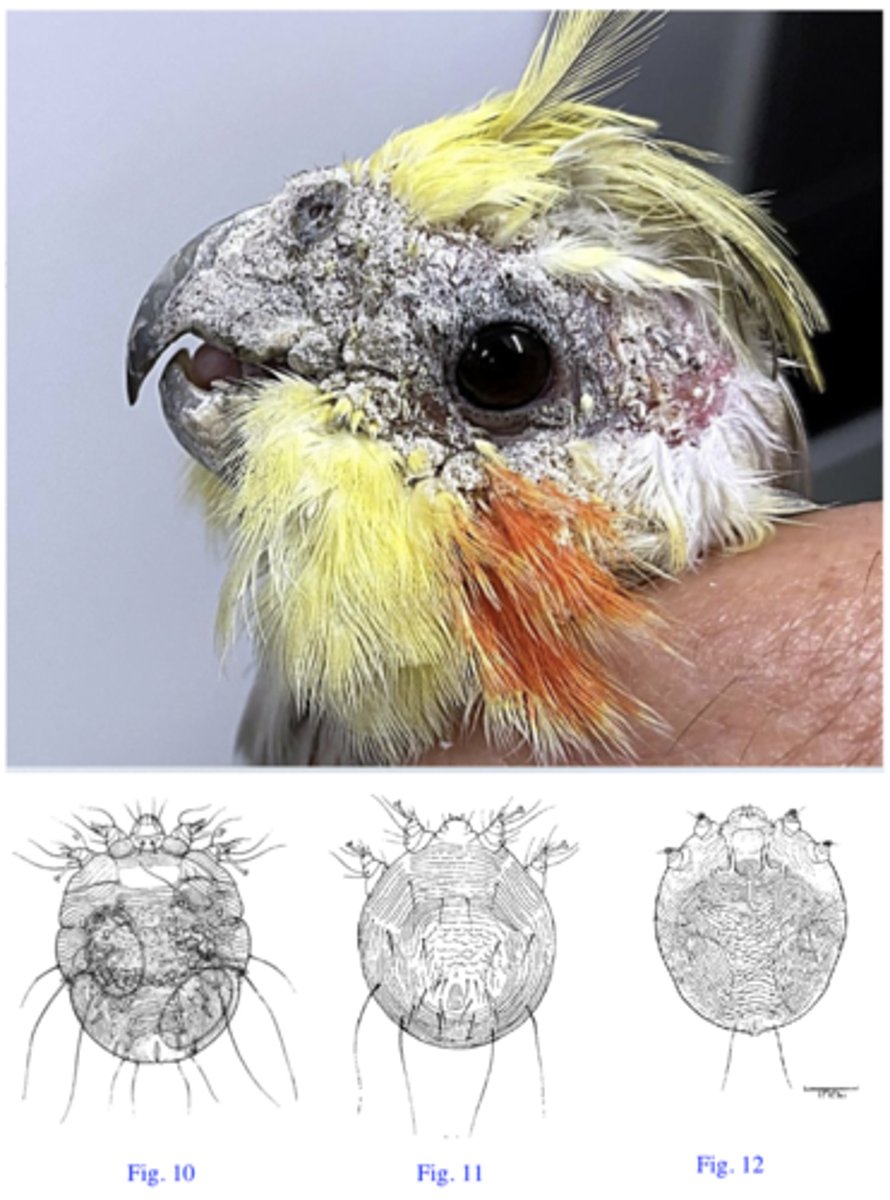
No 12: Cnemidocoptes pilae
- Legs are short and stubby
- Dorsal side of idiosoma: faint striations, no spines
- Anus is terminal;
Station 7:
Q1: Which of the organisms shown in Fig. 10 - 12 is the Vet going to find in the crusts? Name the parasite and describe its most important morphological features used for identification.
Most Likely By Direct Contact
Station 7:
Q2: How did the birds become infested with this parasite?
Ivermectin, Avimax
(orally administered, approved for ornamental caged birds)
Station 7:
Q3: How do you treat these birds?
Fig. 10: Sarcoptes scabiei
Fig. 11: Notoedres cati
Station 7:
Q4: Name the other two organisms
Station 8:
You have been asked to a pheasant farm that is experiencing mortality in young birds. Thebirds show signs of suffocation (gasping for air with outstretched necks andopen mouths). You open the body of a dead bird and in the trachea find the lesions and theparasites shown in Fig. 13. Furthermore in the faeces you find the eggs shown in Fig. 14.
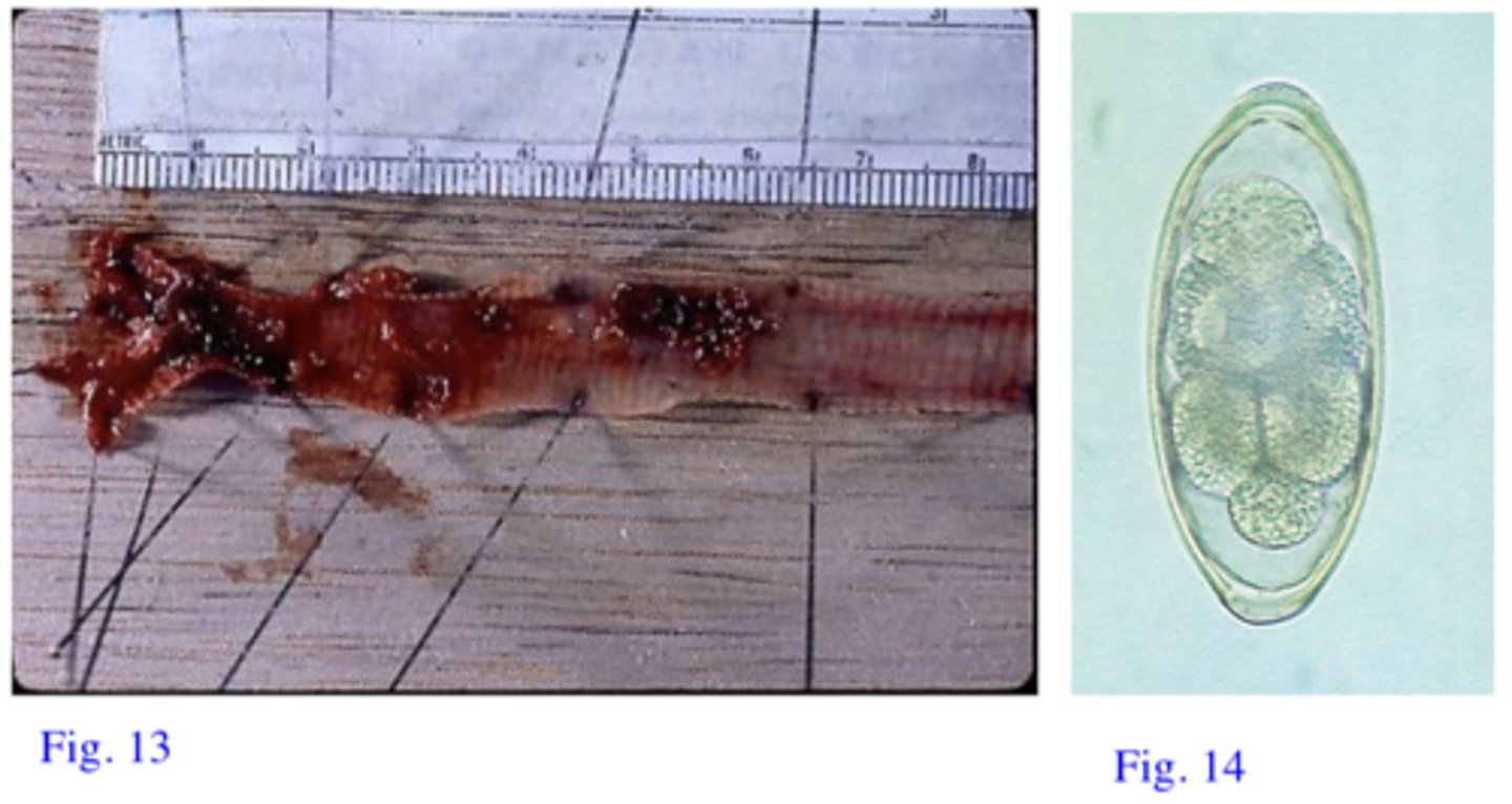
Syngamus trachea
Male and the females are always coupled, the couple has the shape of letter ‘Y”
Station 8:
Q1: Name the parasite (have you noticed anything particular about the morphology of this parasite?)
- Eggs with L3
- Free L3
- Paratenic host containing L3 (earthworms, slugs, snails)
Station 8:
Q2: How did the birds get infected?
Levamisole
(Active against this parasite and approved for use in poultry in Australia but notregistered for this particular parasite)
Benzimidazoles:
Fenbendazole, Mebendazole, Flubendazole work but they are not approvedin Australia for use in poultry.
Station 8:
Q3: How Would you Treat these Birds
Station 9:
The parasites on the microscope slide were found in the caeca of young poultry that were shedding bloody faeces. The farmer is rearing on his farm both poultry and turkey but the turkeys are showing more severe clinical signs: diarrhoea with yellow faeces (Fig. 15),drowsiness, dropping of the wings, closed eyes, emaciation etc. High percentage mortality occurred in the turkeys.
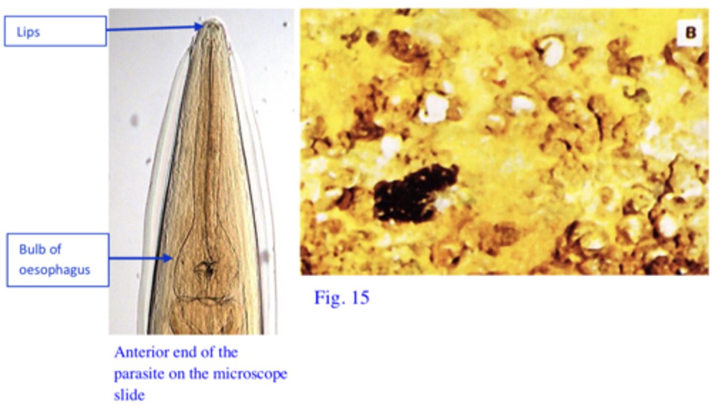
Heterakis gallinarum:
3 lips at the anterior end, oesophagus with a bulb, males with cauda lallae, precloacal sucker and unequal spicules etc.
Station 9:
Q1: Name the parasite on the microscope slide and describe the most important morphological features used for its identification
Most likely not, H. gallinarum has very low, if any, pathogenicity.
H. gallinarum is the vector for Histomonas meleagridis. H. meleagridis cannot survive in the environment more than a couple of hours if not protected within eggs of the H. gallinarum. Furthermore, after oral ingestion H. meleagridis, if not protected by the thick shell of the eggs of H. gallinarum, is very likely to be destroyed by low pH of the stomach!
Station 9:
Q2: Is the parasite on the microscope slide causing the pathology seen in poultry and turkeys? If itis not causing the pathology in the two species of birds why it is found in the caeca of sick birds?
Most likely Histomonas meleagridis is the causative agent of disease in both turkeys in poultry.
Station 9:
Q3: Who is causing the pathology seen in turkeys and poultry, any relationship between thecausative agents of the disease in the two species of birds?
1. Turkey might become infected after ingestion of embryonated eggs of Heterakis gallinarum infected with H. meleagridis,
2. Ingestion of paratenic hosts for H. gallinarum (earthworms)
3. Cloacal drinking.
4. Ingestion of embryonated eggs of H. gallinarum or ingestion of earthworms that have in their body larvae of H. gallinarum infected with H.meleagridis.
Station 9:
Q4: How could the birds become infected with the pathogen causing the disease?
H. meleagridis is more pathogenic for turkeys than for poultry. In turkeys it locates mostly inthe caeca and liver while in poultry it locates mostly in the caeca.
Station 9:
Q5: If both poultry and turkey are infected with the same parasite how is it that the pathology and mortality are significantly different in the two species of hosts?
1. Caeca fill with a mixture of serous and haemorrhagic exudate from the mucosa, inflammatory cells, necrotic debris and the caecal content → caeca become distended with acaseous/cheesy core made up of concentric layers
2. Ulcers → perforation of the caeca → generalized peritonitis;
3. Liver: areas of depressed necrosis (the underlying tissue has been destroyed) that grow up to 1 cm in diameter and are surrounded by a raised ring;In heavy infection lesions may be small, numerous and they may involve a large part of the liver;
Station 9:
Q6: Describe the pathological changes that you expect to see in the dead turkeys.
Growing poultry and turkeys together!
Station 9:
Q7: What management mistake might have caused the severe outbreak of the disease seen in the turkeys?
5-nitroimidazoles (Metronidazole, Dimetridazole, Ipronidazole etc)
All are effective against H.meleagridis but they have been banned for use in food animals in Europe, USA and Australia!!!!
In some countries (not Australia) Histostat® (nitarsone) and Histobloc® (paromomycin)might offer some prophylactic activity.
Station 9:
Q8: How are you going to control this outbreak?
1. Eggs of H. gallinarum are very resistant, they may survive in the environment for up to 3 years.
2. The larvae of H. gallinarum can survive in the earthworms for many years.
Station 9:
Q9: Generally, after the disease is introduced into a farm it will persist for long time. Do you have an explanation for the long persistence of the disease in the farms considering that the causative agent is extremely sensitive to the environmental conditions?
1. Do not grow chickens and turkeys together
2. Do not use chickens sheds for turkeys (Heterakis eggs can survive for a long time)
3. House flocks in concrete or wire floored pens
4. Anthelmintics treatment for Heterakis gallinarum: seems to have some prophylactic activity;
Station 9:
Q10: How are you going to prevent further outbreaks?
Station 10:
The parasites on the microscope slide were found in the deep scrapings of the skin of the bird shown in the attached figure (Fig. 16). The bird belonged to a farmer (backyard) from Toomulla. The bird showed uplifting/upturning of the scales, thick and adherent crusts and deformed legs.
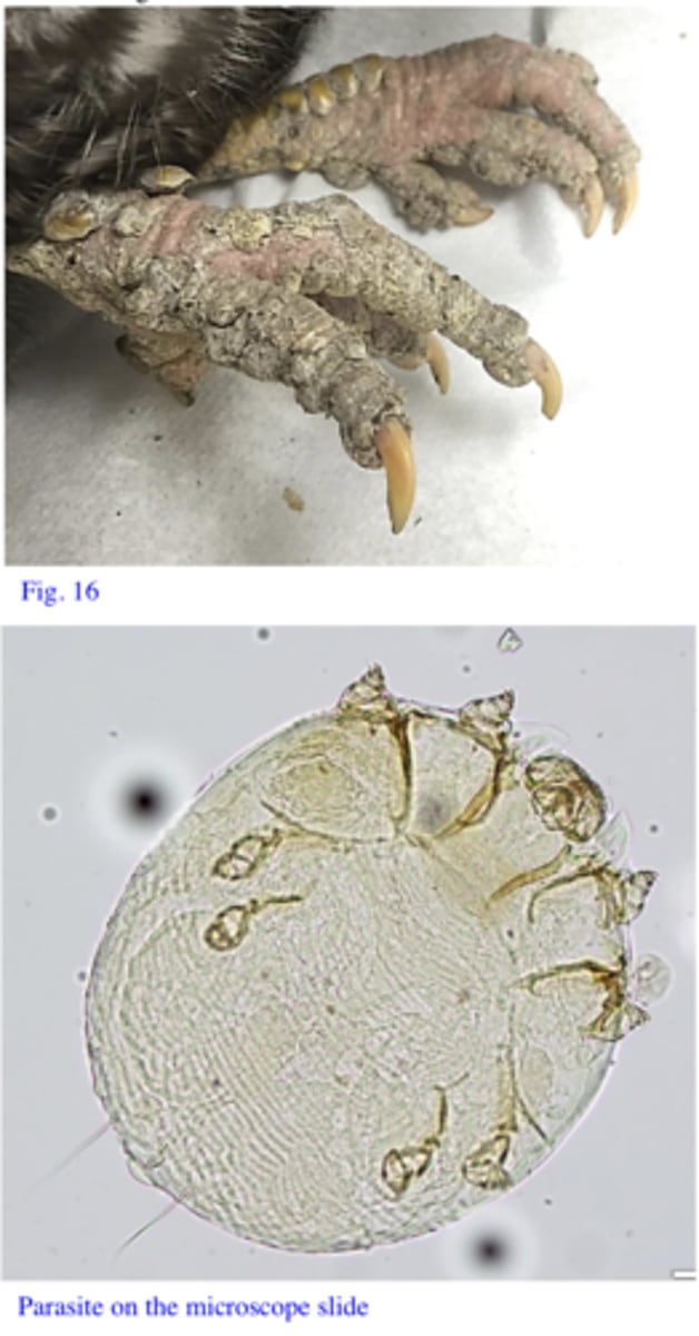
C(K)nemidocoptes mutans
Circular, 215-450 μm in diameter; Gnathosoma is short, wider than longer; In females dorsal surface has a scaly aspect; The legs are short, stuby
Station 10:
Q1: Name the parasite and tell how you identified it (most important morphological features).
Most likely by direct contact. The parasites might be picked up from the ground as well
Station 10:
Q2: How did the birds become infested with this parasite?
The parasites burrow under the epidermal scales of the legs and feet → irritation and inflammation → exudate which hardens to form crusty, proliferative masses.
Station 10:
Q3: How do you explain the thick crusts, deformed legs and the fact that birds are crippled?
Ivermectin and Moxidectin: orally, 2-3 treatments, works but not approved.
Maldison: 2-3 local applications at 10 day intervals of time might work.
Before treatment soak the birds legs in warm/soapy water; Apply the acaricide in a oil based product
Station 10:
Q4: How could the birds like this one be treated?
None
Station 10:
Q.5 What is the significance of this ectoparasite for humans?
Station 11:
Anemia, loss of condition, fall in egg production and irritation were noticed in birds grownunder traditional conditions in a small farm araound Townsville. The parasites on the microscope slide were found on the body of the birds.
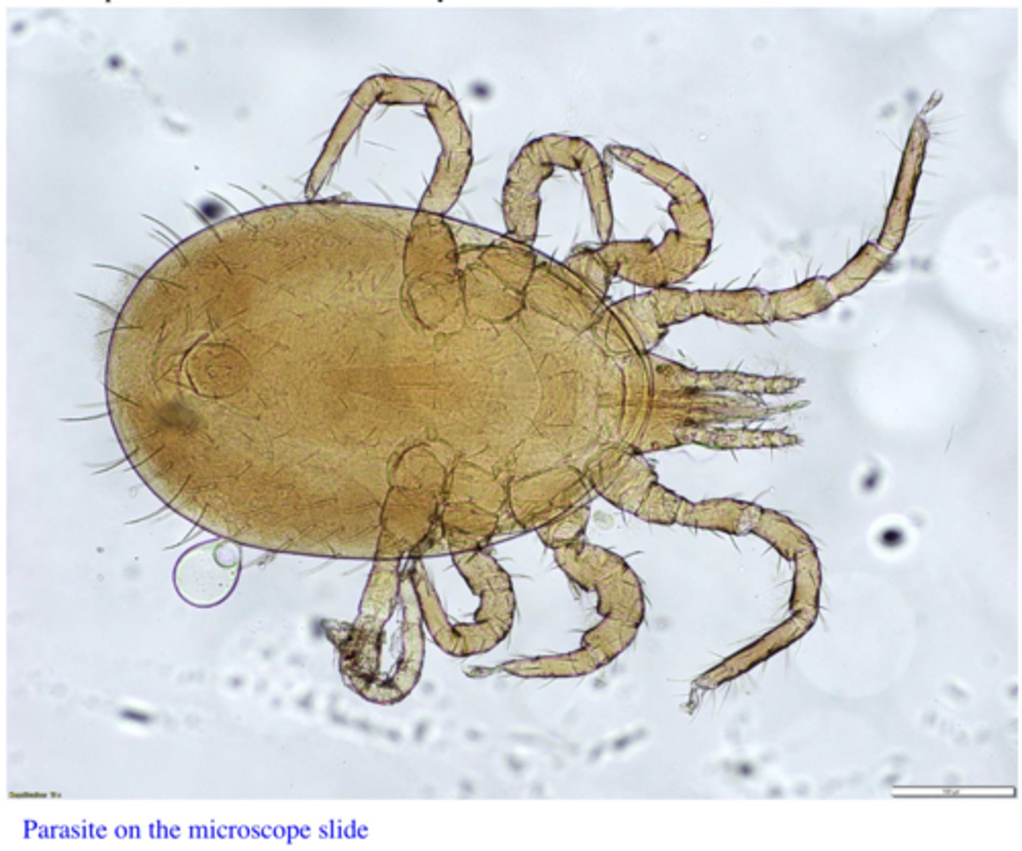
Ornithonyssus spp, Ornithonyssus bursa (Tropical fowl mite)
Size: 0.6-1 mm
Oval body
Colour: grey-whitish before feeding, dark red after feeding;
Anus located in the anterior half of the anal plate
Chelae developed
Dorsal shield tapers gradually to a blunt posterior end
Station 11:
Q1: Name The Parasite and describe the most important morphological features used for its identification
Blood
Station 11:
Q2: What Does this Parasite Feed on?
Direct contact and indirect Contact
Station 11:
Q3: How did these birds become infested with this parasite:
Synthetic pyrethroids
Check if they are approved for birds. Treat the birds and the environment (the mites spend time both on birds and in the environment)
Station 11:
Q4: How would you treat these birds?
Can Bite Humans
Station 11:
Q5: What is the significance of this parasite for human health?
Station 12:
Intense pruritus, hair loss, erythema, papules, nodules and crusts (Fig. 17) are seen in fattening pigs in a farm from Queensland. Scrapings of the areas with crusts, especially the ear crusts, reveal the parasites on the microscope slides 1 & 2. However, the parasite is not found in the skin scrapings from areas with papules or nodules.
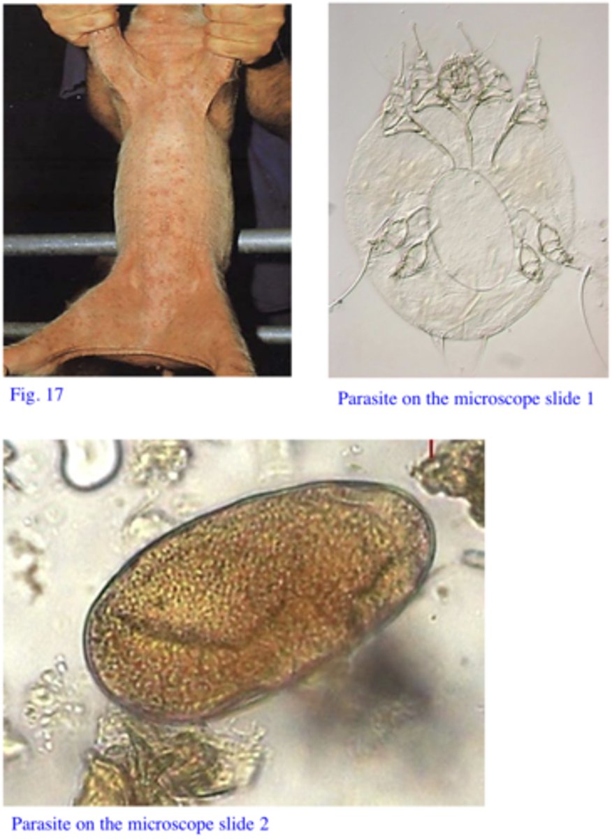
Sarcoptes scabiei var suis
Up to 450 μm, oval/circular body
Gnathosoma: square; short, conical legs
On the dorsal side of idiosoma: transversal striations, spines (3 anterior pairs and 7 posterior pairs) and triangular scales.
Station 12:
Q1: Name the parasite and tell how you identified it (most important morphological features).
The erythema, papules and nodules are the result of a hypersensitivity reaction to parasite faeces and salivary secretions.
Station 12:
Q2: How do you explain the negative skin scrapings from areas with papules or nodules?
Adult animals with chronic/hyperkeratotic lesions (ear, forehead, legs), especially breeding animals: sows and boars (large numbers of mites in the crusts);
Station 12:
Q3: What category of animals serves as the main source of infestation within a herd?
Direct contact (indirect possible but of less importance: Sarcoptes can survive to up to 3 weeks but it seems that it is infective only for few days)
Station 12:
Q4: How are these parasite usually transmitted?
1. S. scabiei var suis can temporarily infest humans (Pig handler's itch)
2. The parasite does not seem to reproduce/complete its life cycle on humans → clinical signsdisappear in 2-3 weeks
3. Constant exposure (pig handlers) → clinical signs last longer;
Station 12:
Q5: What is the significance of this parasite for human health?
Ivermectin (SC injection twice at 14 day intervals or in food)
Doramectin (SC/IM one injection);
Station 12:
Q6: What would you recommend the owner for the control of the disease (name two drugs, waysof administration, number of treatment etc)?
Station 13:
The nematode egg on the microscope plate was found in a faecal float of a sample collected from a group of poorly growing finisher pigs. Some of the pigs from the same group manifest dyspnoea and coughing
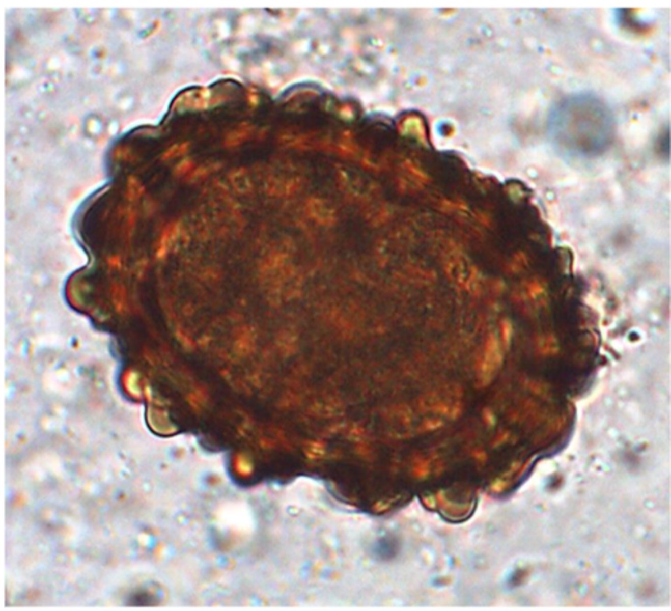
Ascaris suum
Thick shell, yellow brown, mammilated outer layer, one cell inside
Station 13:
Q1: Name the parasite and tell how you identified it (most important morphological features)
Ingestion of embrionated eggs (eggs that contain L3)
Station 13:
Q2: How did the pigs get infected with this parasite (stage and way of entry into the host)?
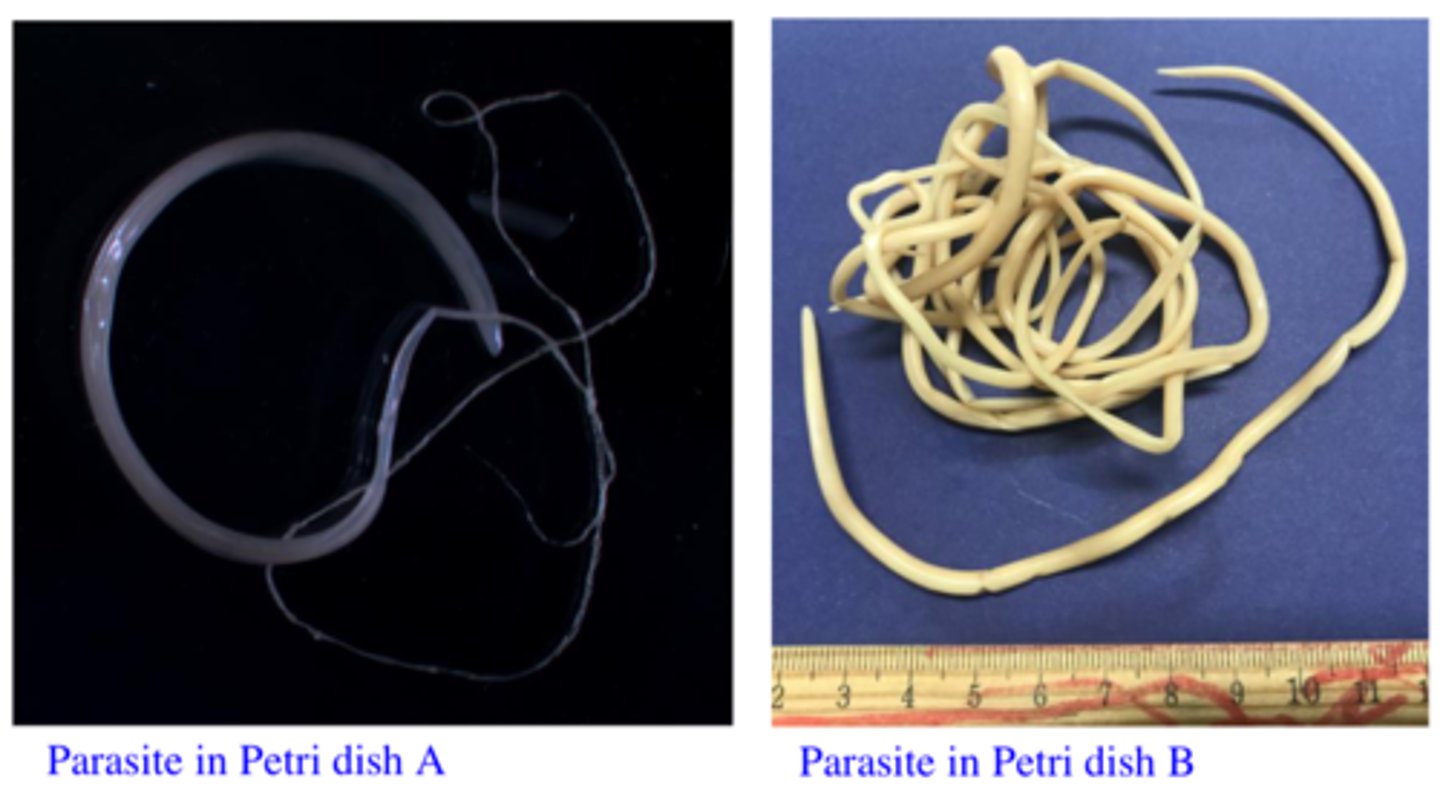
A: Trichuris suis: Small Intestine + Caecum
B. Ascaris suum: Small Intestine. Likely to Shed Eggs
Station 13:
Q3: Select from the parasites in Petri dishes A and B the one that is likely to shed eggs like this and say where it locates in the body of the host.
Because of larval migration through the lungs.
Station 13:
Q4: How do you explain the dyspnoea and coughing?
Milk spots are caused by larval migration through the liver
Station 13:
Q5: The owner of the farm is very upset because a large proportion of the livers from his pigswere condemned to the meat inspection because they presented with ‘milk spots’. Anyrelationship between this parasite and the ‘milk spots’ in the liver?
Eggs are very resistant in the environment - the can survive many years in the environment;The female parasite are very prolific
Station 13:
Q6: Why is this parasite causing problems each year if the piggery operates on an all-in-all-outsystem?
Piperazine – food/water
Levamisole – food/water, injectable
Fenbendazole – administered in food, effective but not registered in Australia
Ivermectin – administered in food or injectable
Doramectin - injectable
Station 13:
Q7: What would you recommend for the treatment of these pigs (2 drugs and ways ofadministration etc)?
Ivermectin – administered in food or injectable
Doramectin - injectable
Station 13:
Q8: The owner of the farm is telling you that some of the pigs suffer from mange and asks you torecommend some drugs that are active against both parasites. What drugs are you going torecommend him?
Can mature in the small intestine of humans (closely related to Ascaris lumbricoides the ascarid that is specific to humans) and can cause VLM
Station 13:
Q9: What is the significance of this parasite for human health?
Station 14:
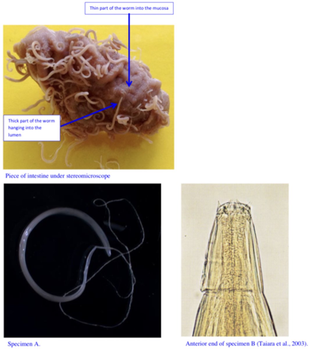
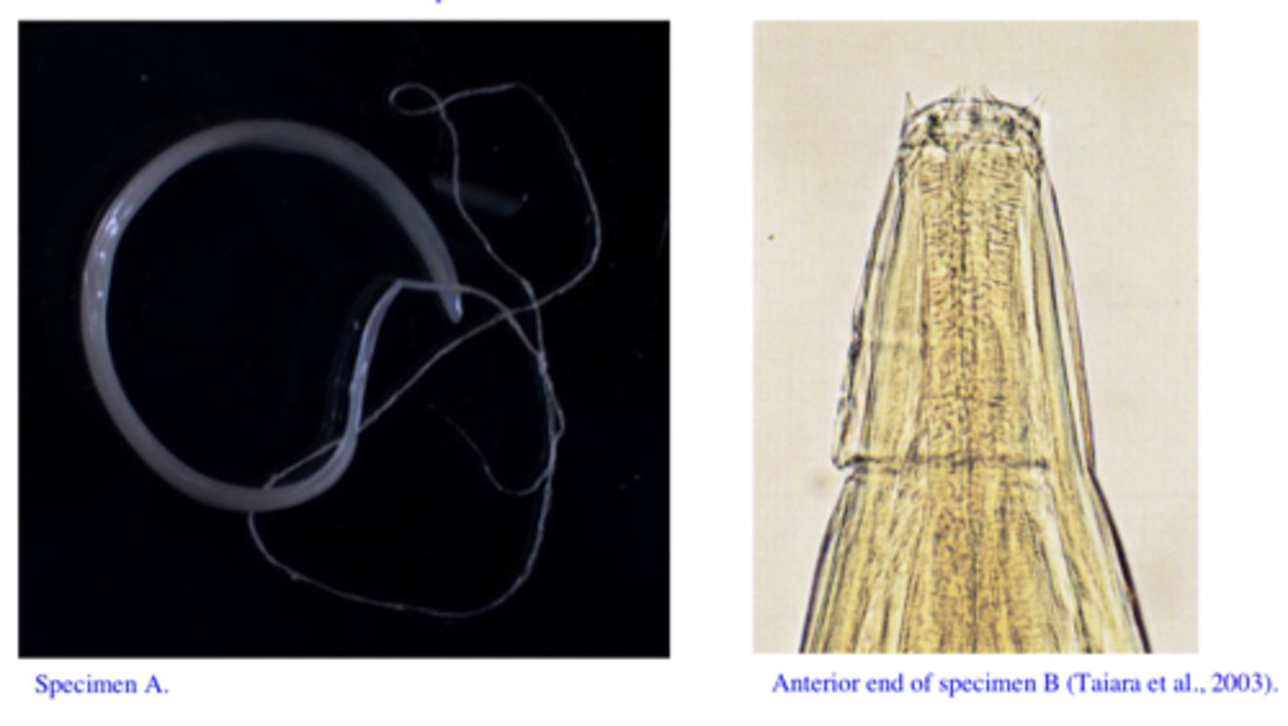
You are called to a free range piggery that has experienced mortalities in finisher pigs. Thepigs die after a short period of diarrhoea; in some pigs this is blood tinged. At necropsy, you can see a thickened, congested large bowel with focal haemorrhages (check the piece of the intestine in jar under the stereomicroscope) and on a closer examination you find two species of parasites (Specimens A and B).
A) Trichuris suis
B) Oesophagostomum spp.
Station 14:
Q1: Name the two different parasites and tell how you identified them (most important morphological features for each of them).
Trichuris suis
Adults of Oesophagostomum cause minimum damage to the mucosa of the large intestine (larvae can cause nodules – nodular worm).
Station 14:
Q2: Which of these two parasites is likely to be responsible for this condition?
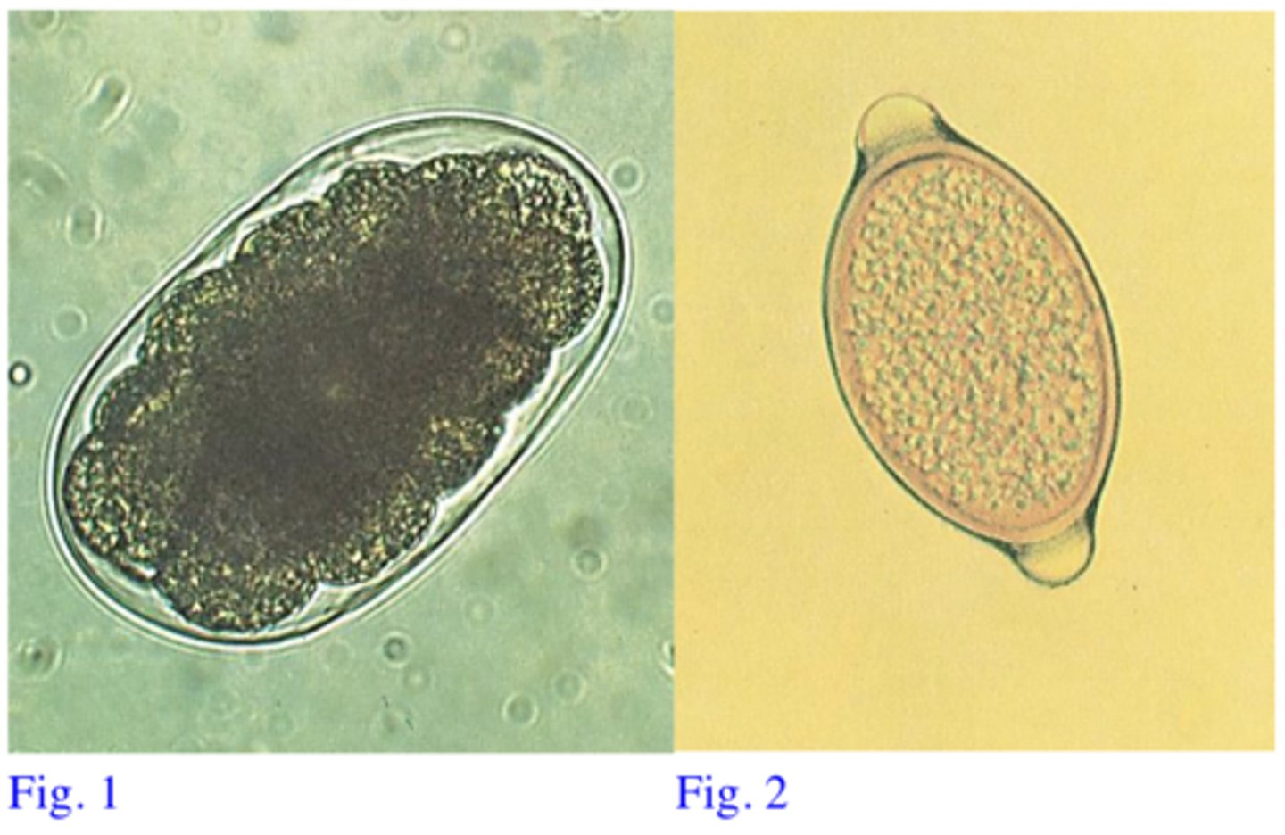
Fig 1: Thin shelled, morula stage: Oesophagostomum spp.
Fig 2: Lemon shaped, thick & smooth shell, yellow-brown, plugs at poles, one cell inside:Trichuris suis
Station 14:
Q3: Match the eggs shown in Fig. 1 and Fig 2 with each of the two parasites
Oesophagostomum: Ingestion of 3rd stage larvae for
Trichuris: Ingestion of embryonated eggs for
Station 14:
Q4: How did the pigs get infected with these parasites (stage, way of entry in the host)?
Benzimidazoles: Fenbendazole, Flubendazole (in food, effective but not registered inAustralia)
MLs: Doramectin (injectable)
Hygiene (eggs of Trichuris can survive long time in the environment)
Station 14:
Q5: How would you treat these pigs?
Female of Trichuris are intermitent egg layers, eggs do not float easily, clinical signs might be caused by larval stages;
False presence of eggs in the faeces (penmates are infected, passage of T. muris eggs etc)
Station 14:
Q6: Are there any problems with detection of the eggs in the faeces of infected animals?
Serpulina (Treponema) hyodysenteriae, Campylobacter and Pseudomonas aeruginosa) andBalantidium coli infections
Station 14:
Q7: Are there any other pathogens involved in the pathogenesis of the disease caused by this parasite?
Can infect humans but it is rare.
Station 14:
Q8: What is the significance of specimen A for humans?
Station 15:
You are enjoying a well deserved vacation in Europe and decide to visit one of yourcolleagues that is working as a vet with a pig farm. He is showing you some parasites that hehas found in the diaphragm muscles of one pig (Fig. 17) by compressor method (Fig. 18) (thesame parasite is on the microscope plate in a histological section (H&E staining).
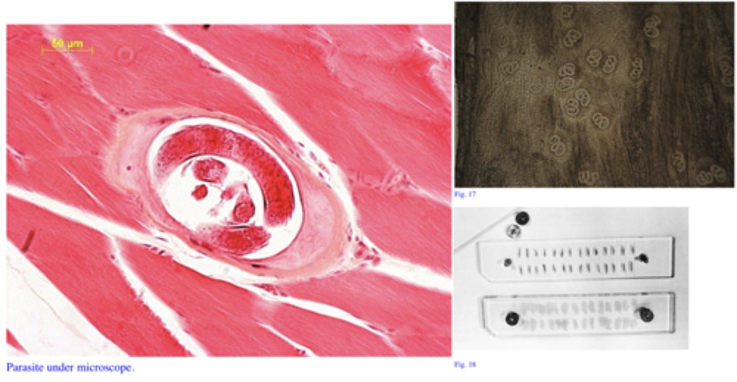
Trichinella spiralis
Station 15:
Q1: Name the parasite
Ingestion of infected rats, carcasses of infected pigs etcLarvae in the musculature of rats, pigs etc
Station 15:
Q2: How did the pig get infected (parasite stage, way of entry)?
Can infect humans and causes a severe disease in humans
Station 15:
Q3: What is the significance of this parasite for humans?