CHAPTER 17: COMPLEX ODONTOGENIC INFECTIONS
 Call Kai
 Learn
Learn Practice Test
 Spaced Repetition
 Match
Match Flashcards
 Knowt Play
1/65

Earn XP
Description and Tags
ppt
Name | Mastery | Learn | Test | Matching | Spaced | Call with Kai | Chat |
|---|
No analytics yet
Send a link to your students to track their progress
66 Terms
complex odontogenic infections
are severe dental infections that spread beyond local areas, involve deep fascial spaces, and may lead to potential systemic or life-threatening complications.
fascia
thin casing of connective tissue beneath the skin
surrounds and holds in place:
organs, bones
blood vessels
nerve fibers, muscles
deep spaces of the head and neck
fascia-lined spaces containing loose areolar connective tissue
these are “potential” spaces
only become actual spaces when invaded by bacteria
primary spaces
vestibular
canine / infraorbital
buccal
infratemporal
submandibular
submental
sublingual
secondary spaces
submasseteric / masseteric
pterygomandibular
superficial / deep temporal
parapharyngeal
retropharyngeal
danger
prevertebral
periorbital
vestibular space
most common site for spread of dental infections that break out of alveolar bone
location:
lies between cortical bone and mucoperiosteum
path of least resistance is determined by
thickness of bone in relation to the root apex
relationship of bone perforation to muscle attachments
its infection may spread to:
canine / infraorbital space → maxilla
space of the body of the mandible → mandible
canine / infraorbital space
infections from:
buccal roots of teeth or teeth with root apices positioned more buccally
usual pathway:
vestibular space → canine/infraorbital space
boundaries:
between levator anguli oris and levator labii superioris muscles
clinical findings:
swelling of vestibule, upper lip, lower eye lid
obliteration of the the nasolabial fold
clinical significance of canine / infraorbital space
most commonly affected by infections from the long root of the maxillary canine
occurs when infection from the mx canine apex perforates the alveolar bone:
superior to the attachment of the levator anguli oris
inferior to the origin of the levator labii superioris
abscesses often drain near the:
medial canthus
lateral canthus
may also develop by extension from an adjacent buccal space infection → may similarly spread directly into the buccal space
dental source of canine / infraorbital space / periorbital
mx canines or incisors
periorbital space
location:
lies between orbicularis oris and orbital septum
manifestation of infection:
redness and swelling of eyelid → obstruct vision
buccal space
location:
lies between buccinator and skin
location of the buccal pad
boundaries:
superficial: overlying skin and subcutaneous tissues
deep: buccinator muscle
manifestation of the infection:
marked cheek swelling
skin irregularities over the zygomatic arch
its infections commonly drain spontaneously through the skin at the inferior border of the mandible.
this produces an orocutaneous fistula / sinus tract.
If untreated, these relatively simple infections may spread to the deep fascial spaces of the neck, resulting in significant patient morbidity.
clinical findings of buccal space
there may be skin irregularities over the zygomatic arch bc:
fascial layers superficial to the arch are tightly bound to the bone
these layers become superficially edematous.
if infection has not spread to adjacent spaces:
zygomatic arch, inferior border of the mandible usually remains palpable.
dental source of buccal space
mx-mn premolars or mx molars
infratemporal space
a potential fascial space that may be involved in the spread of maxillary odontogenic infections
location:
posterior to the maxilla continuous with deep temporal space (so infections involving one may also involve the other)
boundaries:
medial: lateral pterygoid plate of the sphenoid bone
superior: base of the skull
important structures
branches of the internal maxillary artery
pterygoid venous plexus
clinical finding:
temporal fullness may be visible
difficult to examine because of its deep location.
clinical significance of infratemporal space
because veins of the head and neck lack valves, bacteria can propagate retrogradely.
pterygoid plexus
infection reaching the pterygoid plexus may travel directly to the cavernous sinus
the is the vein that pass through foramina in the skull base and connect with intracranial dural sinuses
due to its deep location, infratemporal space infections are difficult to adequately examine clinically.
temporal fullness may be visible.
dental source of infratemporal space
mx 3rd molars
mx sinusitis of odontogenic origin
periapical infections from maxillary teeth may:
erode superiorly → penetrate the floor of the maxillary sinus → maxillary sinusitis → potentially spread to neighboring sinuses
causes:
periapical osteitis
restorative materials
bone grafting materials
endodontic foreign bodies
retained tooth or bone fragments
iatrogenic, traumatic, implant-related
schneiderian membrane
any violation of this membrane may cause mx sinusitis
may occur through:
spread of a periapical infection
penetration by a dental implant
iatrogenic injury from sinus membrane elevation for bone graft placement
common clinical findings
facial pain, congestion
postnasal discharge
common bacteria associated with odontogenic mx sinusitis
anaerobic gram-negative
Streptococcus
Peptostreptococcus
Fusobacterium
aerobic gram-negative
Streptococcus
Staphylococcus
less common but more difficult to treat:
Aspergillus species
spread of odontogenic mx sinusitis
acute odontogenic sinusitis may:
propagate through the ethmoid sinus → spread to the periorbital space
preseptal cellulitis:
infection of the eyelid structures anterior to the orbital septum → progress to orbital cellulitis.
spread to cavernous sinus
infection can spread freely because the veins of the head and neck have no valves.
possible routes include:
infraorbital vein → infraorbital space
inferior ophthalmic vein
via the sinuses → common ophthalmic vein → superior orbital fissure → cavernous sinus
spread to the cavernous sinus can cause cavernous sinus thrombosis.
can be fatal even with optimal medical and surgical management.
tx:
open sinus surgery
functional endoscopic-assisted sinus surgery
perimandibular space
submental
ssublingual
submandibular spaces
described by Grodinsky and Holyoke

submental space
commonly affected by infections from mandibular incisor
it develops as an extension of a submandibular space infection, why?
the anterior belly of the digastric muscle is the main anterior boundary of the submandibular space.
it is not a true barrier between the:
submandibular space-submental space
spread across the midline:
infection from one submandibular space can pass through the submental space and reach the opposite submandibular space
dental source of submental space
mn incisor
sublingual space
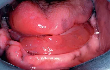
commonly the first deep fascial space involved in mandibular odontogenic infections
boundaries:
floor of the mouth, submucosa, mylohyoid muscle
clinical findings:
elevation of the tongue, floor of the mouth
difficulty with speech, swallowing
there should be no noticeable extraoral swelling bc the infection is limited to an area cephalad/superior to the mylohyoid muscle
note:
has no posterior boundary → freely communicates with the submandibular space
therefore, isolated sublingual infection without submandibular involvement is uncommon!
dental source of sublingual space
mn incisor, canine, premolar or 1st molar (roots cephalad/above mylohyoid muscle)
submandibular space
location:
caudal/below the mylohyoid muscle and superficial layer of deep cervical fascia
the only barriers between the abscess cavity and skin are:
platysma muscle
superficial layer of deep fascia (SLDF)
boundaries:
inferior border of the mandible
anterior & posterior belly of digastric
shape:
triangular space
clinical findings:
almost always presents with visible extraoral swelling (submandibular triangle)
note:
has no posterior boundary.
freely communicates with deeper neck spaces, including:
pterygomandibular space
lateral pharyngeal space
therefore, infection can cause significant morbidity.
dental source of submandibular space
occurs caudal/below to the mylohyoid muscle.
mn 2nd or 3rd molar (roots below mylohyoid muscle)
[unlike the sublingual space, it almost always presents with visible extraoral swelling]
determining the space of spread
infection perforating the lingual cortex cephalad/superior to the mylohyoid attachment:
→ sublingual space
infection perforating the lingual cortex inferior/caudal to the mylohyoid attachment:
→ submandibular space
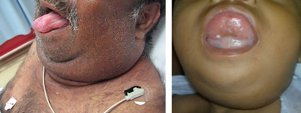
ludwig's angina
a life-threatening cellulitis involving ALL perimandibular spaces
technically involves 5 spaces:
2 sublingual, 2 submandibular, 1 submental
intraoral clinical findings:
elevation of tongue, floor of mouth
bilateral fluctuant swelling (inf. border of mn → hyoid bone)
blocks oropharynx compromises the airway
other clinical findings:
trismus, dysphagia, dysphonia
globus sensation, “hot potato” voice
inability to handle oral secretions
head held in forward “sniffing” position
tx:
hospitalization (surgical intervention with I&D)
aggressive antibiotic therapy (ampicillin-sulbactam)
airway management (tracheal intubation, tracheostomy)
masticator space
deep temporal
pterygomandibular
superficial temporal spaces
masseteric or submasseteric
mnemonic: TIME (temporalis, internal/medial pterygoid, masseter, external/lateral pterygoid)
submasseteric / masseteric space
boundaries:
masseter muscle
ascending ramus of the mandible
dental source:
impacted mn third molars
another common cause:
infected mandibular angle fracture
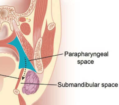
major pathway of spread in ludwig’s angina
submandibular space →
buccopharyngeal gap →
pterygomandibular space →
lateral pharyngeal space →
retropharyngeal space →
danger space →
mediastinum → mediastinitis
an infected mandibular angle fracture
another common cause of infections in submasseteric and masseteric space
superficial & deep temporal spaces
superficial temporal space
presents with lateral “hourglass” facial swelling
formed by the temporalis fascia and temporalis muscle
deep temporal space
formed by the temporalis muscle and calvarium
dental source of superficial & deep temporal spaces
mx second or third molar
pterygomandibular space
boundaries:
medial pterygoid muscle
ascending ramus of mandible
contains:
inferior alveolar nerve, inferior alveolar vessels
critical clinical feature:
trismus → due to involvement of the medial pterygoid muscle
high risk!
due to its close proximity to the lateral pharyngeal space
dental source of pterygomandibular & parapharyngeal space
mn 3rd molars due to pericoronitis
deep cervical fascial spaces of the neck
parapharyngeal
retropharyngeal
danger
prevertebral spaces
in order!
parapharyngeal
aka: lateral pharyngeal space
an inverted triangle-shaped space
location:
between masticator space anteriorly & retropharyngeal space posteriorly
possible routes of infection:
any extension posteriorly from the:
pterygomandibular space
sublingual space
submandibular space
very high risk!
due to its close proximity to the retropharyngeal space
dental source of retropharyngeal & danger
mn molars
manifestation of parapharyngeal space
intraoral:
edema pushes the superior constrictor medially → causes bulging of the lateral oropharyngeal wall toward the midline which is called pharyngeal draping
extraoral:
swelling of the lateral neck (inability to visualize and palpate the angle of the mandible)
[is dangerous bc it is close to the airway, carotid sheath, cranial nerves]
retropharyngeal space
location:
located behind the posterior pharyngeal wall
extends from the base of the skull to the mediastinum
boundaries:
anterior:
superior, middle, and inferior pharyngeal constrictor muscles
retropharyngeal fascia
posterior: alar fascia
superior: base of skull
inferior: usually between C6 and T4
contents:
loose areolar connective tissue, lymph node
very high risk!
acts as a “highway” for infection spread to danger space
complications:
erosion of the carotid artery wall
thrombosis of the internal jugular vein (IJV)
impingement on cranial nerves IX, X, and XII
IX – glossopharyngeal nerve
X – vagus nerve
XII – hypoglossal nerve
danger space
location:
between alar fascia anteriorly & prevertebral fascia posteriorly
boundaries:
anterior: alar fascia
posterior: prevertebral fascia
superior: skull base
inferior: diaphragm
extends from the base of the skull to the posterior mediastinum and diaphragm
extremely high risk!
acts as a “highway” for infection spread from the retropharyngeal space
infection in this space can spread far down toward the mediastinum
remember: lateral pharyngeal → retropharyngeal → danger → mediastinum
prevertebral space
rarely involved in odontogenic infections, why?
its fascia is tightly adherent to the vertebrae
a rare infection may occur with vertebral osteomyelitis
complications of prevertebral space
mediastinitis
neural compression
compression of the heart and lungs
rupture of esophagus, lungs, lower airway
interrupted cardiovascular and respiratory systems
primary goals of management of fascial space infections
medical optimization
airway protection
removal of the source of infection
surgical incision and drainage
adjunctive antibiotic therapy
frequent assessment of response to therapy
specific infections
candidiasis
actinomycosis
cavernous sinus thrombosis
cervical necrotizing fasciitis
osteomyelitis (acute-chronic suppurative, chronic sclerosing, garre’s)
cavernous sinus thrombosis
one of the most severe complications of a maxillary odontogenic infection
a rare, life-threatening condition where a blood clot (thrombus) forms in the cavernous sinus
usually a late complication of:
infection of the central face, paranasal sinus infections
possible route:
inferior ophthalmic vein → superior orbital fissure → cavernous sinus
other causes:
extraction of mx anterior teeth in the presence of an acute infection
tx:
mainstay is aggressive antibiotic administration
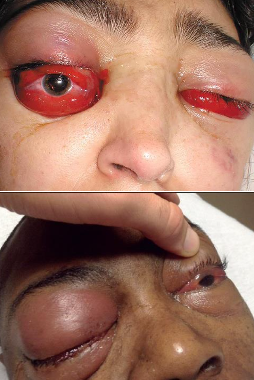
cavernous sinuses
are intracranial compartments that serve as bilateral venous drainage channels for the middle cranial fossa
contents of the cavernous sinus
internal carotid artery
cranial nerves II, III, IV, V2, VI
cervical necrotizing fasciitis
aka: “flesh-eating” bacterial infection
affects the investing layer of the deep cervical fascia, deep to the platysma muscle
a soft tissue infection characterized by widespread tissue death involving:
necrosis of the platysma, skin, underlying soft tissues
note:
does NOT follow the normal fascial planes of the head and neck
polymicrobial, extremely destructive, potentially fatal
tx:
removal of all affected/nonviable tissues, source of infection
aggressive surgical debridement
empiric broad bacteriacidal IV antibiotics
medical optimization: surgical airway, ICU monitoring
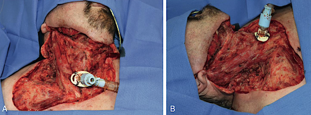
osteomyelitis
inflammation of bone, predominant bacteria → staphylococcus
more common in the mandible, why?
mandible → thicker cortex
blood supply mainly from:
inferior alveolar artery, periosteum
maxilla → thinner cortex
periosteal vessels penetrate the cortex more easily.
maxilla has more porous bone.
how it develops?
begins in the medullary spaces of the jaw
bacteria enter the marrow spaces → marrow edema.
bc the marrow is enclosed by cortical bone → pressure increases.
if pressure becomes greater than the pressure in the feeding arteries:
blood supply decreases, tissue necrosis occur → pain develops
failure of microcirculation prevents delivery of:
oxygen, nutrients, blood-borne immune defenses → allows bacteria to proliferate and spread.
risk factors:
immunocompromised conditions, poorly controlled diabetes mellitus
chronic corticosteroid use, chronic drug abuse, chronic malnutrition
tx:
empiric antibiotic of choice: penicillin
others: fluoroquinolones, clindamycin
general types:
acute / chronic suppurative osteomyelitis
chronic sclerosing osteomyelitis
osteomyelitis with proliferative periostitis (Garre osteomyelitis)

acute suppurative osteomyelitis
infection of the medullary bone with purulence
associated with:
bacteremia, fracture, surgery
extension of periapical abscess
osteo-radio-necrosis (ORN)
medication-related osteo-necrosis of the jaw (MRONJ)
clinical findings:
pain → primary feature
usually no systemic manifestations
lymphadenopathy, edema, erythema, pain
lower lip paresthesia, restricted movement
radiographic findings:
early stage:
no changes bc there is minimal bone loss
later stage:
moth-eaten appearance → radiolucent
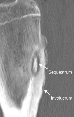
sequestrum → radiopaque within radiolucency
involucrum → surrounding radiolucent
tx:
debridement of necrotic bone
aggressive antibiotic therapy
drainage of pus
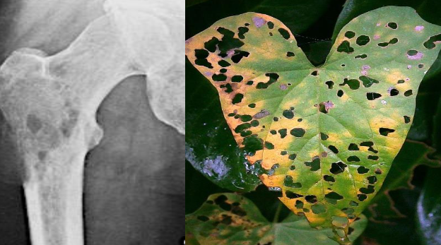
sequestrum
radiopaque within radiolucent
a dead bone surrounded by granulation tissues
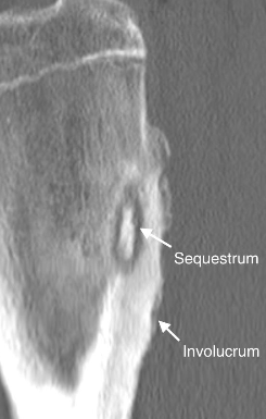
involucrum
reactive new bone formation
surrounding radiolucent area
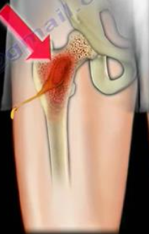
cloaca
the draining sinus
chronic suppurative osteomyelitis
treated in a manner similar to the acute form with removal of the source of the infection
causes:
a sequela of acute osteomyelitis
may represent a long-term, low-grade inflammatory reaction
tx:
culture and sensitivity testing of bone biopsy
aggressive debridement of necrotic bone
may involve large segments of the jaws
high-dose IV antibiotics (6 weeks of outpatient IV antibiotics)
chronic, unresponsive cases may require 6 months or more of IV antibiotics.
why in need of longer treatment?
bc antibiotic penetration into bone is difficult and eliminating bacterial colonization is more challenging.
diffuse chronic sclerosing osteomyelitis (non-suppurative)
causes: bacteria, chronic periodontal disease
a low-grade inflammatory reaction in the maxilla and mandible
chronic sclerosing osteomyelitis (non-suppurative)
aka: focal sclerosing osteitis, bony scar, condensing osteitis
rare form of osteomyelitis.common to young adults
usually seen at the apex of a tooth with long-standing, low-grade pulpitis
bacterias:
Actinomyces & Eikenella corrodens → can cause sclerosis & fibrosis
clinical finding:
chronic dull pain, intense pain may fluctuate with:
acute exacerbations, mandibular expansion, soft tissue edema
no: pus, drainage
symptoms can persist for up to 5 years before diagnosis
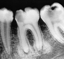
radiographic finding:
increased trabecular bone density → increased radiopacity
tx:
no treatment (physiologic bone reaction)
surgical resection of diseased bone is often required.
osteomyelitis with proliferative periostitis (non-suppurative)
aka: garre’s osteomyelitis
a chronic disease that usually affects children
due to their increased vascularity and regenerative capabilities
causes:
periapical infection of a mandibular tooth
clinical findings:
mandibular expansion, pain
no: purulence, drainage, erythema
radiographic findings:
onion-skin appearance → due to paracortical bone formation from repetitive irritation of the periosteum
note:
sometimes called periostitis ossificans, but this is considered inappropriate → bc the periosteum itself does not become ossified
tx:
removal of the offending infectious source
short course of antibiotic therapy (penicillin, tetracycline, clindamycin)
long-term antibiotic therapy is not indicated.

actinomycosis
a chronic but relatively uncommon infectious process affecting the maxillofacial skeleton
usually affects soft tissue, but can also involve bone → Actinomyces osteomyelitis
clinical findings:
forms a lumpy pseudotumor
induration with nodular fibrosis
intermittent spontaneous drainage tracts
forms a fistula to the cutaneous skin surface
infection does not respect normal anatomical planes.
presence of sulfur granules (small yellow pus)
because it drains spontaneously:
patient often does not experience severe pain.[
bacteria:
Actinomyces israelii
A. naeslundii, A. odontolyticus
A. meyeri, A. viscosus
tx:
surgical removal of the infection
long-term oral penicillin therapy (IV penicillin if acute)
if allergic to penicillin:
doxycycline, clindamycin

actinomyces israelii (anaerobic bacteria)
causative agent of actinomycosis
this organism usually causes no pathologic signs, it must seed itself in an area of susceptibility in order to flourish such as:
injured site
area of fracture
tooth extraction site
other traumatic area
candidiasis
most common fungal disease evaluated by dentists
diagnosis is done thru:
20% potassium hydroxide (KOH) → unreliable, no tissue invasion
microscopy may reveal:
hyphae, pseudohyphae
tissue biopsy + periodic acid–schiff (PAS) staining → more reliable; there’s tissue invasion
risk factors:
immunocompromised states:
AIDS,diabetes mellitus, chemotherapy, leukemia
species:
candida albicans → an endogenous organism normally present in the mouth
others:
C. krusei, C. tropicalis, C. glabrata
C. parapsilosis, C. pseudotropicalis, C. guilliermondii
tx:
elimination of contributing local or systemic factors
topical therapies such as:
nystatin oral suspension
clotrimazole troches → higher toxicity, contains sugar
3 most common forms of candidiasis
pseudomembranous candidiasis
erythematous candidiasis
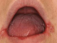
angular cheilitis
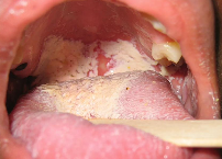
pseudomembranous candidiasis
distinct white patches that can be wiped off easily exposing an underlying erythematous surface
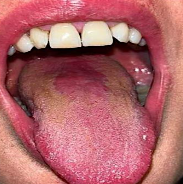
erythematous candidiasis
appears to be a raw surface, such as that seen with the loss of filiform papillae of the tongue
angular cheilitis
aka: perleche
associated with;
px with dentures with inadequate vertical dimension & “freeway” space
reduced vertical dimension → overclosure of lips → chronic moisture
clinical findings:
erythema, white patches at the commissures often bilateral
often accompanied by Staphylococcus aureus infection.
nutritional deficiencies:
folic acid, iron, riboflavin, thiamine, vit. B12
tx:
eliminate contributing factors
nystatin oral suspension