5. Cell Injury: Cellular Accumulations
1/141
There's no tags or description
Looks like no tags are added yet.
Name | Mastery | Learn | Test | Matching | Spaced | Call with Kai |
|---|
No analytics yet
Send a link to your students to track their progress
142 Terms
excessive production, or normal amount, of substance that is accumulating faster than can get rid of
accumulations
Why can’t the body get rid of the accumulations?
due to problems metabolizing, packaging, and excreting (genetic or acquired disabilities)
What are common accumulations? What is an example?
indigestible exogenous substance; carbon
accumulations of a substance within the cell
intracellular accumulations
What are examples of intracellular accumulations?
L
G
P
V
C
L
lipid
glycogen
protein absorption
viral inclusions
crystalline protein inclusions
lead poisoning produced intranuclear inclusions
accumulations that occur outside the cell in the interstitium
extracellular accumulations
What are examples of extracellular accumulations?
A
G
C
F
F
amyloidosis
gout
cholesterol
fibrosis
fatty infiltration
True or false: Some of the accumulation substances are harmless while others, when present in enough quantities, can lead to cell injury and death.
true
What are causes of accumulations?
M
G
I
metabolic abnormalities which result in excessive production of a substance
genetic abnormalities which lead to excessive storage or inability to break down or release a substance (lysosomal storage disease)
indigestible substance where the cell lacks machinery to break down material or transport out of a cell (carbon)
accumulation of lipid within parenchymal cells, which can develop in many organs and tissues
steatosis
Where can you see steatosis most notably? Why?
the liver it is extremely important in lipid metabolism
What are causes of hepatic lipidosis?
I
A
I
increased mobilization of free fatty acids
abnormal hepatocellular metabolism of lipids
impaired release of lipoproteins
Grossly, lipid accumulation in the liver will be visualized as a ________ liver with ________ edges, which is often ________/________-________ with an accentuated ________ pattern. It is also ________ to the touch. This type of liver will also frequently ________ in formalin rather than ________ like a normal liver would.
swollen; rounded; yellow/yellow-brown; centrolobar; greasy; float; sink
True or false: Histologically, lipid accumulation in the liver shows hepatocytes that have one or more colorless punctate cytoplasmic vacuoles which can displace the nucleus and result in degeneration and necrosis if severe enough. The lipid vacuoles are colorless and punctate because the lipids have leached from the cell in the process of processing.
true
What histochemical stains confirm lipid?
Sudan black or Oil-Red-O
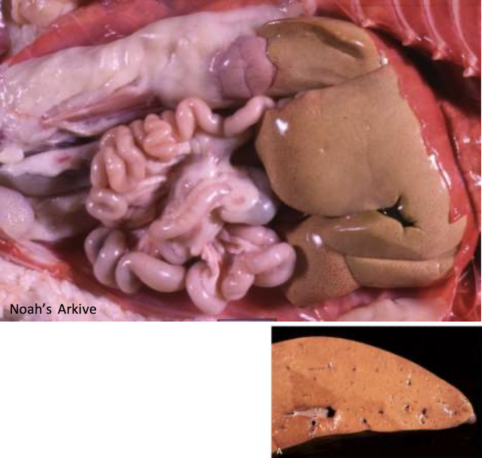
What does this liver show?
lipid accumulation
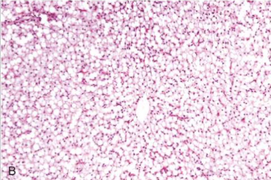
What is this showing of a histologic slide of the liver?
lipid accumulation
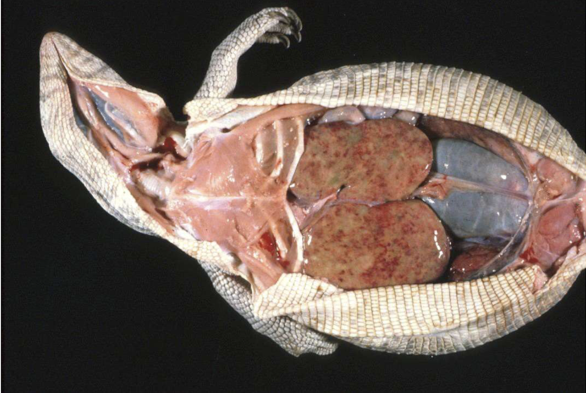
What disease is this monitor lizard suffering from?
hepatic lipidosis
Where is glycogen stored in homeostasis?
liver and skeletal muscle cells
What happens to glycogen stores in sick and starving animals?
they are depleted
When do you often see glycogen accumulations?
in the same sites, such as with storage disease, or alternatively with certain endocrine diseases, such as canine hyperadrenocorticism or diabetes mellitus
Grossly, how will the liver appear with glycogen accumulations?
enlarged with rounded edges, pale/light brown, and mottled in appearance
How will the liver appear on histopathology with glycogen accumulations?
hepatocytes are enlarged and swollen with intracytoplasmic fine, lacy vacuolations
Since cell swelling (hydropic degeneration) can appear the same in hepatocytes, what can pathologysts do to differentiate between cell swelling and glycogen accumulation?
can apply special stains
What stain is used to determine glycogen accumulations?
PAS
To differentiate between cell swelling and glycogen accumulations, 2 slides are stained with ________ and one is ________ treated. In the ________ slide, we expect to see positive ________, which confirms ________ accumulation. If the pink is not present in the ________ treated slide, then that confirms ________.
PAS; diastase; PAS; cytoplasm; carbohydrate; diastase; glycogen

What is this an example of?
glycogen accumulations
Lead poisoning produced intranuclear inclusions, which are a mixture of lead and proteins, are ________ and ________. They stain with ________ ________ stains.
pale; eosinophilic; acid fast
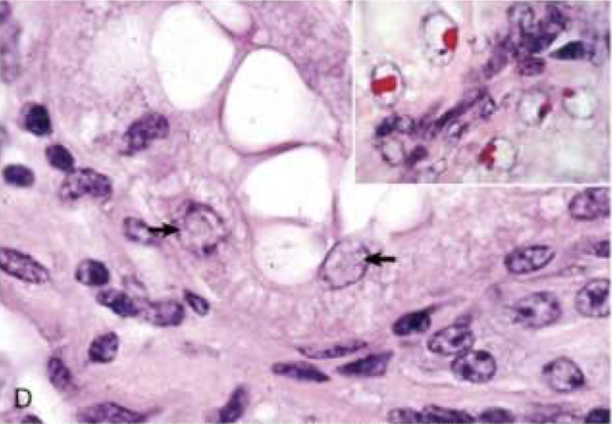
What is this an image of?
lead poisoning produced intranuclear inclusions
rhomboidal inclusions seen in the renal and hepatocyte epithelial cells in dogs that are often seen in increased frequency in aged dogs and their significance is unknown
crystalline protein inclusions
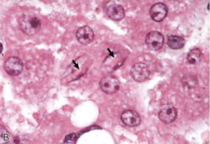
What is this image showing?
crystalline protein inclusions
How do protein often stain in H & E stained tissue sections?
pink to orange
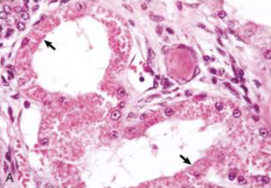
What is this an example of?
protein absorption
Viral inclusion can be ________ or ________ depending on the viral agent.
intranuclear; cytoplasmic
True or false: Some viruses (herpes, adenoviruses, parvoviruses) produce exclusively intranuclear inclusions. Other viruses produce cytoplasmic inclusions (pox viruses, rabies). A few viruses, such as canine distemper, produce both intranuclear and intracytoplasmic inclusions. Inclusions can range from round and eosinophilic to basophilic or amphophilic depending on the viral agent.
true
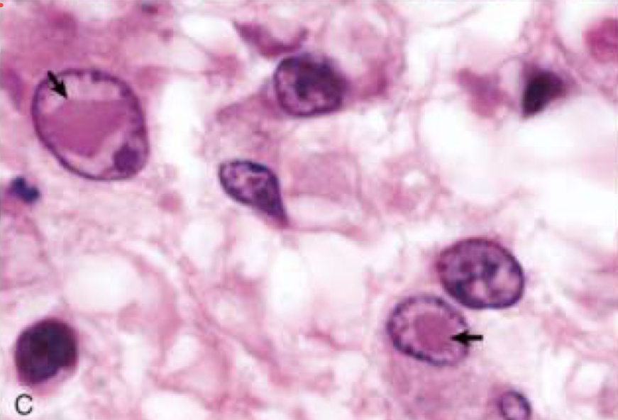
What is this image showing?
viral inclusions
biochemically diverse group of disorders with a common pathogenesis and is a protein misfolding disorder converting them into insoluble non-functional aggregates (rich in beta pleated sheet)
amyloidosis
In what dog breed is amyloidosis common in?
shar pei
Where does amyloid frequently deposit?
B
B
S
blood vessels
basement membrane (liver and kidney)
spleen
Mechanisms of Amyloidosis:
Propagation of ________ proteins, which serve as a ________ for self replication.
Accumulation of ________ ________ ________ with failure to degrade them.
Genetic ________ which promote protein misfolding.
Protein ________ because of abnormality or ________ in synthesizing cells.
Loss of ________ molecules or other essential components of the protein assembly process.
misfolded; template
misfolded precursor peptides
mutations
overproduction; proliferation
chaperoning
What are the types of amyloidosis?
AL amyloid
AA amyloid
hereditary amyloidosis
B-amyloid
secreted in B cell proliferative disorders (plasma cell tumors, multiple myeloma)
AL amyloid
What amyloid does the liver secrete during inflammation?
SAA
What is the AA amyloid synthesized from?
SAA
True or false: Hereditary amyloidosis is not induced by chronic inflammation.
true
In what breeds in hereditary amyloidosis common?
shar pei
abyssinian cats
amyloid present in dogs with cognitive dysfunction and Alzheimer’s disease
B- amyloid
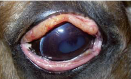
What is this image showing?
amyloidosis
Grossly, what does amyloid do to an organ?
enlarges it, gives it a pale red/tan color, and rounded edges and would feel waxy to the touch
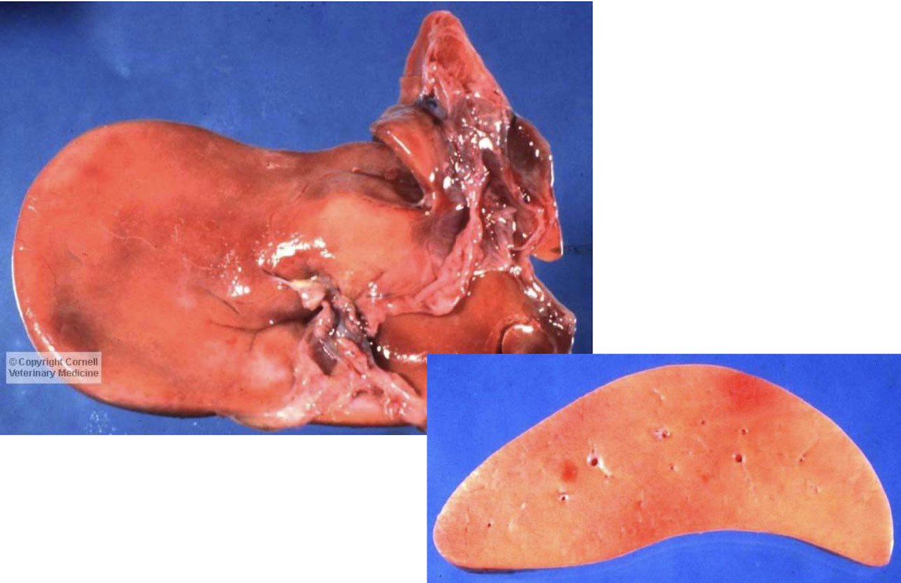
What does this liver have?
amyloidosis
How do you diganose amyloidosis?
I
S
iodine stain on fresh tissue
special stain used on formalin fixed tissue
What is the special stain used with amyloidosis?
congo red
True or false: An organ that appears to have yellow waxy deposits can be stained grossly with iodine. Amyloid is not pure protein and has carbohydrate moities which the iodine binds to.
true
How will amyloid stain with congo red?
bright red
What is significant and unique to amyloid and staining with congo red?
amyloid with congo red stain is the only thing that fluoresces bright green under polarized light

What is this showing?
amyloidosis
deposition of sodium urate crystals (tophi) in tissues extracellularly
gout
What is gout common in?
birds and reptiles
What is gout due to?
D
R
E
dehydration
renal failure
excess protein intake
What are the 2 types of gout?
visceral
articular
chalky white deposits on the surface of viscera
visceral gout
chalky white deposits within and overlying joints
articular gout
Grossly, how does gout appear?
chalky white deposits and feels gritty
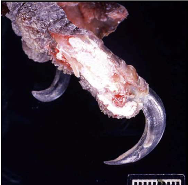
What is this macaw suffering from?
gout
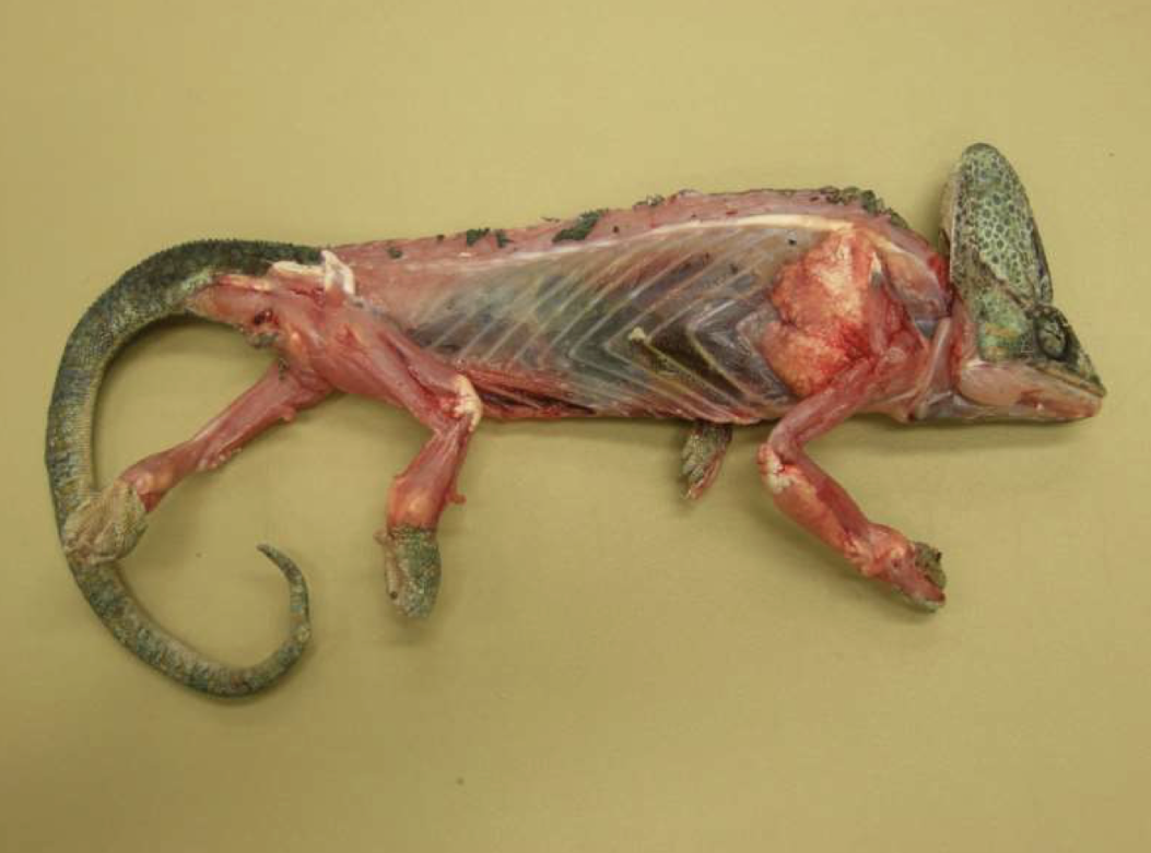
What is this chameleon suffering from?
articular gout
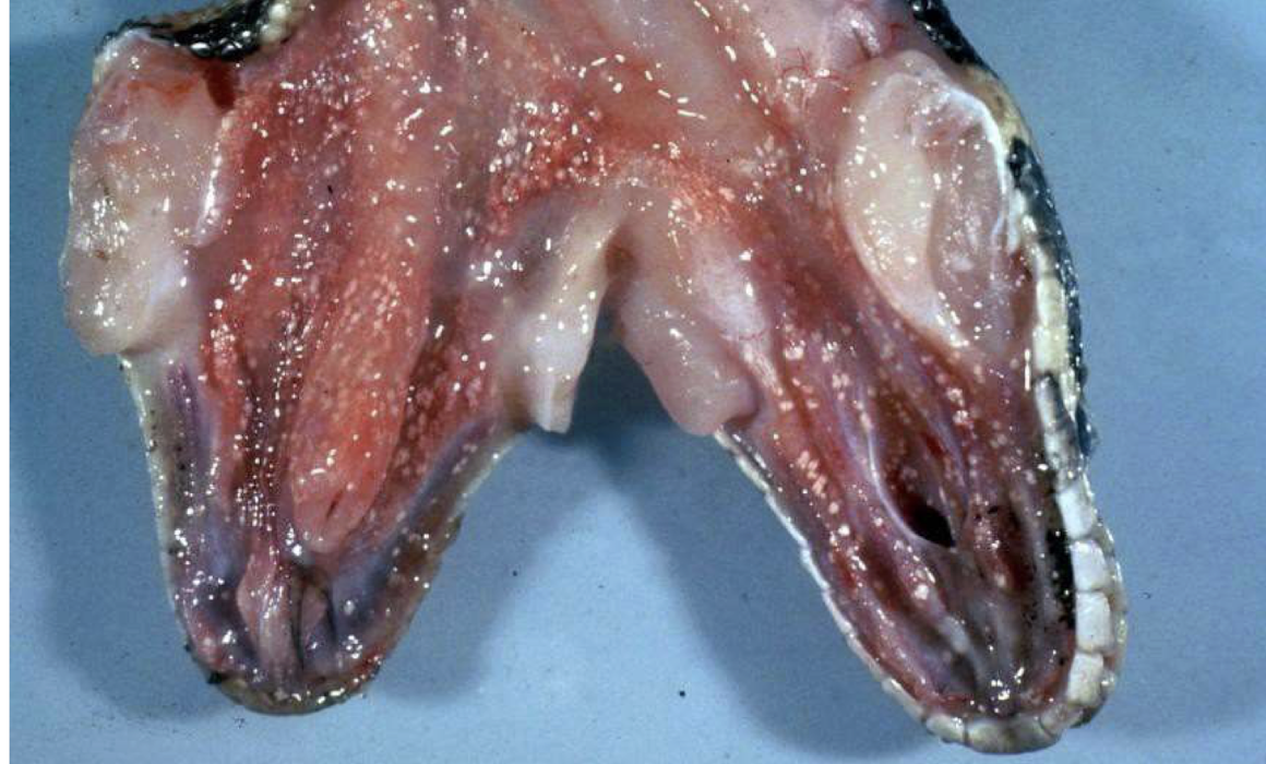
What is this snake suffering from?
visceral gout
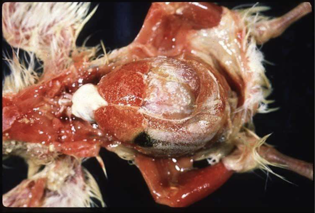
What is this chicken suffering from?
visceral gout
Where do cholesterol crystals form at? What do they typically illicit?
sites of necrosis; granulomatous inflammation
What happens to cholesterol crystal in the histologic processing? What does this mean?
they dissolve; not seeing the actual crystal, just seeing where it was
How do cholesterol crystal appear on histology?
needle shaped clefts
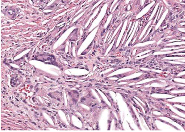
What is this an image of?
cholesterol accummulations
excessive fibrosis with proliferation of small vessels termed neovascularization
granulation tissue
collagen deposition, predominantly type I collagen in the interstitium of organs or tissues
fibrosis
What is fibrosis generally a result of? What is this a part of?
necrosis or inflammation; healing
In general, if there is excessive fibrosis, what can this do to organ function?
can impair organ function
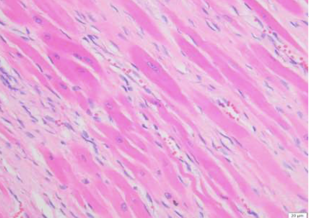
What is this showing?
fibrosis
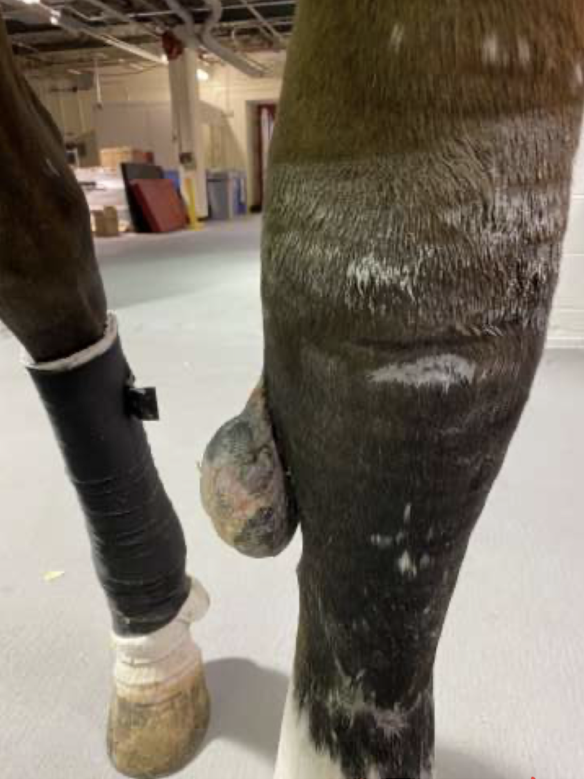
What is this horse suffering from?
exuberant granulation tissue/proud flesh/fibrosis
increase in the number of adipocytes in an organ or tissue
fatty infiltration
Why can fatty infiltration occur?
O
C
obesity
certain mardiomyopathies
What are the types of pathologic calcification?
D
M
dystrophic
metastatic
What does dystrophic calcification result from? What occurs?
dying cells (necrosis); intracellular calcium is released from sequestered places within the cell or into the extracellular space
What does metastatic calcification result from? What is this?
hypercalcemia; calcium/phosphorus imbalance
What can the hypercalcemia be due to?
V
H
C
I
C
vitamin D toxicosis
hyperparathyroidism
chronic kidney disease
iatrogenic supplementation
certain neoplasias
What does metastatic calcification target?
tunica intima and media of blood vessels, particularly in the lungs, pleural surface, kidneys, and stomach
True or false: Both types of calcification look the same.
true
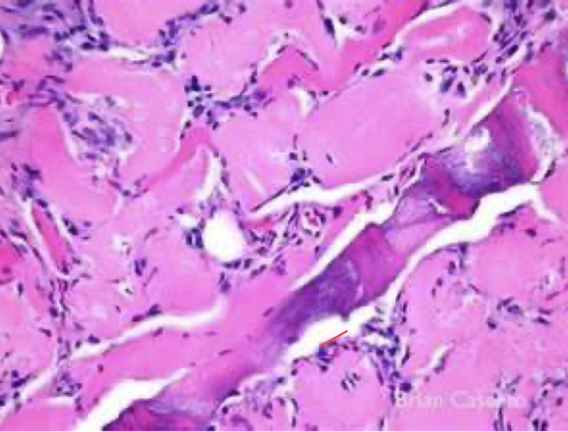
What is this showing?
myocyte degeneration, necrosis, and mineralization
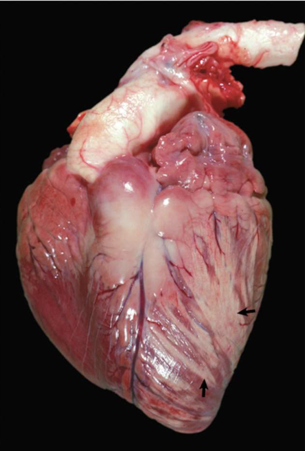
What does this show?
chalky white lesions within the myocardium of the heart of this calf due to dystrophic mineralization due to an underlying mineral deficiency (vitamin E/selenium deficiency) → myocyte degeneration, necrosis, and mineralization
brown black intracellular pigment produced by melanocytes
melanin
congenital lack of pigmentation
albinism
What occurs in multiple diseases conditions?
depigmentation
What are two diseases that lead to depigmentation?
copper deficiency
partial albinism in Chediak-Higashi syndrome
What are symptoms of copper deficiency?
fading of coat (leukotrichia), wiry hair coat, hair loss
LYST mutation affection lysosomal trafficking
Chediak-Higashi syndrome
When does hyperpigmentation occur?
chronic inflammatory states or neoplasia
non-pathologic process that can happen in any organ
congenital melanosis
In what species is congenital melanosis common?
S
C
C
sheep (black faced)
cattle
chow chows
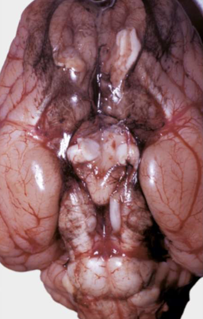
What is this an image of?
congenital melanosis
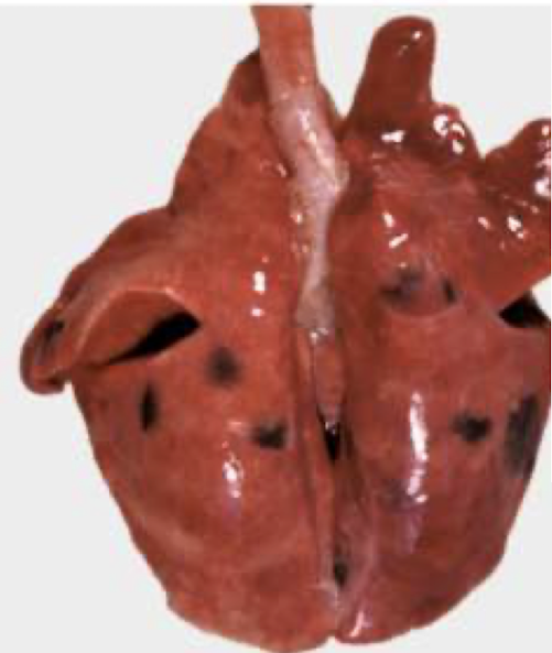
What is this an image of?
congenital melanosis
lipoprotein which accumulates in long lived post mitotic cells (neurons, cardiac myocytes) seen in greater quantities in aged animals and is an intracellular wear and tear pigment that can be age associated
lipofuscin
Where is lipofuscin common?
liver, heart, and CNS