MCAT: Biology: Neurobiology
1/99
There's no tags or description
Looks like no tags are added yet.
Name | Mastery | Learn | Test | Matching | Spaced |
|---|
No study sessions yet.
100 Terms
dendrites
a neuron's bushy, branching extensions that receive messages and conduct impulses toward the cell body
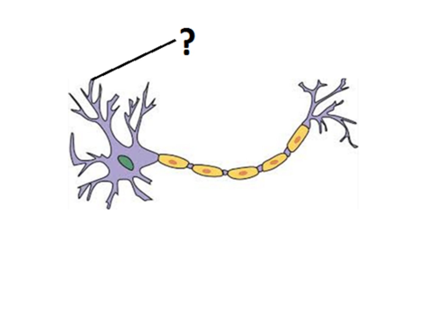
soma
cell body of a neuron
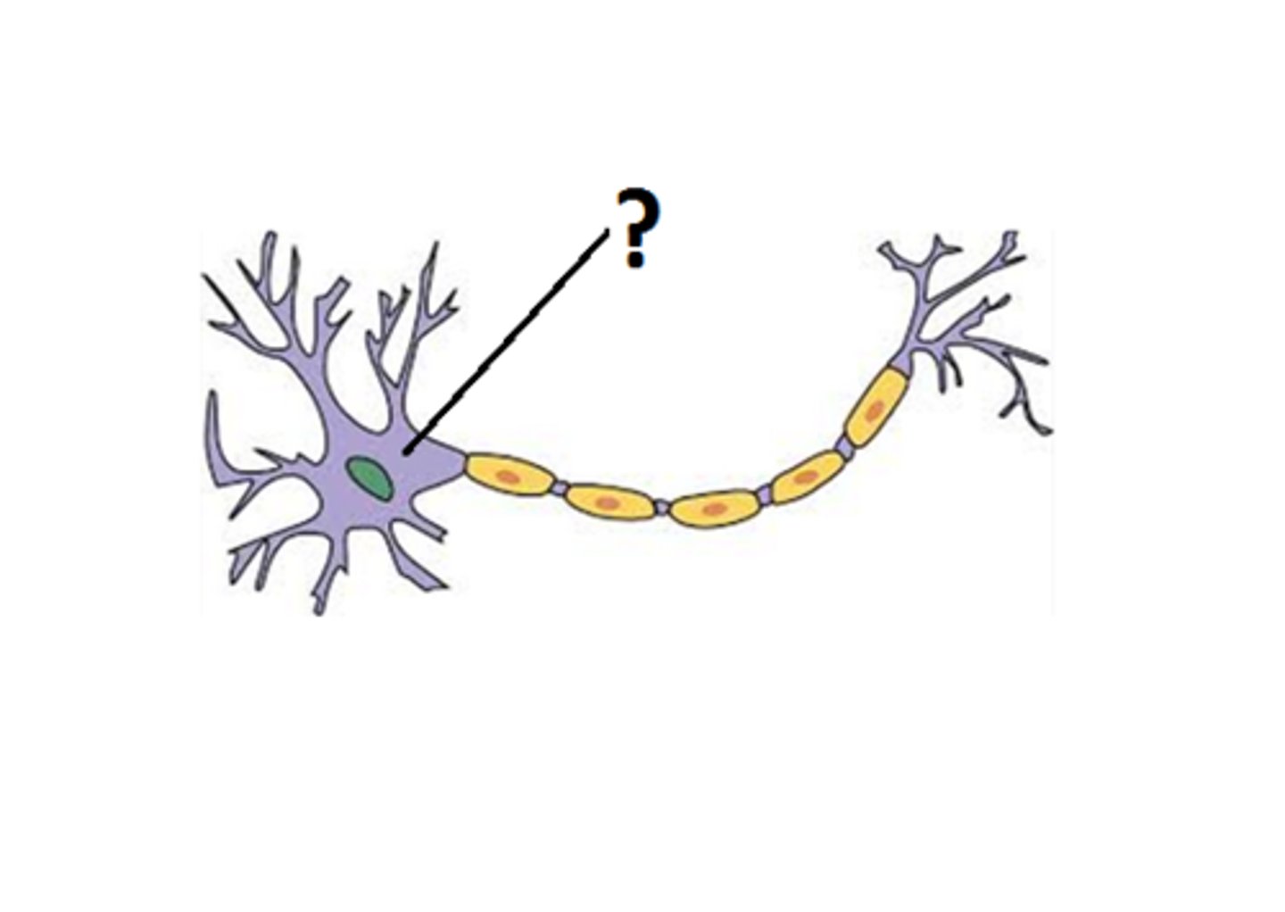
axon hillock
The conical region of a neuron's axon where it joins the cell body; typically, the region where nerve signals are generated.
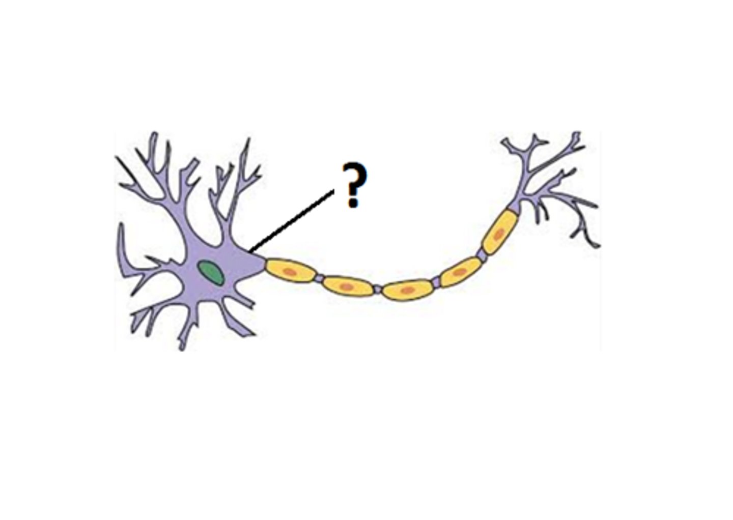
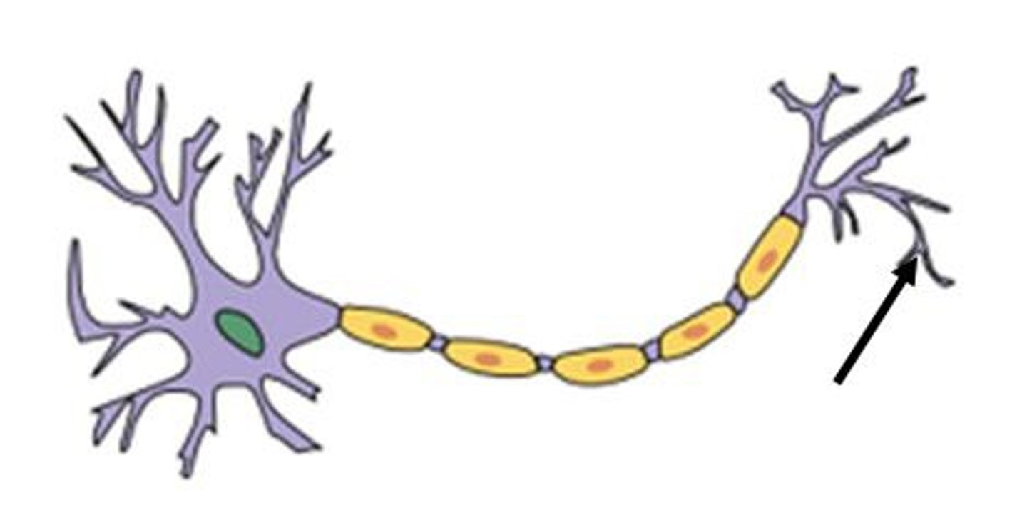
axon
the extension of a neuron, ending in branching terminal fibers, through which messages pass to other neurons or to muscles or glands
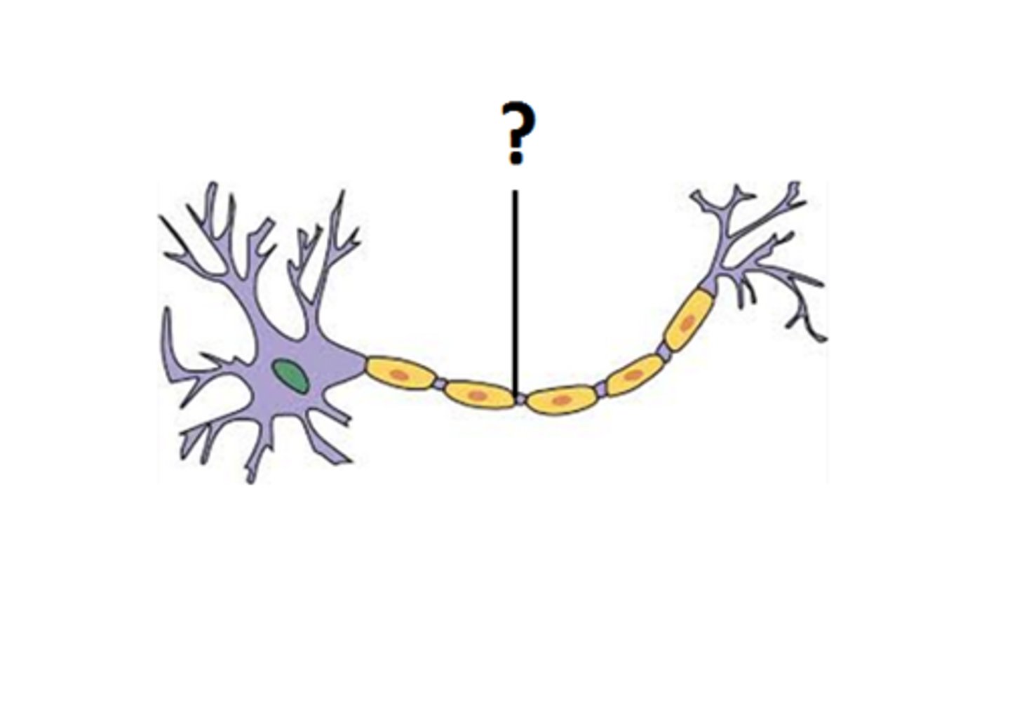
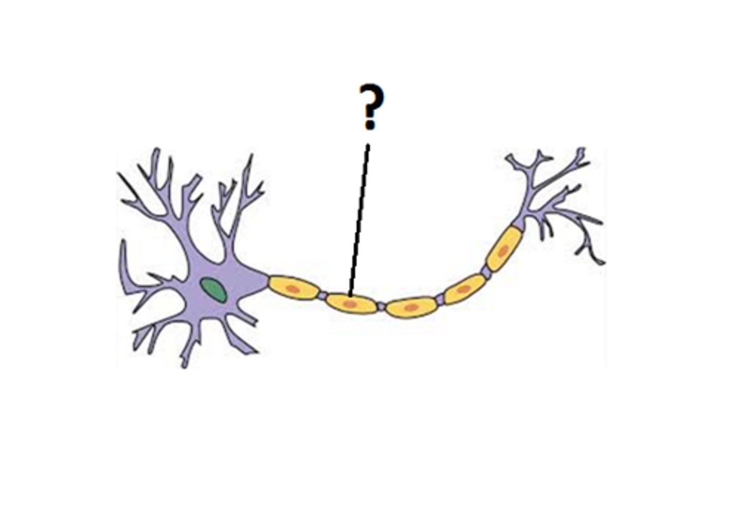
nodes of ranvier
Gaps in the myelin sheath to which voltage-gated sodium channels are confined.

myelin
A layer of fatty tissue segmentally encasing the fibers of many neurons enables vastly greater transmission speed of neural impulses as the impulse hops from one node to the next.
axon terminus
End of axon where neurotransmitters are released.
saltatory conduction
Rapid transmission of a nerve impulse along an axon, resulting from the action potential jumping from one node of Ranvier to another, skipping the myelin-sheathed regions of membrane.
Schwann cells
Supporting cells of the peripheral nervous system are responsible for the formation of myelin.
oligodendrocyte
a type of glial cell that forms myelin in the central nervous system
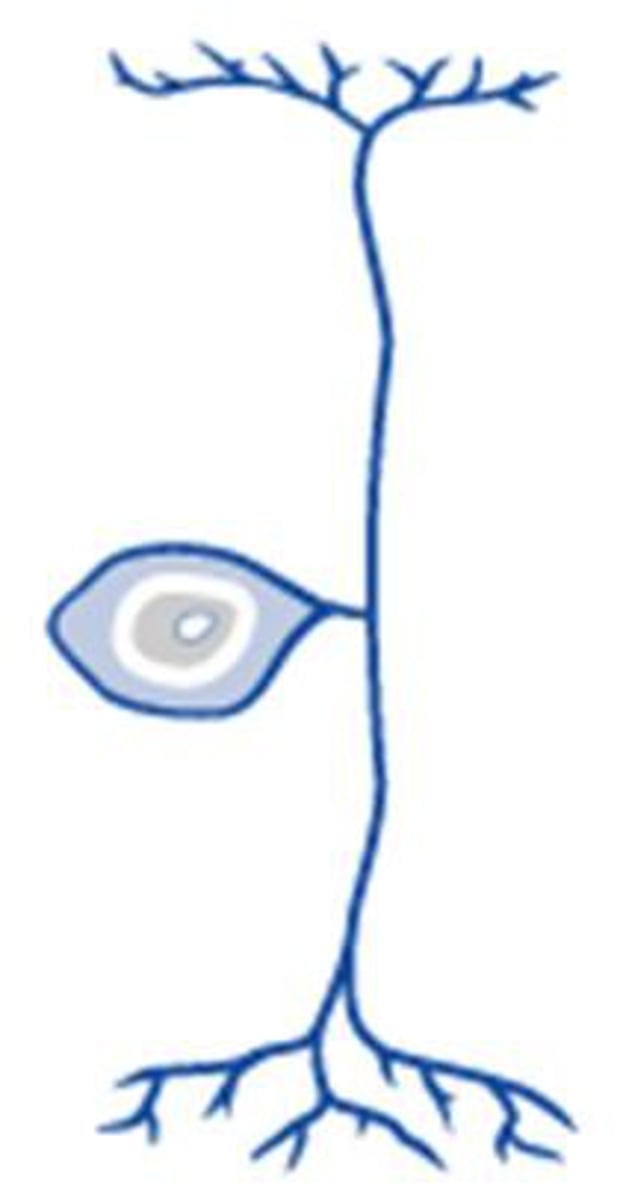
unipolar neuron
A neuron with one axon attached to its soma; the axon divides, with one branch receiving sensory information and the other sending the information into the central nervous system.
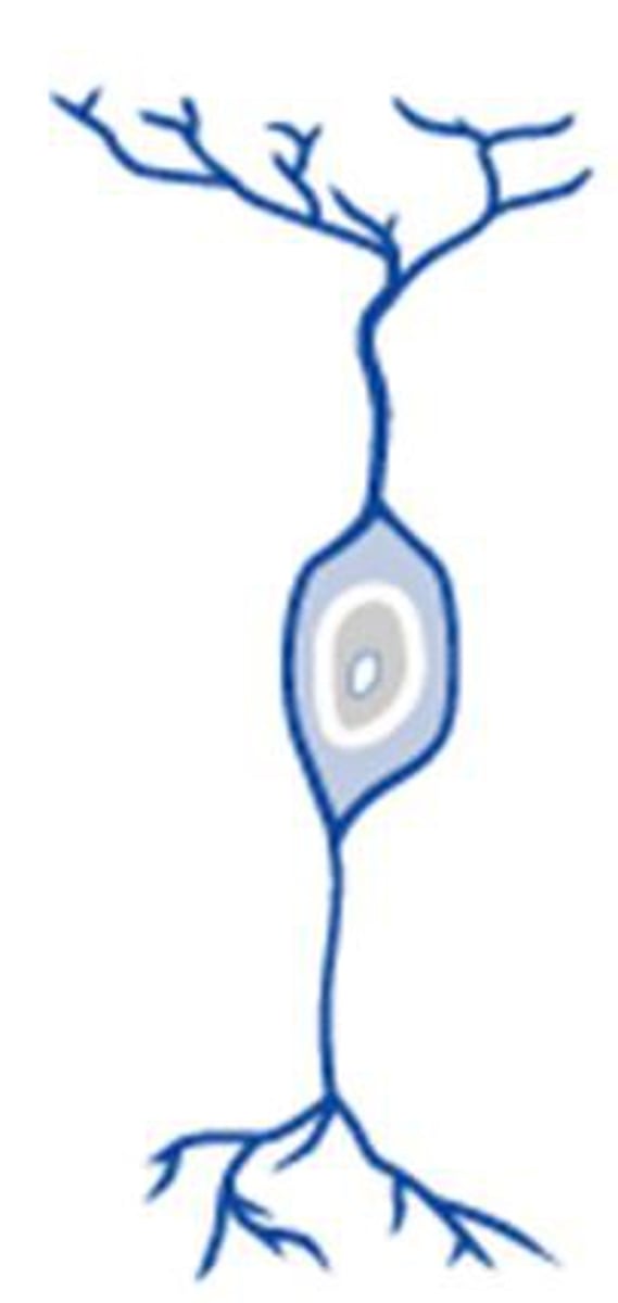
bipolar neuron
a nerve cell that has a single dendrite at one end and a single axon at the other end found in the retina
resting membrane potential
An electrical potential is established across the plasma membrane of all cells by the Na+/K+ ATPase and the K+ leak channels. -70 mV in most cells
Na+/K+ ATPase
A protein found in the plasma membrane of all cells in the body that uses the energy of an ATP (hydrolyzes ATP) to move three Na+ ions out of the cell and two K+ ions into the cell, thus establishing concentration gradients for these ions across the cell membrane. Pumps out one net positive ion
K+ leak channels
allow K+ to leak out across the membrane
charge on the inside of a cell
-70mV
depolarization
movement away from resting potential in the positive direction
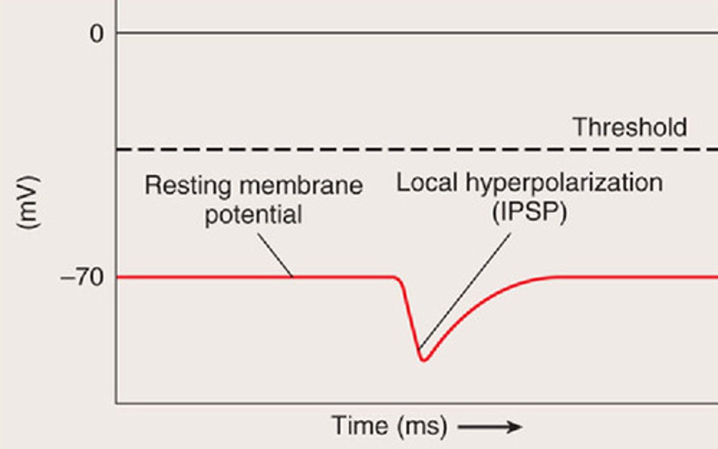
hyperpolarization
move away from the rest potential in the negative direction
repolarization
return to rest potential
equilibrium potential
potential at which there is no driving force on an ion, equal concentration on either side of the membrane
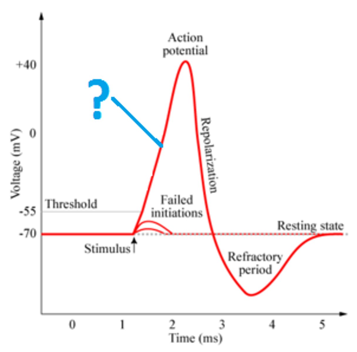
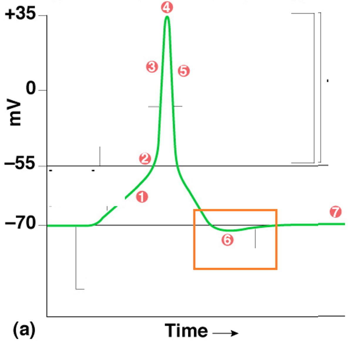
what is happening during depolarization
Input pushes the potential past the threshold, and voltage-gated Na+ and K+ channels open. The voltage-gated Na+ channel opens quickly, and Na+ influx depolarizes the cell
What is happening during the peak of an action potential
The voltage-gated Na+ channel is inactivated, and the voltage-gated K+ channel is fully open
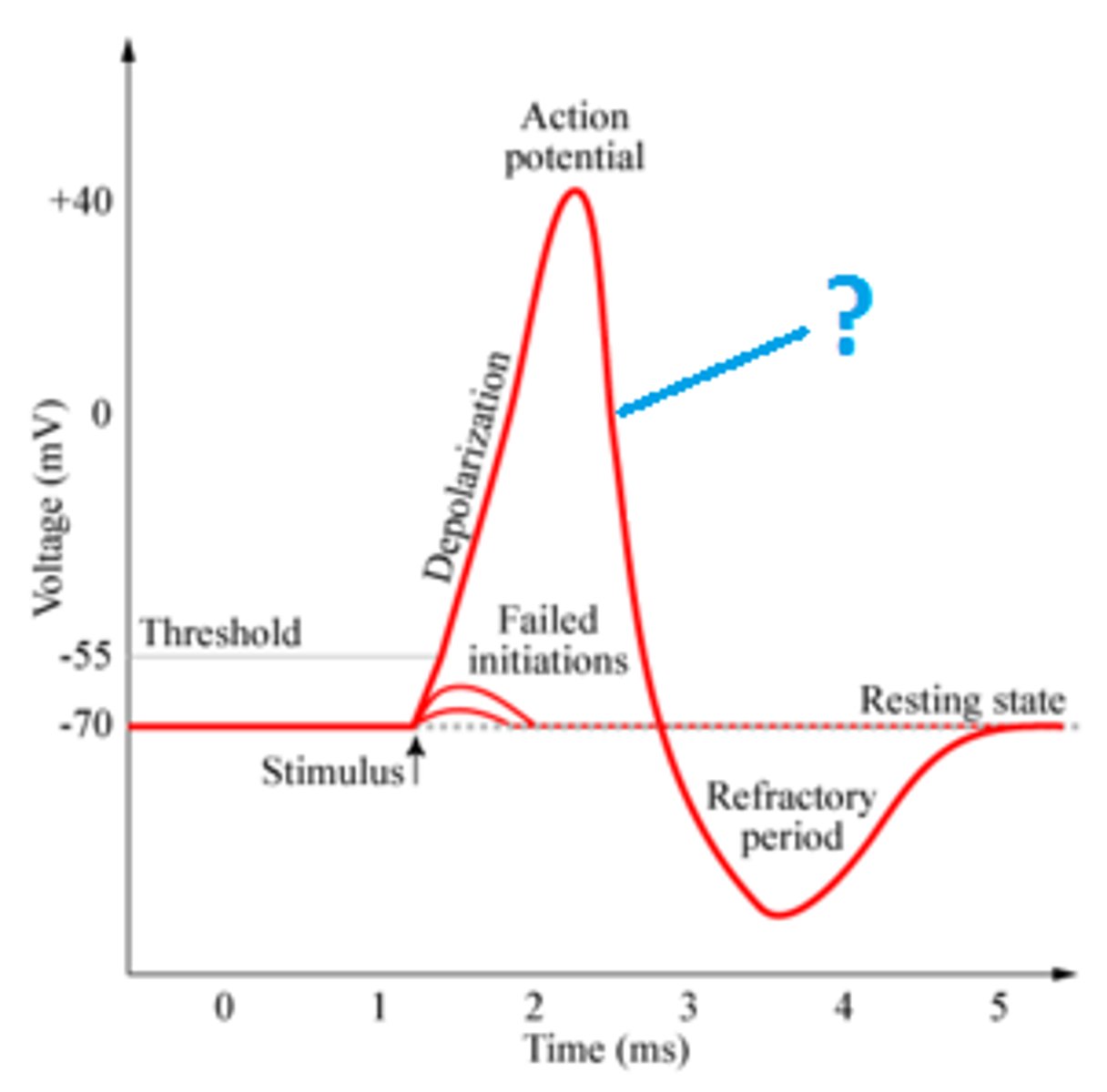
What is happening during repolarization
influx of K+ repolarizes the cell
what is happening during hyperpolarization
The voltage-gated K+ channels close slowly, and extra K+ leaves to hyperpolarize the cell
absolute refractory period
time during which another action potential is impossible; Na+ channels are inactivated and the cell is too positive, near the Na+ equilibrium potential
relative refractory period
The period of time following an action potential, when it is possible, but difficult, for the neuron to fire a second action potential, Na+ channels are closed, and the cell is too negative. It is further from the threshold and near the K+ equilibrium potential
how long does an action potential take
2-3 ms
presynaptic neuron
conducts impulses toward the synapse
postsynaptic neuron
the neuron on the receiving end of the synapse
synapse
the junction between the axon tip of the sending neuron and the dendrite or cell body of the receiving neuron
electrical synapses
ions flow directly from one cell to the next in gap junctions, always an excitatory signal that causes an action potential in the postsynaptic cell, bidirectional flow of ions, and the process is unregulated. seen in CARDIAC MUSCLE CELLS
chemical synapse
a type of synapse at which a chemical (a neurotransmitter) is released from the axon of a neuron into the synaptic cleft, where it binds to receptors on the next structure (either another neuron or an organ)
steps of chemical synapses
1. The action potential arrives at the voltage-gated Ca2+ channel, and the entering Ca2+ breaks the synapse
2. Neurotransmitter vesicles migrate to the membrane, fuse with it, and release neurotransmitters into the synapse
3. Neurotransmitters diffuse across the synapse and bind to receptors on postsynaptic cells
4. Neurotransmitter-gated ion channels open, and ions flow according to the gradient.
5. The flow continues until the neurotransmitters are removed. Acetylcholine is broken down by acetylcholinesterase. NT are reuptake into the presynaptic cell
flow of ions in chemical synapses
Salty C surrounds our cells: Na+, Cl-, and Ca2+ are higher outside the cell and flow in when ion channels open. Therefore, K+ flows out of the cell.
neurons only make
one type of neurotransmitter but can respond to many
leftover neurotransmitters in synaptic cleft
can be recycled or broken down
how medication affects neurotransmitters
change the amount of time neurotransmitters spends in the cleft to adjust responses
the response of the postsynaptic cells depends on
ions and receptors, not the neurotransmitters
synapsin
tethers neurotransmitter vesicles to cytoskeleton
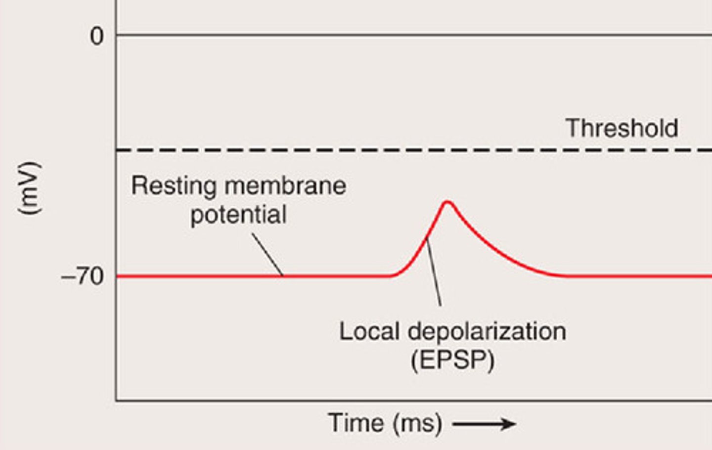
excitatory post synaptic potential
input that pushes potential towards threshold, like Ca2+ and Na+ influx
inhibitory post synaptic potential
input pushes potential away from threshold, influx of K+ or efflux of Cl-
summation
adding EPSP and IPSP at axon hillock, if enough input to push past threshold and trigger and action potential
spatial summation
add up inputs from multiple sources
temporal summation
add up frequent impulses from a single source
greater effect of summation occurs
closer the synapse is to the axon hillock
afferent neurons
Carry sensory input, Approaching the CNS from the PNS
interneurons
CNS neurons that internally communicate and intervene between the sensory inputs and motor outputs; integration
efferent neurons
carry out commands sent out to the body
reflex
rapid integration to avoid potential injury
patellar tendon stretch reflex
aka knee jerk reflex; patellar ligament stretched, quads contract/knee extends and hamstring stays relaxed
hindbrain
medulla, pons, cerebellum
pons
balance
medulla
3 Bs: Breathing, Blood pressure, and Barfing (gag reflex)
spinal cord
reflexes, walking, urination, sex organs
cerebellum
hand-eye coordination
midbrain
relay of vision and hearing, wakefulness
limbic system
emotions
diencephalon
thalamus, hypothalamus, epithalamus
epithalamus
controls the pineal gland which makes melatonin which regulates circadian rhythms
thalamus
relay for conscious sensation except for smell
hypothalamus
controls the pituitary gland for homeostasis
white matter
myelinated axons
tract white matter
myelinated axon bundle in brain/CNS
nerve
bundle of myelinated axons in PNS
gray matter
cell bodies, dendrites, and unmyelinated axons
cortex gray matter
outer layer of cell bodies, dendrites and short axons in brain
frontal lobe
complex decision making, voluntary motion
parietal lobe
general sensation, taste
occipital lobe
visual processing
temporal lobe
smells, hearing, memory, emotions
ganglion
collection of unmyelinated nerve cell bodies in the peripheral nervous system
central nervous system
brain and spinal cord
peripheral nervous system
the sensory and motor neurons that connect the CNS to the rest of the body
somatic nervous system
The division of the peripheral nervous system that controls the body's skeletal muscles/voluntary movement. One neuron from the CNS connects to muscles and uses acetylcholine. Always excititory.
autonomic nervous system
The division of the PNS that controls the involuntary control of glands and smooth muscle. 2 neurons from the CNS connect to the organ, may be excitatory or inhibitory
sympathetic nervous system
The division of the autonomic nervous system that arouses the body, fight or flight. Uses norepinephrine
parasympathetic nervous system
The division of the autonomic nervous system that calms the body, rest, and digest. uses acetylcholine
chain ganglion
Ganglia from the sympathetic nervous system are linked together in the spine and fire at the same time to different organs.
affect of the parasympathetic nervous system
increase blood flow and activity to the GI tract and kidneys. Decrease heart rate, blood pressure, and respiratory rate
affect of the sympathetic nervous system
decrease in blood flow and activity of the GI tract and kidneys. Increase in heart rate, blood pressure, and respiratory rate. Increase in blood flow to the brain, skeletal muscles, and liver. Directly stimulates the adrenal medulla to release epinephrine to prolong the effects
mechanoreceptors
stimulated by physical shape changes; touch receptors
chemoreceptors
respond to chemicals
thermoreceptors
respond to changes in temperature
nociceptors
pain receptors
photoreceptors/electromagnetic receptors
stimulated by light
absolute threshold
the minimum stimulation needed to detect a particular stimulus 50 percent of the time
difference threshold
The minimum amount of difference that can be detected between two stimuli
sensory adaptation
ignore unchanging stimuli, can be retriggered if stimulus changes
bottom up processing
sensory receptors register info, sensory neurons send info to the brain, brain identifies the information
top-down processing
brain applies prior knowledge and experience and forms a holistic view of whats going on
iris
colored part of the eye. regulates the diameter of the pupil
lens
biconvex structure that focuses light on the retina
cornea
external transparent layer of the eye
pupil
black opening in the middle of the eye
ciliary muscles
regulate the curvature of the lense
fovea
The central focal point in the retina, around which the eye's cones cluster. responsible for extreme visual activity
retina
layer at the back of the eye sensitive to light
optic disc
blind spot, place on retina where optic nerve forms
optic nerve
bundle of axons leaving the eye towards the brain
lens at rest
pulled flat by ligaments allowing for far vision