GI 3 - Biliary pathology
 Call Kai
 Learn
Learn Practice Test
 Spaced Repetition
 Match
Match Flashcards
 Knowt Play
1/52

There's no tags or description
Looks like no tags are added yet.
Name | Mastery | Learn | Test | Matching | Spaced | Call with Kai | Chat |
|---|
No analytics yet
Send a link to your students to track their progress
53 Terms
Most normal biliary anomalies (3)
Low insertion of cystic duct
Aberrant right posterior duct
Long common channel (pancreaticobiliary maljunction)
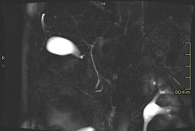
Name variant and the importance
Low insertion av cystic duct. Surgery needs to take extra care not to ligate common bile duct (choledochus / CBD)
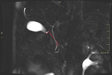
Name variant and importance
Aberrant right posterior duct. Important to know if potential donor has this, as both right hepatic ducts require seperate anastomoses in recipient.
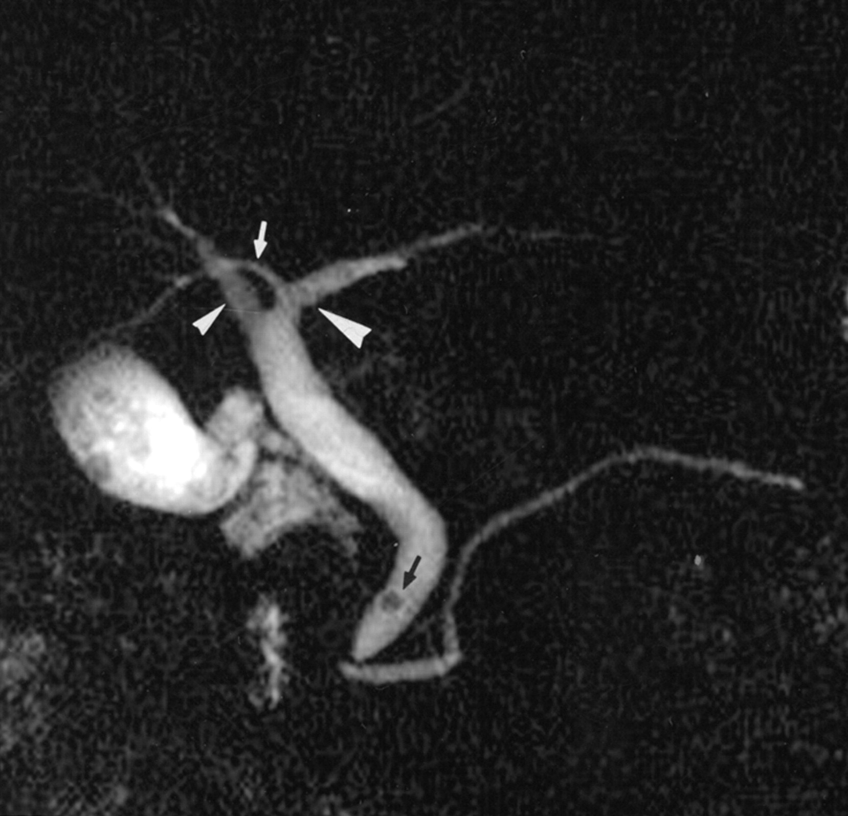
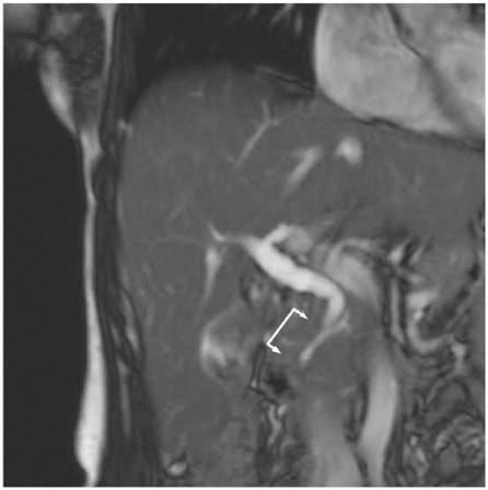
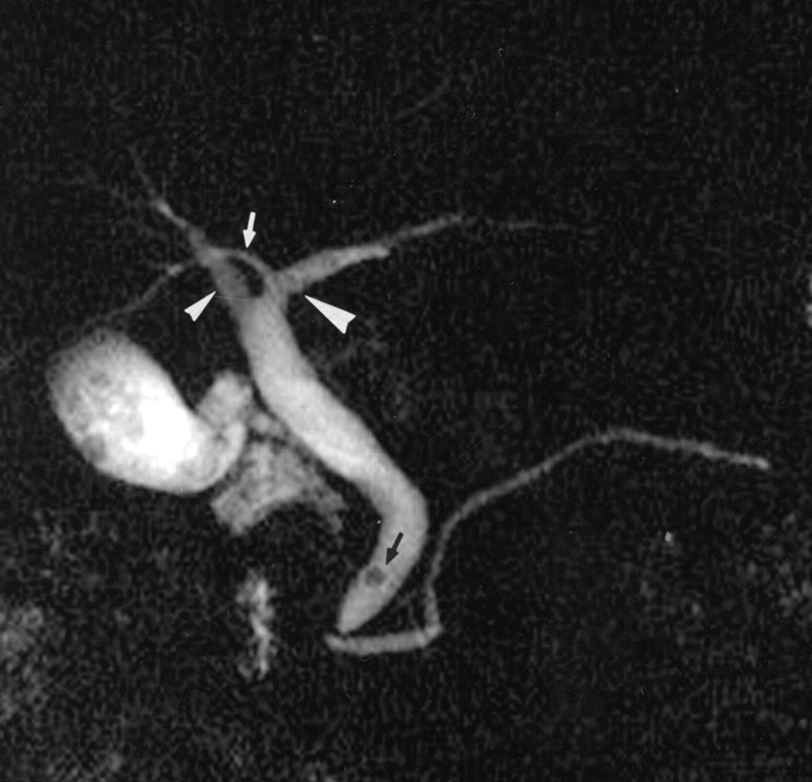
Name variant, the proper definition of the variant and assosiated conditions.
Long common channel (pancreaticobiliary maljunction). Defined as common duct >15 mm. Associated with choledochal cyst, biliary stricture and increase risk of biliary cancer.
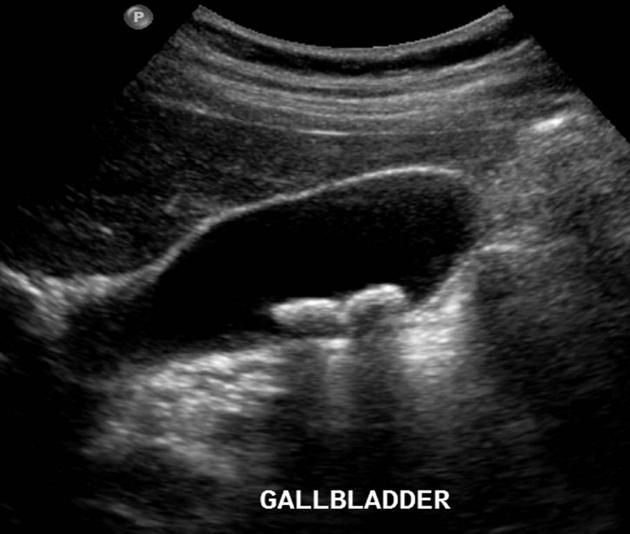
What is the pathology and its risk factors?
Gallbladder stones. RF: female, obesity, pregnancy, middle-ages, diabetes.
Important differentials with ECHOGENIC FOCI in gallbladder lumen/wall?
Stones - shadowing, mobile.
Sludge - non-shadowing, mobile.
Polyp - non-shadowing. immobile.
Hyperplastic cholecystosis - multiple polyps
Porcelain gallebladder - Calcification in gallbladder wall, causes shadowing.
Nearby bowel - Bowel wall and “dirty” shadowing.
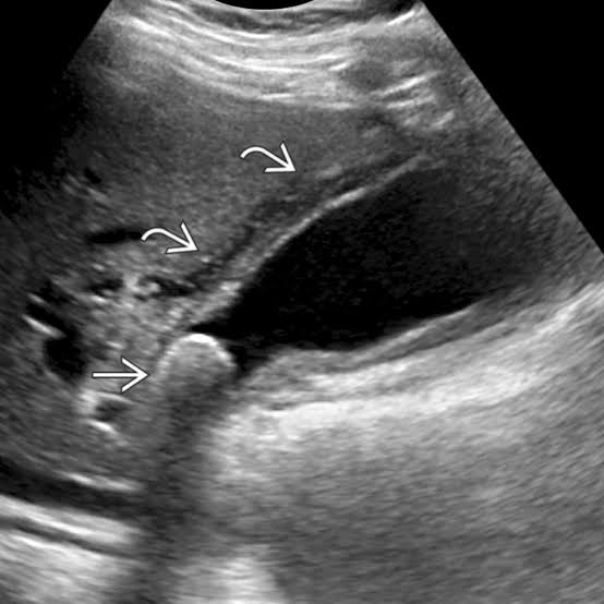
Name findings and diagnosis
Acute calculous cholecystis. Gallbladder wall thickening (>3 mm), hydrops, pericholecystic fluid and echogenic fat. Hyperemia in gallbladder wall mayb be present.
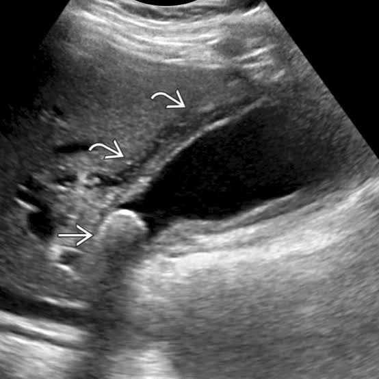
Calculous cholecystitis, name pathomechanism
Obstructing stone in infundibulum or d. cysticus →increased pressure in gallbladder → infection or inflammation
Name complications to acute cholecystitis
Emphysematous cholecystitis, gangernous/necrotizing cholecystitis, perforasion.
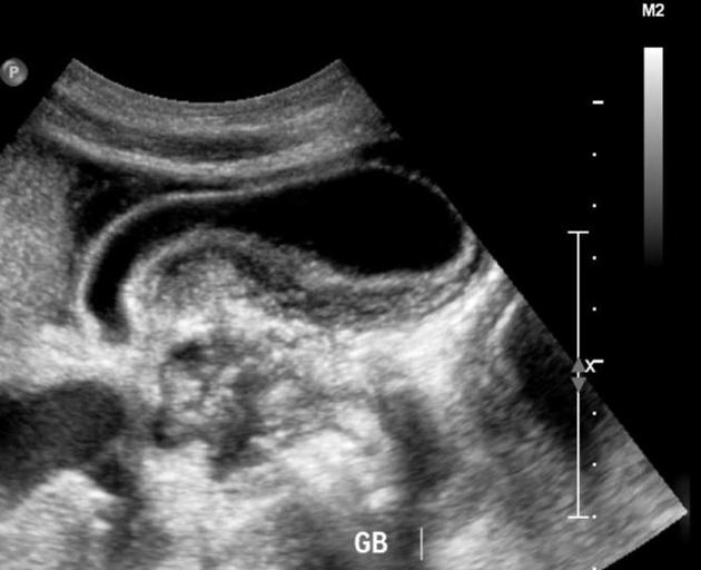
Name findings and suspected pathology
Inflammed gallbladder with no sure stones. Imaging suggests acalculous cholecystitis.
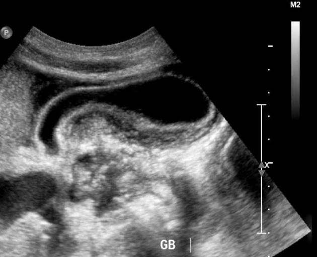
Which patients are at risk for acalculous cholecystitis and what is the suspected pathomechanism
Very sick pts, with certain exceptions (eg EBV). In general, pt with long stays at ICU, parenteral nutrition, trauma, sepsis. Suspected mechanism is biliary statis and hypoperfusion.
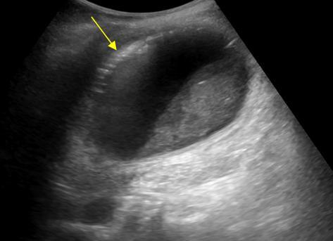
Name finding and diagnosis
Inflamed gallbladder with hyperechoic foci in gallbladder wall suspect of air. Sludge in lumen. Suspected diagnosis is emphysematous gallbladder
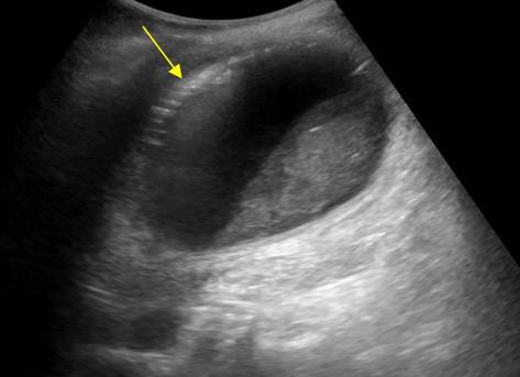
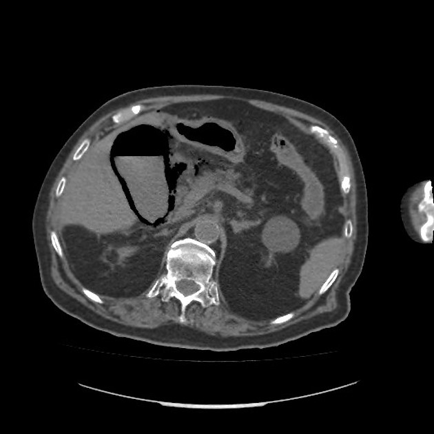
Etiology of emohysematous cholecystitis and at-risk patients.
Rare. Usually due to infection with gas-forming bacteria. Elderly diabetics are at risk.
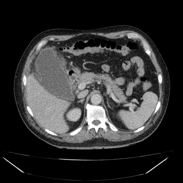
What are imaging findings and what do you suspect
Distended inflamed gallebladder with irregular attenuation in gallbladder wall. Findings consistent with gangernous cholecystitis.
Pathomechanism of gangernous cholecystitis.
Increas intraluminal pressyre → ichemia in gallbladder wall→ necrosis → perforasjon end-point
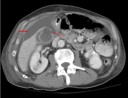
List findings and diagnosis
Defect in gallbladder wall and free fluid. Findings consistent with gallbladder perforation with resultant biliary peritonitis.
Complication of SUBacute perforasion
Pericholecystic - or liverabscess.
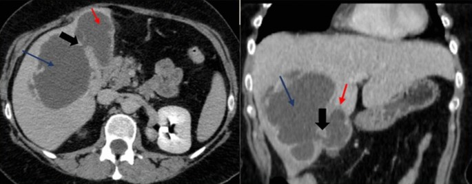
Complication of CHRONIC perforasion
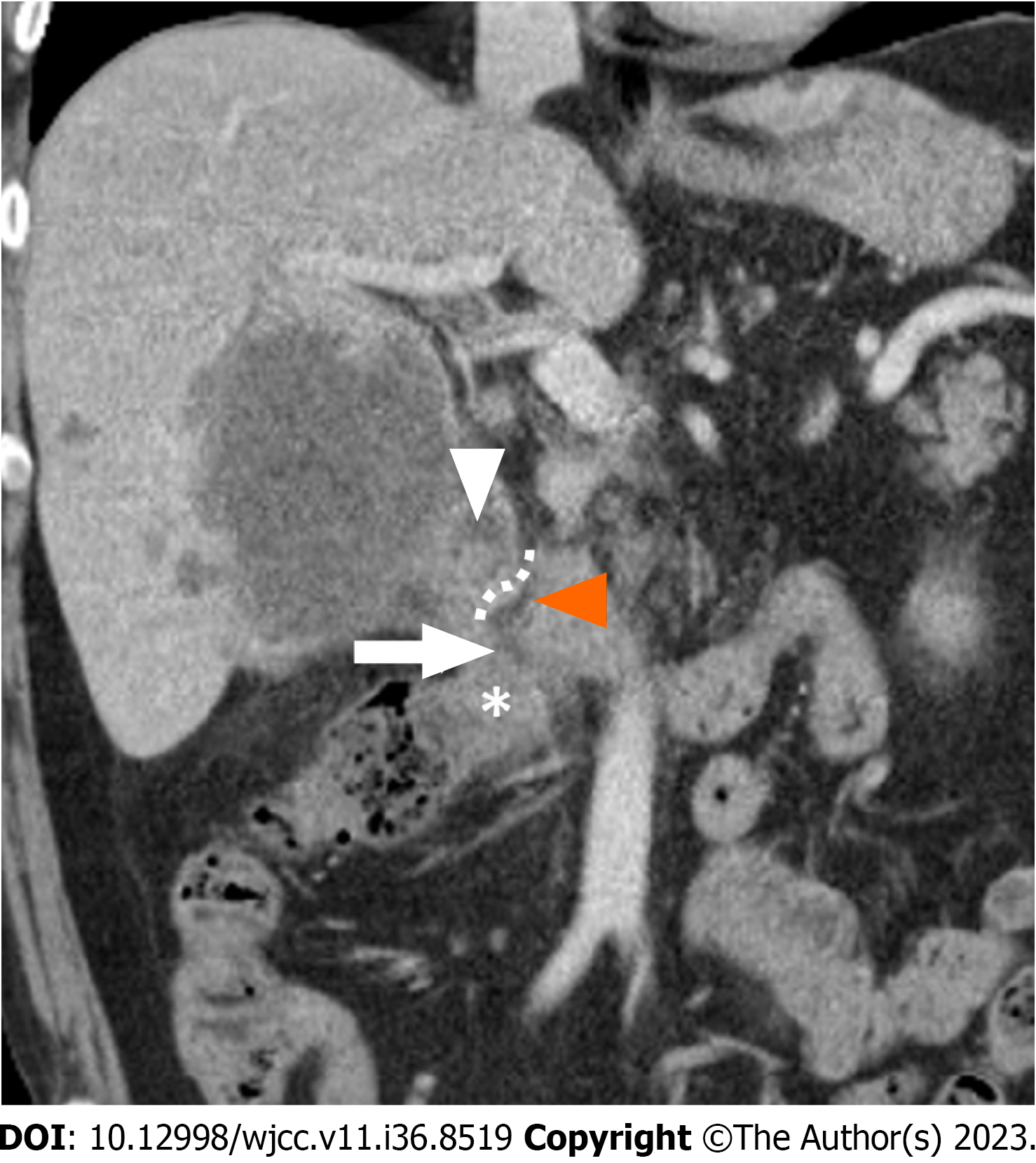
Cholecystoenteric fistula. Here a pt with gallebladder cancer
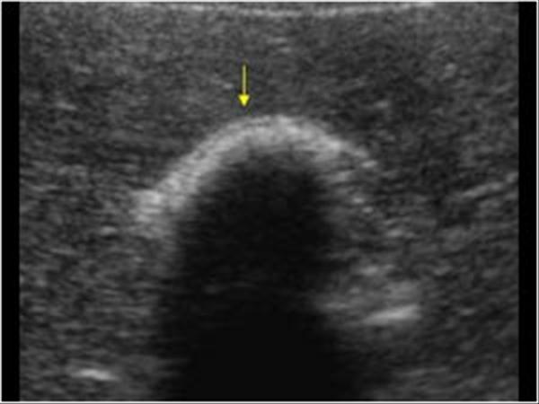
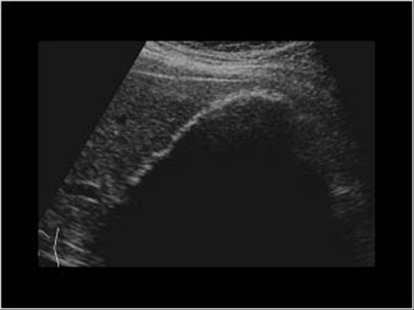
Name findings. What is a normal pitfall?
Image shows big stone, regognized by positive WES-triad (wall-echogenicity-shadow). Can be mistaken with porcelain gallbladder or air in lumen
Pathomechanism behind porcelain gallbladder
Chronic cholecystitis (chronic irritasion → supersaturated bile or recurrent biliary obstructions). Controversial assosiation with gallbladder carcinoma.
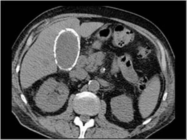
Name finding and pathology
Hyperechoic along gallbladder wall with some shadowing. This was a porcelain gallbadder. Porselensgalleblære. Can be difficult to differentiate from emfysematous gallbladder. No WES-triad so large stone unlikely.
Almost every pt with porcelain gallbladder have stones.
Diff.diagnosis in HYPERECHOGENICITY gallbladder wall
Porcelain gallbladder
Packet with stones / BIG stones (WES-triade)
Emphysematous cholecystitis (intramural gass with “dirty shadowing”)
Adjacent bowel (bowell wall with “dirty shadowing”)
Define Courvoisier gallbladder
Dilated gallbladder due to obstruction from malignant tumor. NO INFLAMMATION.
Can be due to obstructed cystic duct og CBD, last one may lead to dilated intrahepatic biliary tree.
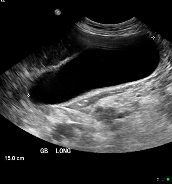
Define hyperplastic cholecystosis
Non-neoplastic proliferation disordser resulting in deposition of holesterol-laden kke-neoplastis proliferasjonsforstyrrelser som et resultat av deposit av kolesterol-fylte makrophages in gallbladder wall. Primariliy composed of adenomyomatosis og cholesterolosis Some include polyps in these groups, if cholesterol based.
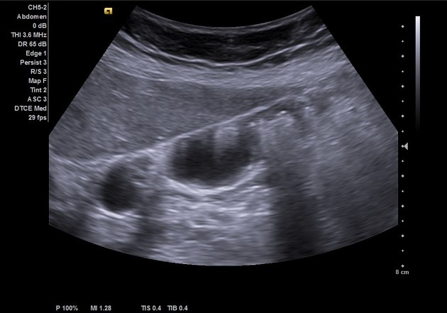
List findings and suspected diagnosis
Echogenic foci in gallbladder wall with “comet tail” artefact. Findings consistent with adenomyomatose.
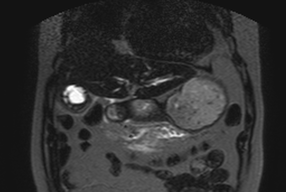
Name MRI sequence and findings
T2 with fat suppression. Focal changes in fundus of gallbladder with high T2 signal. “Pearl necklace sign” - distribution, fitting with adenomyomatosis.
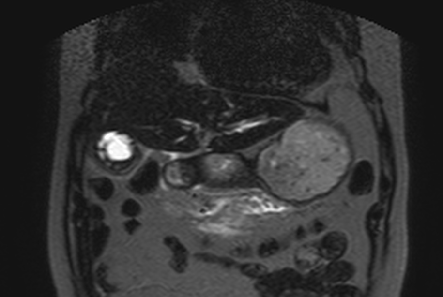
Mechanism of adenomyomatosis
Cholesterol deposits in mural Rokitansky-Aschoff sinuses in gallbladder wall. Can be diffuse or focal, typically in fundus if the latter. Focal findings might be difficult to differentiate from malignancy
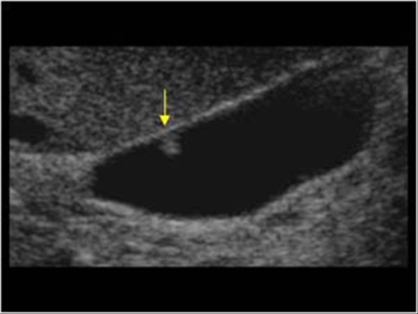
Name findings and diagnosis
Hyperechoic structure seemingly adherrant to gallbladder wall. Findings consistent with polyp.
What are gallbladder polyps
Most of them benign cholesterol polyps, which are a part of hyperplastic cholecystosis.
RARELY may an individual polyp be a pre-malignant adenoma (<5%), there are the reason for controlls.
Name ris factors for malignancy in regards to gallbladder polyp
6 S-es:
Size. >10 mm OR fast growth rate. CHOLECYSTEKTOMI!!
Single. Singular is more suspect for malignancy
Sessile (aka. broad based). Broad based polyp is suspicious of malignancy. Pedunculated polyps are usually benign.
Stones. Gallbladder stones increase risk of (chronic) inflammation, and therefore malignancy.
PSC. Increased risk of malignancy, and some are removed in these pt even if <10 mm.
Sixty og above in age, increased risk of malignancy.
How often are the polyps controlled in Norway and the EU
Norge: If <10 mm, we controll these x1/year in 3 years. After 3rd controll, follow up ends.
EU: Imaging
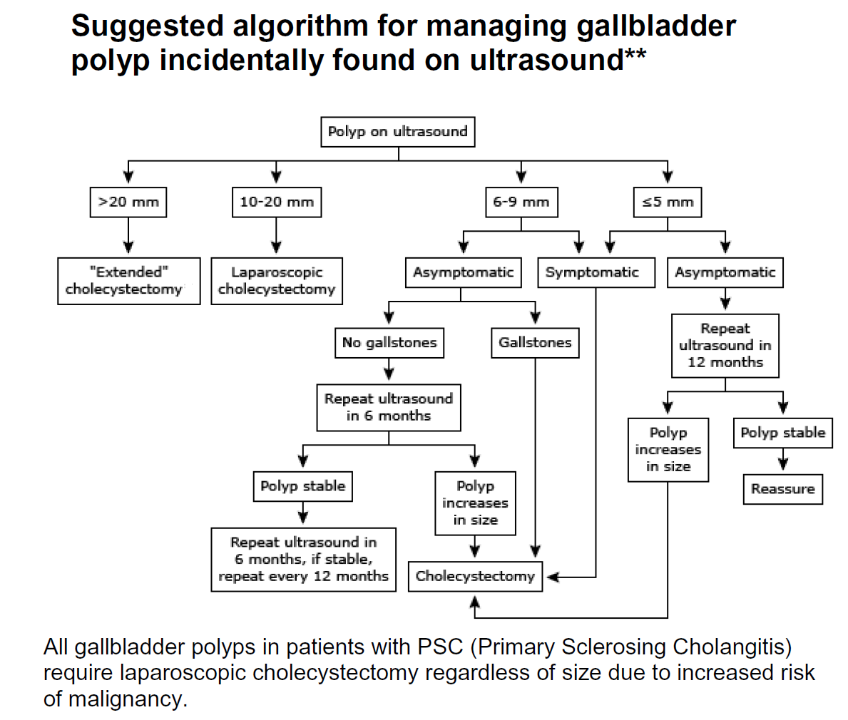
How often are the polyps controlled in USA
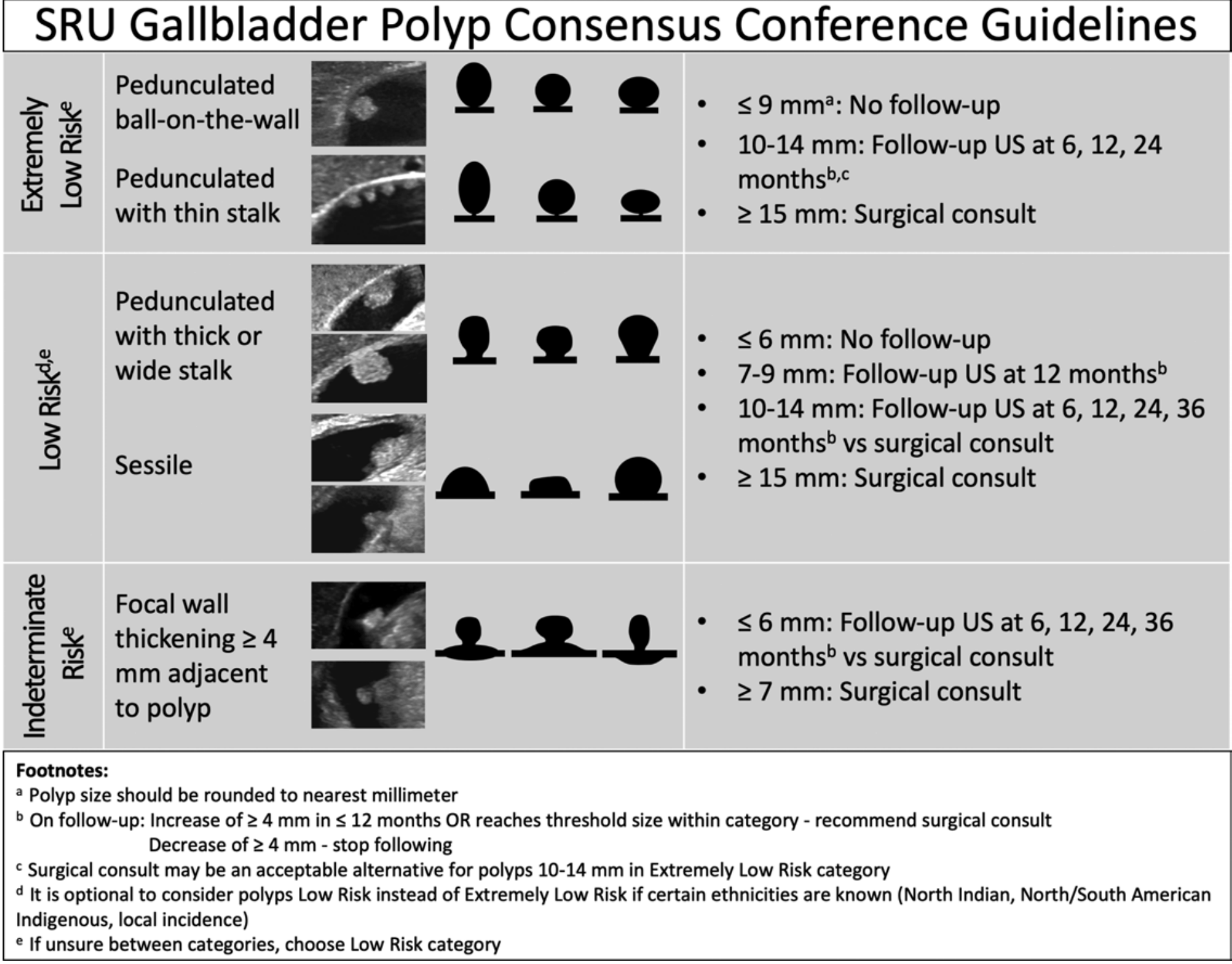
Differenisal in diffuse thikening of gallbladder wall (>3 mm)
Fluid overload/edema: Cirrhosis, heart failure, protein-wasting nephropathy.
Inflammation, infection: Cholecystitis, hepatitis pankreatitis
Infiltrating diseases: gallbladder cancer, metastases (rare)
Post-prandial
Differenisal in focal thikening of gallbladder wall
Hyperplastic cholecystosis: adenomyomatose, kolesterol polypper
Vaskular: Varices:
Neoplastic disease: Adenom-polypper, gallbladder cancer, adjacent liver cancer, metastasis (rare)
Differential in intraluminal contents WITHOUT shadowing
Tumefactive sludge: mobile
Blood/pus: mobile
Polyp: immobile
Cancer: immobile
Differential in increased echogenicity of gallbladder wall.
Porcelain gallbladder
Packed with stones (WES-triad)
Emfysematous cholecystitis
MRCP primarily uses which sequences
T2. Fast-spin echo, incl with breathing techniques. May supply with contrast enhanced T1 sequence with fat saturation.
Choledocal cysts
Caroli disease
Choledocholithiasis
Mirizzi syndome
Pneumobilia
Biliary ductal dilation
Ascending cholangitis
PSC
PSC
PBC
AIDS cholangiopathy
Recurrent pyogenic cholangitis
Biliary cystadenoma / cystadenocarcinoma
Cholangiocarcinoma
Gallbladder carcinoma
Gallbladder metastases