Anatomy Module 3: Muscular System
 Call Kai
 Learn
Learn Practice Test
 Spaced Repetition
 Match
Match Flashcards
 Knowt Play
1/47

There's no tags or description
Looks like no tags are added yet.
Name | Mastery | Learn | Test | Matching | Spaced | Call with Kai | Chat |
|---|
No analytics yet
Send a link to your students to track their progress
48 Terms
myology
the study of the muscular system
functions of muscle
Motion — enables movement of joints for ambulation, reaching, and activities of daily living
Protection — muscles overlie (are superficial to) bone, providing a cushioning effect
Circulation support — skeletal muscle contraction helps pump venous blood back to the heart against gravity (no separate "pump" exists for venous return)
Example: the soleus muscle (calf) is nicknamed the "peripheral heart" because its contraction is essential for venous return from the lower leg
Clinical relevance: bedridden patients are instructed to perform ankle pumps to promote circulation, since standing/walking muscle activity isn't available to assist venous return
Insulation / thermoregulation
Goosebumps = muscular contraction around hair follicles (attempt to retain heat)
Shivering = muscle activity generating heat energy
(Note: the integumentary system is the primary insulator, but muscle contributes significantly)
Pumping blood (cardiac muscle) — the heart itself is composed entirely of cardiac muscle
Vascular/visceral wall function (smooth muscle) — forms the walls of arteries/veins and the GI tract (small/large intestine)
Enables vasoconstriction/vasodilation of blood vessels
Enables peristalsis (propulsion of food/waste through the GI tract)
sympathetic nervous system
("fight-or-flight"): constricts blood vessels to GI tract, redirects blood to skeletal muscle/extremities for action
This constriction/dilation is only possible because vessel walls are muscular (smooth muscle) — connective tissue like ligaments or cartilage cannot actively constrict/dilate
parasympathetic nervous system
("rest-and-digest"): dilates central/GI blood vessels, constricts peripheral vessels, prioritizing digestion\
This constriction/dilation is only possible because vessel walls are muscular (smooth muscle) — connective tissue like ligaments or cartilage cannot actively constrict/dilate
skeletal muscle
Fibers arranged in parallel
voluntary control
Biceps, triceps, quadriceps, hamstrings, etc.
fatigable
uses aerobic glycolysis (oxygen/hemoglobin dependent) energy pathway
cardiac muscle
Network/ridge-like (branching) arrangement
involuntary control
heart only
not fatigable
oxygen-dependent energy pathway but does not fatigue
smooth muscle
Fusiform (spindle-shaped) cells
involuntary control
Blood vessel walls, GI tract (small/large intestine)
not fatigable
oxygen-dependent energy pathway but does not fatigue
shared properties of all muscle tissue
Tone — maintains a baseline resting length/structure even when relaxed
Controlled by the central nervous system
Hypertonicity/spasticity — abnormally increased tone (seen after stroke, in spastic cerebral palsy, etc.)
Extensibility — ability to stretch (like a rubber band); contrasted with rigid tissues like cartilage/ligament, which would tear/break if stretched similarly
(Passive) Elastic recoil — ability to passively return to resting length after being stretched (distinct from active contraction)
Contractility — ability to actively contract (e.g., biceps flexing the elbow; heart contracting to pump blood)
tone
maintains a baseline resting length/structure even when relaxed
Controlled by the central nervous system
Hypertonicity/spasticity — abnormally increased tone (seen after stroke, in spastic cerebral palsy, etc.)
extensibility
ability to stretch (like a rubber band); contrasted with rigid tissues like cartilage/ligament, which would tear/break if stretched similarly
(passive) elastic recoil
ability to passively return to resting length after being stretched (distinct from active contraction)
contractility
ability to actively contract (e.g., biceps flexing the elbow; heart contracting to pump blood)
Layers Superficial to Deep (Skin → Muscle)
Epidermis (skin)
Dermis (skin)
Superficial fascia — yellowish, adipose (fatty) tissue layer
Deep fascia — denser connective tissue layer
Muscle
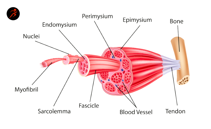
myofibril/muscle fiber
Smallest unit of the muscular system
fascicle
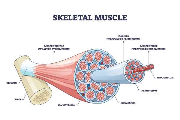
Bundle of muscle fibers
covered by perimysium

endomysium
Innermost layer; surrounds individual muscle fibers
perimysium
Surrounds bundle of 10-100 fibers called fascicles
muscle
a bundle of fascicles
covered by epimysium
epimysium
Outer connective tissue layer; surrounds entire muscle
tendon
how muscle connects to bone which anchors into periosteum
deep fascia in muscles
covers individual muscles and separates groups of muscles into compartments
Each compartment contains its own muscles plus the blood vessels and nerves that supply them
Example — thigh cross-section (3 compartments):
Anterior compartment — quadriceps
Posterior compartment — hamstrings
Medial compartment — adductors
compartment syndrome
During exercise, muscles hypertrophy (temporarily swell with increased blood flow) within their fixed-volume fascial compartment
Because the compartment is a closed space, swelling muscle → increased intra-compartmental pressure → compression of blood vessels and nerves within that compartment
Symptoms can include tingling (e.g., in the toes during prolonged running)
Treatment for severe/acute compartment syndrome: fasciotomy — surgically opening the fascia to relieve pressure
Will be revisited in detail when covering the lower leg (shin splints, compartment syndrome)
myofascial release
a treatment technique targeting adhesions that form between muscle and fascia
neuromuscular junction
the connection point between a nerve ending and a single muscle fiber
motor unit
one nerve fiber + all the muscle fibers it innervates (the smallest functional unit of contraction)
Each muscle (e.g., biceps) contains thousands of muscle fibers organized into many motor units
Action Potential & the All-or-None Law
Nerve impulse → creates a potential difference (imbalance of positive/negative ions) across the muscle cell membrane
This potential difference triggers an influx of ions (sodium, calcium, potassium) into the muscle cell, initiating contraction
Action potential = the threshold of potential difference required to trigger a muscle contraction
All-or-none law: all muscle fibers within a motor unit contract simultaneously, or none do — because they all receive the nerve impulse at the same time
action potential
the threshold of potential difference required to trigger a muscle contraction
all-or-none law
all muscle fibers within a motor unit contract simultaneously, or none do — because they all receive the nerve impulse at the same time
sarcomere
the repeating contractile unit of a muscle fiber, bounded by two "light lines"; many sarcomeres arranged in series make up one muscle fiber
Within each sarcomere:
Actin filament — thin filament (protein)
Myosin filament — thick filament (protein), with a mobile "head"
Troponin — protein with affinity for calcium; normally bound to actin
Tropomyosin — winding, cord-like protein that holds troponin in place along the actin filament
actin filament
thin filament (protein)
myosin filament
thick filament (protein), with a mobile "head"
troponin
protein with affinity for calcium; normally bound to actin
tropomyosin
winding, cord-like protein that holds troponin in place along the actin filament
contraction sequence
Nerve impulse → potential difference → calcium influx into the muscle cell
Troponin (bound to tropomyosin) has a strong affinity for calcium → releases its hold on actin and binds to calcium instead
With troponin/tropomyosin no longer blocking actin, the myosin heads bind to the now-exposed actin
Myosin heads "walk" along the actin filament, pulling the actin and myosin filaments to slide past each other
As filaments slide together, the sarcomere shortens → repeated across thousands of sarcomeres/fibers = whole-muscle contraction
Contraction continues as long as the nerve impulse (and calcium influx) continues
When the nerve signal stops: calcium is removed, troponin/tropomyosin re-bind to actin, and the muscle returns to its resting (relaxed) state
energy requirements for contraction
Muscle contraction requires ATP (adenosine triphosphate), produced via cellular respiration (Krebs cycle) — not covered in detail in this course, as it belongs to physiology
Hemoglobin (in blood) and myoglobin (in muscle) both bind oxygen for transport/storage — proteins are fundamental building blocks for these processes
fast twitch skeletal muscle fiber
short bursts of high-intensity power
Whitish (lower hemoglobin/myoglobin content)
fatigues quickly
Sprinting (100m dash), weightlifting
slow twitch skeletal muscle fiber
Sustained, endurance-based activity
Darker (higher hemoglobin/myoglobin content, more blood supply)
resists fatigue
Marathon/distance running
intermediate twitch skeletal muscle fiber
Combination of power + endurance
intermediate color
moderate fatigability
Brisk walking, general daily activity
Clinical/rehab application — Multiple Sclerosis (MS) example
MS is a demyelinating disorder — nerves lose their myelin sheath (the "insulation" that allows efficient electrical signal transmission)
Without myelin, nerves must expend more energy to transmit the same impulse → contributes to the hallmark symptom of fatigue in MS flare-ups
Rehab approach: focus on energy conservation — train patients to recruit slow-twitch fibers more (lower energy cost, sustained output) rather than fast-twitch (high energy cost, quick fatigue)
biomechanics: muscles as levers
Muscle-joint systems function like seesaws/levers, involving:
Fulcrum (F) — the pivot point (typically the joint)
Effort (E) — the muscular force
Resistance (R) — the weight/load being moved
First-order arrangements can offer the most mechanical efficiency, but the human body cannot be redesigned to always favor this arrangement
What can be adjusted clinically: the distance between resistance and fulcrum (or effort and fulcrum)
Example: External rotation strengthening — start with the arm/resistance close to the body (shorter lever arm = easier), then progress by moving the resistance farther from the body (longer lever arm = harder) as the patient improves
This principle is used to progress rehab exercises by manipulating leverage/distance, since the underlying tissue anatomy itself can't be changed
first-order level muscle lever
Fulcrum in the middle, effort and resistance on either side
Head/neck extension — posterior neck muscles (effort) pull the head up; the resistance (head's weight) is anterior to the spinal fulcrum
second-order level muscle lever
Resistance in the middle, fulcrum and effort on either end
Standing on tiptoes (calf raise) — fulcrum = ball of foot, effort = calf muscles, resistance = body weight (in the middle)
third-order level muscle lever
Effort in the middle, fulcrum and resistance on either end
Bicep curl — fulcrum = elbow joint, effort = biceps (middle), resistance = dumbbell (farthest from fulcrum)
agonist muscle
The primary muscle(s) actively producing a given motion
Biceps during elbow flexion

antagonist muscle
The opposing muscle group that must lengthen/relax (and may control the motion eccentrically) to allow the agonist's action
Triceps lengthening (controlling the descent) during a slow, controlled elbow extension after a bicep curl

synergist muscle
Muscles that assist/support a movement without directly producing the primary action themselves
Rotator cuff muscles (e.g., supraspinatus) and scapular stabilizers assisting shoulder abduction alongside the deltoid

stabilizer muscle
Muscles that stabilize a joint/region to allow the prime movers to act efficiently
(Scapular stabilizers during arm elevation)

Primary vs. Secondary (Synergistic) Muscle Actions
Many muscles cross more than one joint and therefore have both a primary action and secondary/synergistic actions
Example: Quadriceps group (4 muscles total)
Rectus femoris — a two-joint muscle: primary action = knee extension; secondary action = assists with hip flexion
Vastus muscles (3 of the 4 quadriceps muscles) — one-joint muscles: knee extension only
Clinical relevance: When a muscle is weakened (e.g., due to nerve injury), both its primary action AND its secondary/synergistic actions will be affected — important for understanding the full functional impact of nerve injuries or muscle weakness