Head and Neck 1
1/19
Earn XP
Description and Tags
Describe the functions and clinical relevance of the muscles of mastication in the canine and equine • Discuss the path of the trigeminal nerve and it's relevance to canine dental work • Describe the blood supply to and from the head, including vessels of clinical concern • Describe the blood supply of the dental arcades and nasal cavity • Describe the locations of the lymph nodes and salivary glands of the head and neck and discuss in a clinical context • Identify structures of the head with respect to aural surgery • Describe important species differences in soft tissues of the head
Name | Mastery | Learn | Test | Matching | Spaced |
|---|
No study sessions yet.
20 Terms
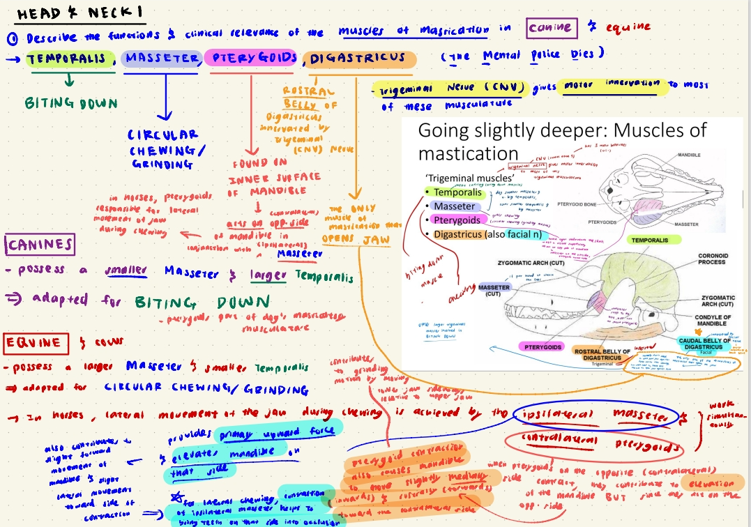
“Trigeminal Muscles”
Motor Innervation to “trigeminal muscles”: Trigeminal Nerve (CNV), except caudal belly of Digastricus: motor innervation supplied by Facial Nerve (CNVII)
Temporalis: for Biting down
Masseter: for circular chewing/grinding
Pterygoids: assists ipsilateral Masseter in circular chewing in horses, just another masticatory muscle in dogs
Digastricus: for OPENING jaw
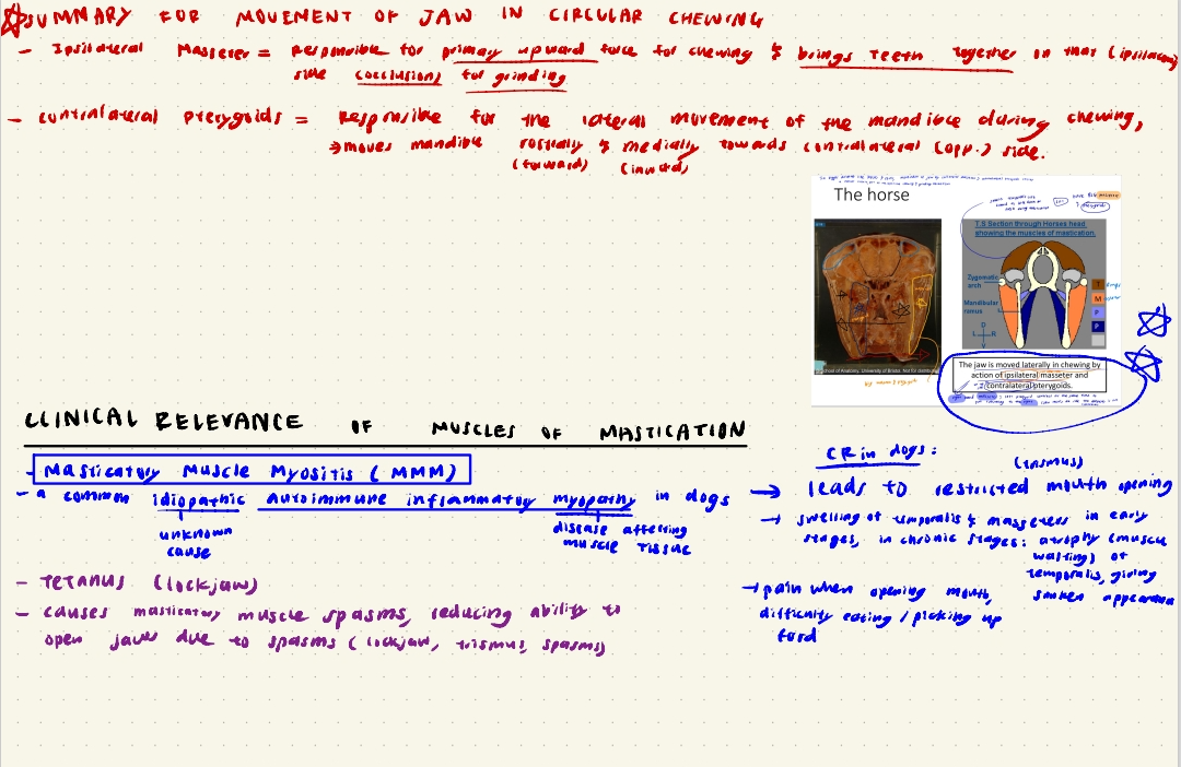
Masticatory Muscle Conditions
Masticatory Muscle Myositis (MMM)
an idiopathic autoimmune inflammatory myopathy affecting the muscles of mastication, leading to pain and difficulty in chewing, swelling and difficulty opening the mouth.
early stage symptoms: swelling of temporalis and masseter muscles, along with pain when opening the jaw.
chronic stage symptoms: sunken appearance of head due to atrophy of temporalis muscles
Tetanus
bacterial infection (Clostridium tetani) affecting jaw muscles, causing masticatory muscle spasms which lead to stiffness and lockjaw (trismus: restricted opening of jaws).

How do Ipsilateral Masseter and Contralateral Pterygoids work together?
Ipsilateral Masseter helps to provide primary upward force for grinding, and brings teeth together on that side for occlusion.
Contralateral pterygoids assist in lateral jaw movement by moving mandible rostrally and medially towards contralateral (opposite) side.
i.e. Ipsilateral Masseter on right side and Contralateral Pterygoids on Left side contract simultaneously to pull everything to the right (side Masseter is on)
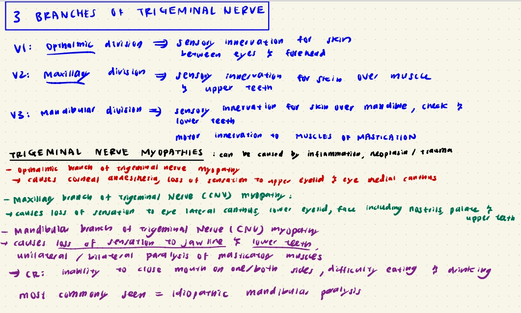
Trigeminal Nerve Myopathies can be caused by inflammation, neoplasia or trauma
When Opthalmic Branch is affected, it leads to:
Corneal Anaesthesia, Loss of sensation to upper eyelid and eye medial canthus
When Maxillary Branch is affected, it leads to:
Loss of sensation to lower eyelid, eye lateral canthus, face incl. nostril, palate and upper teeth.
When Mandibular Branch is affected, it leads to:
loss of sensation to jaw line and lower teeth.
Unilateral/Bilateral paralysis of masticatory muscles, leading to inability to close mouth on one or both sides, diffculty eating and drinking.
most common: idiopathic mandibular paralysis
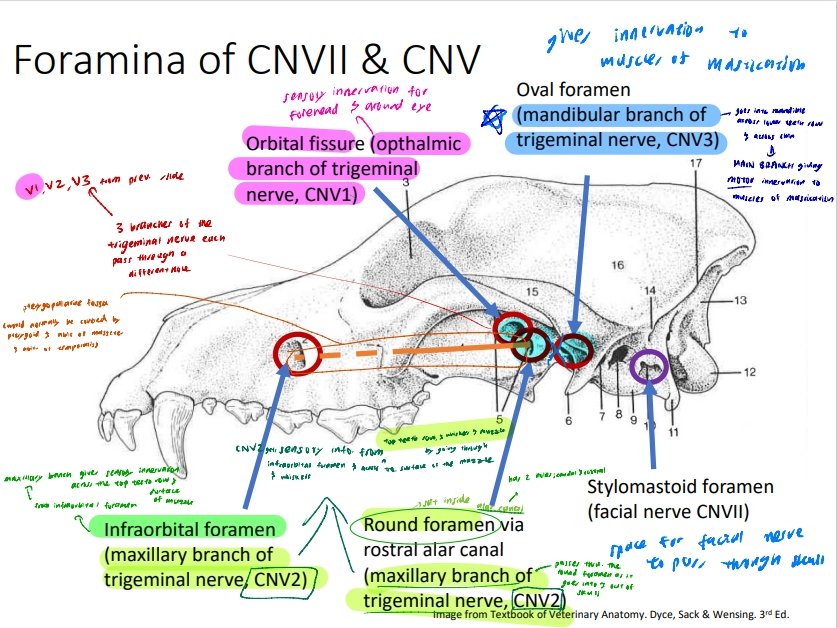
Trigeminal Nerve (CNV) Path for each branch
V1 (Opthalmic Branch):
Passes through the orbital fissure to supply sensory innervation to the forehead, upper eyelid, & skin between eye and forehead, eye medial canthus
V2 (Maxillary Branch):
Passes through infraorbital foramen, and round foramen via rostral alar canal (alar canal has 2 holes, rostral and caudal)
V2 provide sensory innervation for upper teeth row, palate, muzzle nostrils and whiskers, lower eyelid & eye lateral canthus by passing through the infraorbital foramen.
As V2 goes into and out of the skull, it also passes through the round foramen via the rostral alar canal. (round foramen sat inside rostral alar canal)
V3 (Mandibular Branch):
Passes through the oval foramen to provide sensory innervation to the skin over mandible and cheek, lower teeth.
Provides motor innervation for muscles of mastication.
Trigeminal Nerve (CNV) Relevance to Canine Dental work
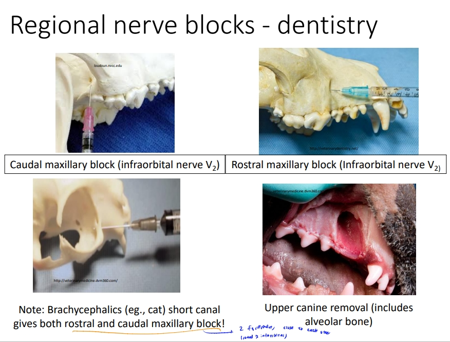
Caudal Maxillary block
Targets infraorbital nerve, a branch of V2 (maxillary branch of trigeminal nerve)
local anaesthetic technique desensitising the 1st & 2nd upper molar teeth of maxilla, all premolars, canine and incisors on that side of maxilla. Also desensitises the nose, cheek and upper lip skin, bone & soft tissue of the maxilla.
Rostral Maxillary block
Targets infraorbital nerve, a branch of V2 (maxillary branch of trigeminal nerve)
local anaesthetic technique desensitising upper canine and incisor teeth (maxilla), 1st 2nd & 3rd premolars and associated soft tissues on the side injected.
Note: in Brachycephalic breeds, either a caudal or rostral maxillary block give rise to a block for both.
Caudal Mandibular Block:
Targets inferior alveolar nerve, a branch of V3 (Mandibular branch of Trigeminal Nerve CNV) via mandibular foramen
local anaesthesia technique desensitising all lower teeth, associated labial tissues and rostral lower lip.
Rostral Mandibular Block:
Targets inferior alveolar nerve, a branch of V3 (Mandibular branch of Trigeminal Nerve CNV) via mental foramen
desensitises lower canine and incisor teeth on that side, possible first premolar, and soft tissues of rostral lower lip and chin
Blood Supply To and From Head
Head is highly vascularised.
2 COMMON CAROTID ARTERIES (Right & Left)
main blood supply to the head, branching into internal and external carotid arteries that supply various structures.
BRAIN ARTERIAL BLOOD SUPPLY
2 Vertebral Arteries
Ventral Spinal Artery
join to form Basilar Artery, which contributes to the Cerebral Arterial Circle (Circle of Willis); a loop of arteries around teh base of the brain.
Internal Carotid Artery
also contributes branches to the Cerebral Arterial Circle
CLINICAL RELEVANCE:
cannot inject IV drugs into internal carotid artery since it goes directly to the brain and can cause spasms
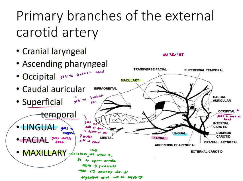
Primary branches of the EXTERNAL CAROTID ARTERY
Old Angry Cats Love Fish, Cheese, Sausages & Mice (O, A, C, L, F, C, S, M) which represent the following branches:
Occipital → goes to back of head
Ascending pharyngeal
Cranial laryngeal
Lingual → goes to tongue
Facial → goes across face
Caudal auricular → goes to back of ear
Superficial temporal → goes up side of face in front of ear & across side of head.
Maxillary (terminal branch) → goes to upper arcade, teeth and structures that CNV2 (Maxillary Branch of Trigeminal Nerve CNV) innervates
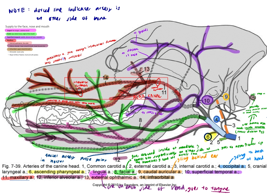
SUPPLY TO FACE NOSE AND MOUTH
Lingual Artery
Supplies Tongue & Palatine tonsil, one on each side of head
Facial Artery
Supplies upper and lower Lips, Lateral Nose, angle of Mouth.
Superficial Temporal Artery
Supplies Eyelids, Dorsum of Nose, Masseter
Maxillary Artery
runs through infraorbital canal to reach snout and whiskers.
Supplies
External Opthalmic (the orbit)
Inferior alveolar: Lower Alveoli & lower teeth, Lower lip and chin (One branch also runs along with inferior alveolar artery and provides blood supply for inferior alveolar NERVE)
Infraorbital: Upper Alveoli and upper teeth
Ethmoidal: Nasal Cavity
Major and Minor Palatine: Hard and Soft Palates
Venous Drainage of Head
Veins mostly satellite = sits next to artery & has same name, drain away tgt.
Variations: External Jugular vein (the vein equivalent of common carotid artery) & Linguofacial Vein
Veins generally drain from deep to superficial, but in head of horse, dog, cow, veins drain from SUPERFICIAL → DEEP
clinical consequence: superficial veins more likely to pick up infections / get damaged cos more likely for something to go wrong at surface→ since drains from superficial to deep, infection from around the eye is more likely to track DEEPER into head and closer to brain, nerves, & arterial fibres
3 VENOUS PLEXUS OF THE HEAD:
Pterygoid venous plexus
Opthalmic venous plexus
Pharyngeal venous plexus.
→ These venous plexuses drain the more superficial veins (e.g. angularis oculi, a vein at the angle of the eye)
Linguofacial vein is a major vein in dogs
In COWS, HORSES & PIGS, the tongue also drains to th e maxillary vein, not just the lingual vein
Jugular vein sits in the jugular furrow between the brachiocephalicus and sternocephalicus
Vessels of Clinical Concern (Horses)
Guttural Pouch Mycosis (fungal infection)
Guttural Pouch involved in cooling blood during high-intensity exercise
External & Internal Carotid Arteries run through membrane in wall guttural pouch → Mycosis / damage in guttural pouch can easily damage arteries and lead to nose bleeds (fatal haemorrhage!)
Vessels used as Pulse Points, and Blood Sampling
Pulse point for Horses: Facial Artery at point of jaw
Good blood sample sites: sinuses (swellings) on deep facial, buccal and transverse facial veins because each vein has a sinus to control the amount of blood in the head
Venous sample: Transverse Facial Vein for Horses
Arterial sample: Lingual, Facial & Common Carotid Artery
Structures at Risk during TECA
Total Ear Canal Ablation: Treatment to resolve chronic changes to ear canal (recurrent inner and middle ear infections that lead to bony changes in ear canal that progressively make infections worse) from OTITIS INTERNA (inner ear infectoin) & OTITIS MEDIA (middle ear infection)
Structures @ risk:
Facial Nerve
Retroauricular vein
Parotid Salivary Gland
Parotidoauricularis muscle
Blood Supply of Dental Arcades & Nasal Cavity
Primary source of blood supply for dental arcades:
Maxillary Artery (Infraorbital & Inferior alveolar arteries are branches of the maxillary artery)
Infraorbital artery supplies the upper alveoli and upper teeth
inferior alveolar artery supplies the lower alveoli, lower teeth and lower lips and chin
Source of blood supply to Nasal Cavity
Ethmoidal Artery (branch of Maxillary Artery)
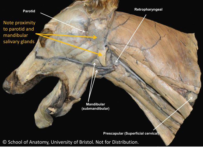
Locations of Lymph Nodes (Head)
Lymph Nodes of Head:
3 Lymphocentres:
Parotid
Mandibular (submandibular nodes palpable in canine)
Retropharyngeal
Parotid Lymph Node:
Receives lymph from dorsal structures (skin, bone, orbit and some muscles of mastication)
Mandibular Lymph Node (Palpate Submandibular Lymph Node in Dogs)
Receives lymph from muzzle, salivary glands, tongue, intermandibular space and muscles of mastication
Retropharyngeal Lymph Node
Receives lymph from deep structures: Pharynx, Larynx and other nodes
Locations of Lymph Nodes (Neck)
Prescapular centre (Superfical cervical) palpable in canine
Cranial to shounder joint, drains superficial neck, upper trunk and proximal forelimb
Deep cervical centre
drains chains of nodes in neck
cranial, middle, caudal groups along length of trachea
drain deep and ventral neck structures
Clinical Significance of Lymph
Lymph nodes act as filters for antigens and release competent immune cells (T & B cells)
In clinical exam:
Indicates regional problems (e.g. infection or neoplasia) examples: Caseous lymphadenitis in ruminants, strangles in equine (can affect guttural pouch)
Indicates Systemic problems (e.g. neoplasia, spread of infection)
Important in disease surveilance
How does Lymph Drain?
Via Tracheal Ducts
Tracheal ducts are lympathic vessels, one on each side of the trachea
Arise in retropharyngeal lymph node
End in lymphatic duct (Right side) or Thoracic duct (Left side), or may drain into external jugulars at thoracic inlet
Facial Muscles
Orbicularis oculi: Circular/round series of muscle fibers that help close the eye tightly
Platysma: Tensions underneath skin of neck
Buccinator: Covers over cheek and keeps food in the mouth.
Equine Facial muscles are more important for specific functions like quidding (keeping food in mouth), and Nostril flaring.
IMPLICATION: care must be taken when putting on headcollars due to superficial location of facial nerve.
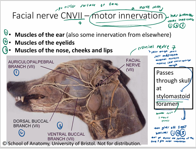
What is the Facial Nerve (CNVII) Responsible for?
Passes through stylomastoid foramen of skull
MOTOR INNERVATION to
Muscles of the ear
Muscles of the eyelids
Muscles of nose, cheek, lips
Branches of Facial Nerve
Auriculopalpebral Branch
→ gives motor innervation to eyelids, ear and forehead
→ damage results in drooping of ear, narrowing of palpebral fissure (space between upper and lower eyelids) and inability to close eye
Dorsal & Ventral Buccal Branches
→ gives motor innervation to lips and cheeks
→ damage results in paralysis of the lip and cheek muscles, allowing spillage of food during mastication and possible deformation of the muzzle
Caudal auricular and coli branches of facial nerve
→ caudal auricular branch innervates caudal auricular muscles move pinna (ear) caudally
→ coli branch innervates caudal platysma and helps neck muscle movement (cutaneous reflex)
ALSO INNERVATES CAUDAL BELLY OF DIGASTRICUS!!!!
Digastricus is a mixed muscle, rostral belly innervated by trigeminal nerve and caudal belly innervated by facial nerve